
Competencies
- AN68.1 Describe and identify multipolar and unipolar neuron, ganglia, peripheral nerve.
- AN68.2 Describe the structure–function correlation of neuron.
- AN68.3 Describe the ultrastructure of nervous tissue.
Introduction
- Nervous tissue is a specialized tissue characterized by excitability (irritability) and conductivity, enabling it to receive, process, and transmit information throughout the body.
- Anatomically, the nervous system is divided into the Central Nervous System (CNS) and Peripheral Nervous System (PNS).
- The CNS consists of the brain and spinal cord, which serve as the primary centers for integration and coordination.
- The PNS includes cranial nerves, spinal nerves, peripheral nerves, and ganglia.
- A ganglion is a collection of neuronal cell bodies located outside the CNS.
- Functionally, the nervous system is classified into the somatic and autonomic nervous systems.
- The somatic nervous system controls voluntary activities, whereas the autonomic nervous system (ANS) regulates involuntary functions.
- The ANS is subdivided into the sympathetic and parasympathetic divisions.
- The ANS controls smooth muscle, cardiac muscle, and glandular secretions.
Constituents of Nervous Tissue
- Nervous tissue is composed of two main cell types: neurons and neuroglial cells (neuroglia).
- Neurons are the structural and functional units of the nervous system. They consist of a cell body, dendrites, and an axon, and are responsible for receiving and transmitting nerve impulses.
- Neuroglial cells provide support, protection, and maintenance for neurons and do not primarily conduct nerve impulses.
- In the CNS, neuroglia include astrocytes, oligodendrocytes, microglia, and ependymal cells.
- In the PNS, neuroglia include Schwann cells and satellite cells.
- Astrocytes contribute to the maintenance of the blood-brain barrier.
Neurons
- Neurons are the structural and functional units of the nervous system.
- They receive, integrate, and transmit information to other neurons or effector cells.
Structure of Neuron
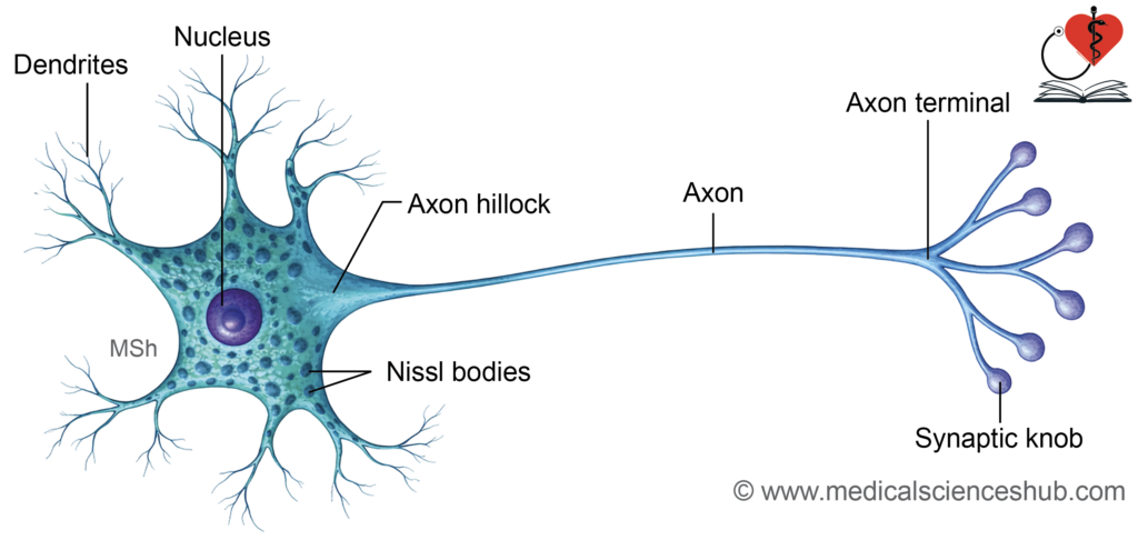
- Each neuron consists of a cell body (soma), dendrites, an axon, and synaptic terminals.
Cell Body or Soma
- The soma contains a large, vesicular nucleus with a prominent nucleolus.
- Most neurons have a centrally placed nucleus, while some (e.g., in sympathetic ganglia) show an eccentric nucleus.
- In females, a Barr body may be seen beneath the nuclear membrane.
- Mature neurons are amitotic and do not divide.
- The cytoplasm contains Nissl bodies, mitochondria, rough endoplasmic reticulum, and Golgi apparatus.
- Nissl granules are basophilic structures involved in protein synthesis.
- They are present in the soma and dendrites but are absent in the axon and axon hillock.
Neurites
Neurites are processes arising from the soma and include dendrites and the axon.
Dendrites
- Dendrites are multiple, short, tapering processes that receive incoming signals.
- Their branching forms a dendritic tree.
- They contain most organelles, including Nissl bodies, but lack a well-defined Golgi apparatus.
- Some neurons show dendritic spines, which increase surface area for synaptic input.
Axon
- The axon is a single, long process with a uniform diameter that conducts impulses away from the soma.
- Its cytoplasm is called axoplasm, and its membrane is the axolemma.
- The axon hillock is the origin of the axon and lacks Nissl bodies.
- The initial segment (preaxon) extends from the hillock to the beginning of myelination.
- Axons may give collateral branches, and terminal branches are called telodendrites.
- Terminal enlargements, known as synaptic boutons, form junctions with target cells.
- In skeletal muscle, the terminal forms a motor end plate.
Axonal transport:
- Axonal transport is the system of transport of substances within the axon.
- Axonal transport moves materials within the axon.
- Anterograde transport carries substances away from the soma.
- Retrograde transport carries substances toward the soma.
- For example, neurotransmitters such as dopamine are transported along axons, and substances taken up at terminals can return to the cell body.
CLINICAL NEUROANATOMY
- The rabies virus is transmitted through the saliva of infected animals during a bite. After entry, it reaches nerve endings and enters axons at the site of injury. It travels toward the central nervous system by retrograde axonal transport.
- The herpes zoster virus remains dormant in sensory ganglia. On reactivation, it moves from neuronal cell bodies to the skin by anterograde axonal transport, producing characteristic lesions.
Classification of Neurons
Neurons are classified according to the number of their processes, their size, and their functional properties.
Classification Based on Number of Processes
Neurons can be structurally categorized according to the number of processes arising from the cell body as unipolar, pseudounipolar, bipolar, and multipolar. In current understanding, unipolar and pseudounipolar types are often grouped together.
- Unipolar neurons possess a single process, typically resembling a dendrite. They are seen in the mesencephalic nucleus of the trigeminal nerve and in certain stages of embryonic development.
- Pseudounipolar neurons have one process that bifurcates into peripheral and central branches. These are characteristic of neurons in dorsal root ganglia and other sensory ganglia.
- Bipolar neurons exhibit two processes—one axon and one dendrite—arising from opposite poles of the cell body. They are found in the retina, olfactory epithelium, and vestibular and spiral ganglia.
- Multipolar neurons contain one axon and multiple dendrites. Common examples include pyramidal, Purkinje, and stellate neurons.
Classification Based on length of axon
Neurons are also categorized according to the length of their axon into Golgi type I and Golgi type II.
Golgi type I neurons
These neurons possess a single, long axon that may extend over considerable distances, sometimes exceeding a meter. Typical examples include motor neurons of the spinal cord, pyramidal neurons of the cerebrum, and Purkinje cells of the cerebellum.
Golgi type II neurons
These neurons have short axons, which may be minimal or even absent. They are mainly involved in local circuit functions. Examples include stellate cells and granule cells of the cerebellar cortex.
Physiological Classification of Neurons
According to function, neurons are grouped into sensory, interneurons, and motor types.
- Sensory (afferent) neurons
These neurons detect stimuli such as pain, touch, and temperature, and convey this information to the central nervous system. - Interneurons
They form connections between sensory and motor neurons. Located within the central nervous system, they are responsible for processing and integrating incoming signals. They constitute the majority of neurons in the body. - Motor (efferent) neurons
These transmit impulses from the central nervous system to muscles, glands, and viscera. They are further divided into:
A. Somatic motor neurons
They innervate skeletal muscles.
- Upper motor neurons: Situated in the motor cortex; their axons project to cranial nerve nuclei or anterior horn cells of the spinal cord.
- Lower motor neurons: Located in cranial nerve motor nuclei and anterior horn cells; they directly supply skeletal muscles.
B. Visceral (autonomic) motor neurons
They control smooth muscle, cardiac muscle, and glands.
- Preganglionic neurons: Found in autonomic nuclei of cranial nerves (e.g., Edinger–Westphal, salivatory, lacrimal, and dorsal motor nucleus of vagus) and in the lateral horn of the spinal cord.
- Postganglionic neurons: Located in autonomic ganglia and relay impulses to target organs.
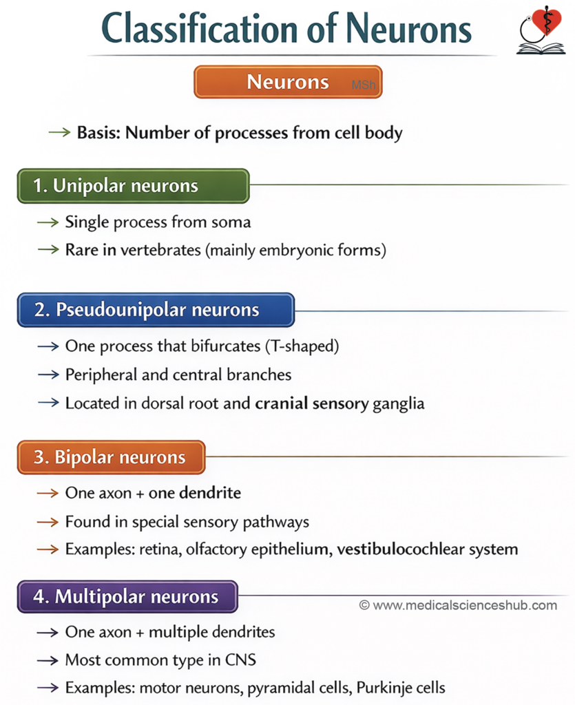
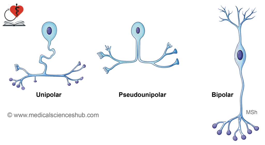
Table 9.1: Differences between axon and dendrites
| Feature | Axon | Dendrites |
|---|---|---|
| Number | Usually a single process arises from the neuron. | Multiple processes arise from the neuron. |
| Length and Thickness | Typically long and slender. | Generally short and relatively thicker near their origin. |
| Direction of Impulse Transmission | Conducts nerve impulses away from the cell body. | Receives signals and conducts them toward the cell body. |
| Branching Pattern | Branches infrequently and terminates as terminal arborizations or axon terminals. | Extensively branches to form a dendritic tree. |
| Diameter | Maintains a relatively uniform diameter throughout its length. | Gradually tapers toward the distal end. |
| Nissl Bodies | Nissl bodies are absent. | Nissl bodies are present in the proximal cytoplasm. |
| Primary Function | Responsible for transmitting output signals to other neurons, muscles, or glands. | Primarily involved in receiving and integrating incoming signals. |
Neuroglia
Neuroglial cells provide structural and functional support to neurons. They contribute to insulation and play a key role in forming the blood–brain barrier. Unlike neurons, they are non-excitable and retain the ability to divide.
They are broadly classified into:
- Central neuroglia (within the central nervous system)
- Astrocytes
- Oligodendrocytes
- Microglia
- Ependymal cells
- Peripheral neuroglia (within the peripheral nervous system)
- Schwann cells
- Satellite cells
Central Neuroglia
- Neuroglia are the supporting cells of the central nervous system.
- The main types include:
- Astrocytes
- Oligodendrocytes
- Microglia
- Ependymal cells
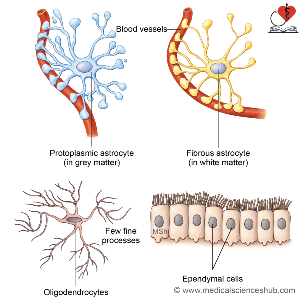
Astrocytes
- Astrocytes are large, stellate cells with numerous branching processes.
Classification
- Fibrous astrocytes
- Located mainly in white matter
- Possess fewer, long, slender processes
- Terminal ends of processes expand to form vascular end feet
- Protoplasmic astrocytes
- Predominantly found in grey matter
- Have abundant cytoplasm with many short, thick processes
- Contribute significantly to the formation of the blood–brain barrier
Histological Identification
- Both types express glial fibrillary acidic protein (GFAP)
- They can be demonstrated using immunohistochemical staining with anti-GFAP antibodies
Functions of Astrocytes
- Provide structural and metabolic support to neurons and capillaries
- Regulate the extracellular environment by controlling ions, metabolites, and neurotransmitters
- Serve as an energy reserve by storing glycogen
- Participate in blood–brain barrier formation by surrounding capillaries with their end feet
- Form the glia limitans, a thin layer beneath the pia mater and ependyma, acting as a boundary between the CNS tissue and cerebrospinal fluid
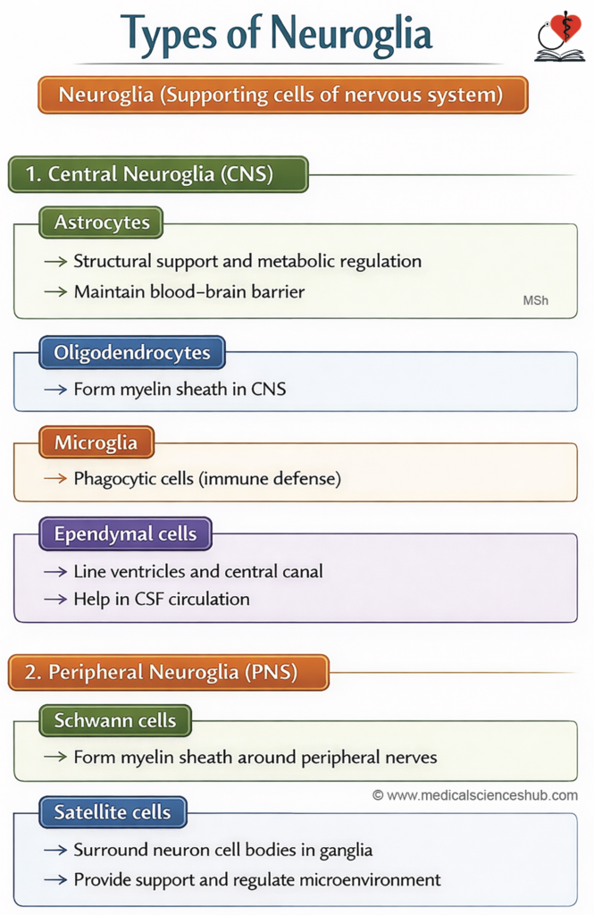
Table 9.2: Glial cells
| Cell Type | Origin | Location | Functions |
|---|---|---|---|
| Central Neuroglia | |||
| Fibrous astrocytes | Neural tube | White matter of CNS | Provide structural support and help maintain the blood–brain barrier |
| Protoplasmic astrocytes | Neural tube | Grey matter of CNS | Participate in repair processes and form glial scar after injury |
| Oligodendrocytes | Neural tube | Predominantly in white matter of CNS | Produce myelin sheath around CNS axons |
| Microglia | Mesoderm (bone marrow lineage) | Throughout CNS | Act as phagocytic cells for immune defense |
| Ependymal cells | Neural tube | Line ventricles of brain and central canal of spinal cord; form choroid plexus | Contribute to cerebrospinal fluid production and form the brain–CSF interface |
| Peripheral Neuroglia | |||
| Schwann cells | Neural crest cells | Peripheral nerves | Form myelin sheath around peripheral axons; support regeneration of damaged nerves |
| Satellite cells | Neural crest cells | Sensory and autonomic ganglia | Provide support, insulation, and regulate the microenvironment of neuronal cell bodies |
Oligodendrocytes
- Oligodendrocytes are small, round neuroglial cells with only a few cytoplasmic extensions (“oligo” means few).
- A single oligodendrocyte can form myelin around segments of several nearby axons.
- Node of Ranvier: These are small interruptions between successive myelin segments along an axon.
- Function: They are responsible for forming and maintaining the myelin sheath in the central nervous system (brain and spinal cord).
Microglia
- Microglia are the smallest glial cells in the central nervous system, comprising roughly 5% of the total glial population. They act as resident phagocytes and are part of the mononuclear phagocyte system.
- Origin: They arise from granulocyte–monocyte progenitor cells in the bone marrow and reach the CNS via the bloodstream.
- Structure: These cells are small with elongated nuclei and possess short, irregular, branched processes. Their cytoplasm is rich in lysosomes, inclusions, and vesicles.
Functions:
- Microglia act as phagocytes, clearing:
- Dead and damaged cells
- Cellular debris
- Infectious agents such as bacteria
- Abnormal or neoplastic cells
- They are actively involved in inflammatory responses and contribute to various degenerative disorders of the CNS.
Ependymal Cells
Ependymal cells form a lining over the fluid-filled spaces of the brain and spinal cord. They are arranged in a single layer and appear cuboidal to columnar in shape.
They develop from the neural tube during early embryonic life.
Types of Ependymal Cells:
- Ependymocytes: These cells line the ventricles of the brain and the central canal of the spinal cord.
- Choroid epithelial cells: These cells are present in the choroid plexus and are responsible for producing cerebrospinal fluid (CSF).
- Tanycytes: These are specialized cells located in the floor of the third ventricle. They help monitor and respond to changes in metabolite levels, especially glucose.
Ependymal cells have tight junctions near their apical surface. Their apical surface shows cilia and microvilli, while the basal surface has many infoldings.
Functions:
- Choroid epithelial cells produce CSF in the choroid plexus.
- Ependymocytes contribute to the barrier between CSF and brain tissue.
- Tanycytes help regulate and monitor metabolite levels in the brain.
Peripheral Neuroglia
- These are present in the PNS. These are of two types:
- Schwann cells
- Satellite cells
Schwann Cells (Neurolemmocytes)
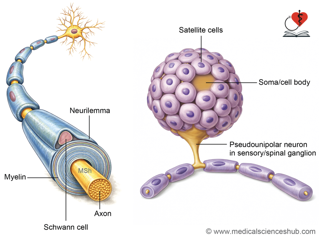
- Schwann cells are found only in the peripheral nervous system (PNS). They are flattened cells with a flattened nucleus and a relatively large amount of cytoplasm. These cells develop from neural crest cells.
- Schwann cells form the myelin sheath around axons in the PNS. Along a myelinated nerve fiber, gaps are present between adjacent Schwann cells. These gaps are called nodes of Ranvier, where the axon is not covered by myelin.
- Functions:
- Schwann cells produce myelin, which helps in rapid transmission of nerve impulses by saltatory conduction.
- They also surround and support non-myelinated nerve fibers.
- Schwann cells play an important role in the repair and regeneration of damaged axons in the PNS.
Satellite Cells
- Satellite cells are found in peripheral ganglia, including both sensory and autonomic ganglia. They are small, cuboidal cells arranged in a single layer around the cell body of neurons.
- In sensory ganglia, satellite cells completely surround the neuron and form a capsule. This capsule allows the nerve fibers to pass through. In autonomic ganglia, the covering formed by satellite cells is incomplete because synapses are present in these ganglia.
Functions:
- They protect and support the neurons in peripheral ganglia.
- They help maintain a stable environment around neurons and assist in exchange of nutrients and waste.
- They provide insulation to the neuronal cell body.
Synapse
- A nerve impulse is an electrical signal that travels along a neuron. This signal is passed from one neuron to another at a specialized junction called a synapse.
- A synapse is a specific site of contact between two or more neurons. It can also occur between a neuron and a muscle cell or a gland cell.
- At a synapse, the impulse is transmitted across a small gap from one cell to another. Therefore, conduction occurs by contact (contiguity) and not by direct continuity.
Classification of Synapse
Structural Classification of Synapse
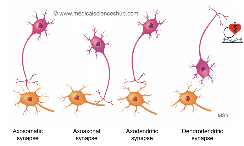
Synapses can be classified based on the parts of neurons that come in contact with each other:
- Axodendritic synapse: The axon of one neuron connects with the dendrite of another neuron.
- Axosomatic synapse: The axon of one neuron connects with the cell body (soma) of another neuron.
- Axoaxonic synapse: The connection occurs between the axons of two neurons.
- Dendroaxonic synapse: The dendrite of one neuron connects with the axon of another neuron.
- Dendrodendritic synapse: The connection occurs between the dendrites of two neurons.
- Somatosomatic synapse: The cell bodies of two neurons are in contact.
- Somatodendritic synapse: The cell body of one neuron connects with the dendrite of another neuron.
Chemical and Electric Synapses
Synapses can also be classified based on how nerve impulses are transmitted:
- Chemical synapse: In this type, the nerve impulse is transmitted by releasing chemical substances called neurotransmitters from one neuron to another.
- Electrical synapse: In this type, the electrical signal passes directly from one cell to another through gap junctions without the use of neurotransmitters.
Structure of Chemical Synapse
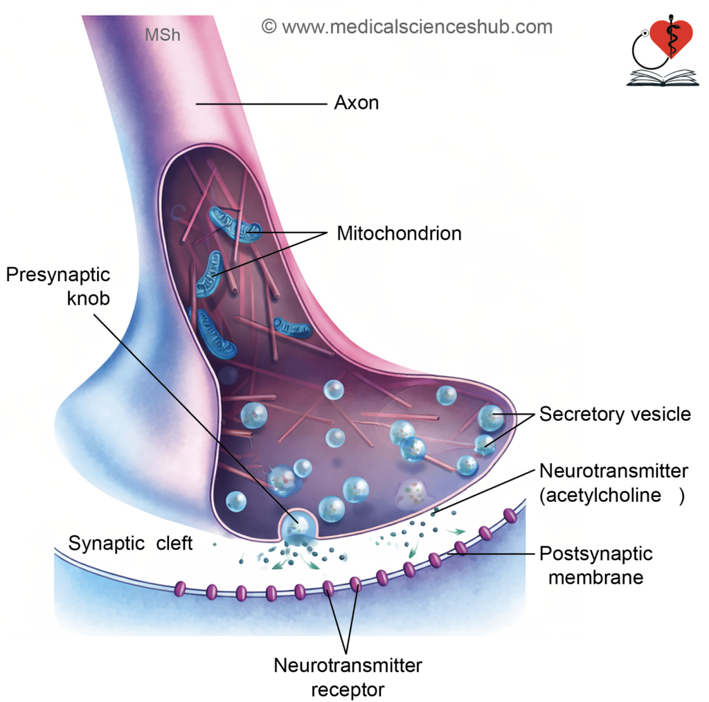
Chemical synapses are the most common type of synapse. In these synapses, nerve impulses are transmitted by the release of chemical substances called neurotransmitters.
Parts of a Chemical Synapse
A chemical synapse has three main parts:
- Presynaptic knob
This is the terminal end of the sending neuron. It contains mitochondria and many synaptic vesicles. These vesicles store neurotransmitters such as acetylcholine, adrenaline, noradrenaline, and dopamine. The vesicles are formed from the Golgi apparatus or smooth endoplasmic reticulum, and some are recycled by endocytosis. When an impulse arrives, these vesicles release neurotransmitters by exocytosis. - Synaptic cleft
This is a small gap (about 20–30 nm wide) between the presynaptic and postsynaptic cells. Neurotransmitters diffuse across this gap. - Postsynaptic membrane
This is the receiving surface of the next cell. It is thickened and contains specific receptor proteins. These receptors bind to neurotransmitters and help continue the transmission of the nerve impulse.
Properties of Synapses
- Multiple connections of a neuron
A single neuron can form synapses with one or many other neurons (up to about 5500). For example, bipolar cells in the retina connect with a single ganglion cell, which helps maintain precise vision. - Feed-forward inhibition
In this type of circuit, one excitatory neuron sends signals to two neurons. At the same time, it also sends a branch to an inhibitory interneuron. This interneuron suppresses one of the target neurons. As a result, one pathway is activated while the other is inhibited. This mechanism helps in coordinated actions, such as activating an agonist muscle and relaxing its antagonist. - Feedback inhibition
This mechanism prevents excessive activity in neurons. A good example is the Renshaw cell in the anterior horn of the spinal cord. It receives a branch from a motor neuron and then inhibits the same neuron. This control helps avoid overactivity of muscles, ensures smooth and coordinated movements, and regulates reflexes like the knee jerk. - Synaptic delay
Synaptic delay is the time taken for an impulse to pass from one neuron to another. It includes the release of neurotransmitter, its movement across the synaptic cleft, and its binding to receptors. The average delay is about 0.5 milliseconds. This delay allows the postsynaptic neuron to process signals, including summation or inhibition of inputs.
CLINICAL NEUROANATOMY
In tetanus, the bacterium Clostridium tetani releases a toxin at the site of a wound. This toxin blocks the function of Renshaw cells in the spinal cord, which normally inhibit motor neurons. When this inhibition is lost, motor neurons become overactive. As a result, muscles remain continuously contracted, leading to stiffness and painful spasms.
Peripheral Nerve
- The PNS connects body organs with the central nervous system (CNS).
- It enables transmission of sensory input and motor output.
Components
- Somatic nerves control voluntary activities.
- Autonomic nerves regulate involuntary functions and include sympathetic and parasympathetic divisions.
Constituents
- The PNS consists of:
- 12 pairs of cranial nerves
- 31 pairs of spinal nerves
- Autonomic (visceral) nerves
Types of Nerve Fibers
- Somatic afferent fibers carry sensory signals from skin, muscles, bones, and joints to the CNS.
- Somatic efferent fibers transmit motor impulses to skeletal muscles.
- Visceral afferent fibers convey sensory information from organs and blood vessels.
- Visceral efferent fibers supply glands, smooth muscle, and cardiac muscle.
- In the CNS, bundles of nerve fibers are termed tracts.
Structures of Peripheral Nerve
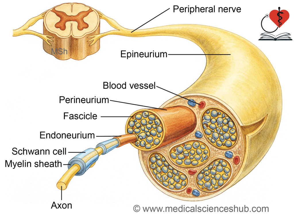
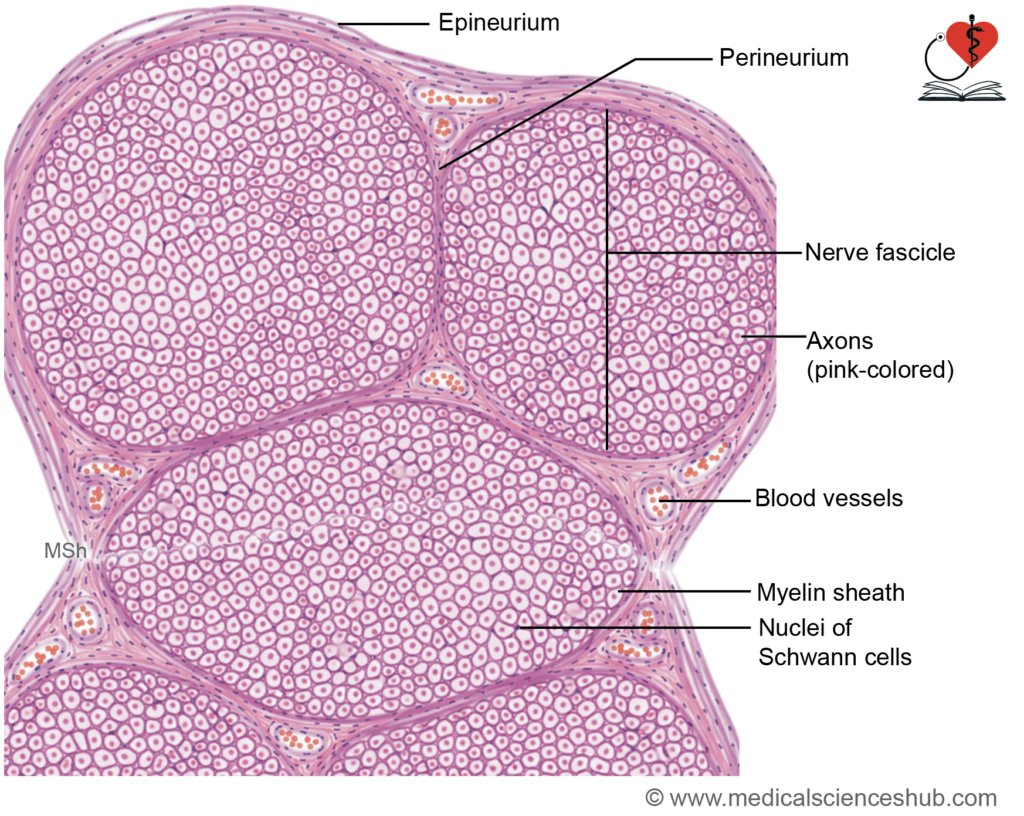
- A peripheral nerve is a bundle of multiple nerve fibers enclosed by connective tissue.
- The term nerve fiber refers to a neuronal process, not a connective tissue fiber.
Types of Nerve Fibers
- Motor (efferent) fibers are axons that transmit impulses from the CNS to muscles or glands.
- Their cell bodies are located in the grey matter of the spinal cord and brainstem.
- Sensory (afferent) fibers carry impulses toward the CNS from peripheral structures.
- Their cell bodies lie in sensory ganglia.
Ganglion
- A ganglion is a cluster of nerve cell bodies situated outside the CNS.
- Examples include the dorsal root ganglion and autonomic ganglia.
Basic Structure of Peripheral Nerve
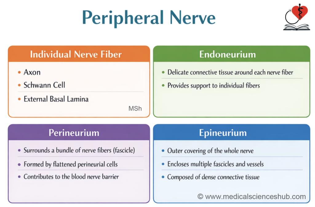
- A nerve fiber, along with Schwann cells and a basal lamina, forms the structural unit of a peripheral nerve.
Connective Tissue Coverings
- Peripheral nerves have three layers:
1. Endoneurium
- Each nerve fiber is enclosed by delicate loose connective tissue.
- It contains collagen fibers and a few supporting cells.
- Most nuclei seen in sections belong to Schwann cells.
2. Perineurium
- Surrounds a bundle of fibers (fascicle).
- Formed by flattened cells arranged in layers with tight junctions.
- Creates a protective blood–nerve barrier.
3. Epineurium
- Encloses multiple fascicles forming the whole nerve.
- Composed of dense connective tissue with blood vessels.
- In H&E staining, myelin appears clear, while osmium tetroxide stains it black.
Myelin Sheath
- Myelin is a specialized insulating covering that surrounds the axons of certain nerve fibers.
- It is formed by oligodendrocytes in the central nervous system (CNS).
- In the peripheral nervous system (PNS), myelin is produced by Schwann cells.
- The primary function of myelin is to insulate and protect axons.
- It significantly enhances the speed of nerve impulse conduction along the axon.
Formation of Myelin Sheath
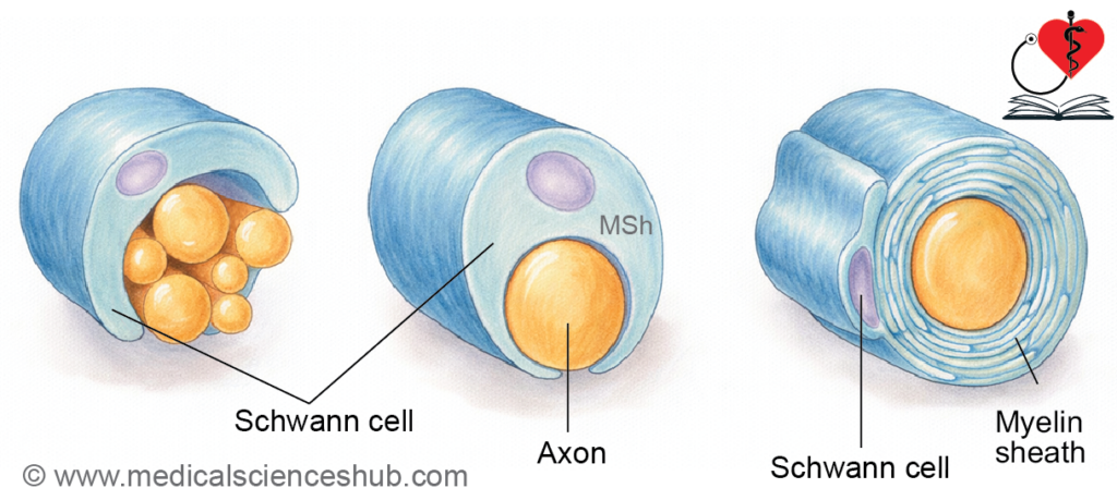
- During myelination, the axon initially lies within a groove on a Schwann cell.
- The Schwann cell membrane differentiates into three regions:
- Abaxonal membrane, which faces the external environment.
- Adaxonal (periaxonal) membrane, which is in direct contact with the axon.
- Mesaxon, a double-layered fold connecting the two membranes.
- The mesaxon elongates and wraps concentrically around the axon in a spiral manner.
- As wrapping progresses, most of the cytoplasm is displaced, forming a compact lipid-rich myelin sheath.
- A thin outer layer of Schwann cell cytoplasm remains, known as the neurilemma (neurilemmal sheath).
Composition of Myelin
- Myelin is mainly composed of lipids (about 80%), along with proteins and water.
- Schmidt–Lanterman clefts are small cytoplasmic pockets retained within the layers of the myelin sheath.
- These clefts represent areas where cytoplasm is not completely expelled during myelin formation.
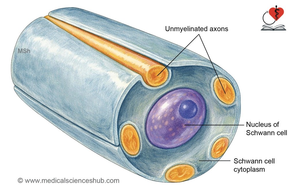
- Non-Myelinated Fibers: In the PNS, non-myelinated axons are also enclosed by Schwann cells. A single Schwann cell can envelop multiple non-myelinated axons, without forming a true myelin sheath.
Functions of Myelin Sheath
- The myelin sheath provides protection and structural support to axons.
- It acts as an insulator, preventing current loss.
- It enhances impulse transmission via saltatory conduction.
- The neurilemma is essential for nerve regeneration.
TABLE 9.3: The difference between myelinated and unmyelinated fibers
| Feature | Myelinated Fibers | Unmyelinated Fibers |
|---|---|---|
| Myelin sheath | It is present, formed by Schwann cells (PNS) or oligodendrocytes (CNS). | It is absent; axons are only partially enclosed by Schwann cells. |
| Nodes of Ranvier | They are present as gaps between myelin segments. | They are absent, forming a continuous membrane. |
| Conduction speed | Impulse conduction is fast due to saltatory conduction. | Impulse conduction is slow and continuous. |
| Diameter | Fibers have a larger diameter (1–20 μm). | Fibers are smaller (0.2–1.5 μm). |
| Energy efficiency | They are more efficient, as depolarization occurs only at nodes. | They are less efficient, as the entire membrane depolarizes. |
| Location | Found in motor and sensory pathways. | Found in autonomic fibers and some pain/temperature pathways. |
| Impulse propagation | Impulses jump between nodes, increasing speed. | Impulses travel along the entire length, reducing speed. |
CLINICAL NEUROANATOMY
Enuresis (bed-wetting).
- Enuresis is the involuntary passage of urine, commonly seen in early childhood.
- It is typically observed during the first 3–6 years of life.
- This occurs until full myelination of corticospinal tract fibers that regulate bladder control is achieved.
- Embryological Basis: Myelination begins before birth and continues for several years after birth. Therefore, enuresis is considered physiological (normal) in infants and young children.
Classification of Nerve Fibers
- Nerve fibers are categorized according to their diameter and degree of myelination.
Group A Fibers
- These are highly myelinated fibers.
- They have a large diameter and conduct impulses rapidly.
Group B Fibers
- These fibers are thinly myelinated.
- Their diameter is up to about 3 μm.
- Conduction is slower than Group A fibers.
Group C Fibers
- These are non-myelinated fibers.
- Their diameter ranges from approximately 0.2–1.5 μm.
- They conduct impulses slowly.
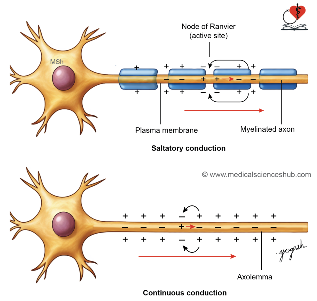
Table 9.4: Classification of peripheral nerve fibres
| Group & Type | Example / Function | Diameter | Myelin | Velocity (m/s) |
|---|---|---|---|---|
| Group A (Afferent) | ||||
| Type Ia | Muscle spindle (annulospiral endings) | 20 µm | Present | 80–120 |
| Type Ib | Golgi tendon organ | 20 µm | Present | 80–120 |
| Type II | Muscle spindle (flower-spray endings) | 12 µm | Present | 33–75 |
| Type III | Cutaneous receptors (touch, pressure) | 5 µm | Thin | ~30 |
| Type IV | Free nerve endings (pain, temperature) | — | Absent | — |
| Group A (Efferent) | ||||
| Alpha (α) | Extrafusal muscle fibres | 20 µm | Present | ~120 |
| Beta (β) | Extrafusal + spindle fibres | 12 µm | Present | ~70 |
| Gamma (γ) | Intrafusal muscle fibres | 8 µm | Present | ~24 |
| Group B | Preganglionic autonomic fibres | 5 µm | Present | ~15 |
| Group C | Postganglionic autonomic fibres | 1.5 µm | Absent | ~2 |
Nerve Injuries
- The rate of nerve regeneration is about 3.5–4.5 mm/day, and adequate nutrition supports faster recovery.
Classification (Based on Severity)
- Neurotmesis: Both axon and myelin sheath are completely damaged.
- Axonotmesis: The axon is damaged, but the myelin sheath remains intact.
- Neuropraxia: Both axon and myelin sheath are preserved, with temporary functional loss.
Regeneration
- Regeneration occurs in peripheral nerves in neuropraxia and axonotmesis.
- No regeneration occurs in neurotmesis due to complete structural disruption.
Nerve Degeneration and Regeneration
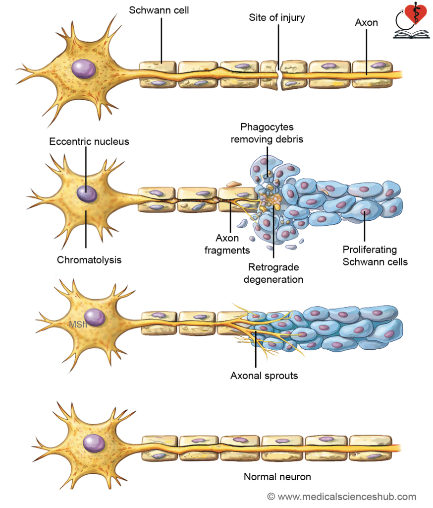
- Injury to an axon with its myelin sheath leads to degeneration followed by regeneration.
Degeneration of Nerve Fiber
- It refers to structural changes occurring after nerve injury.
- Changes involve both the nerve fiber and the cell body.
Changes in Nerve Fiber
- Wallerian degeneration: The distal segment degenerates, with breakdown of axon and myelin.
- The proximal segment degenerates up to the nearest node of Ranvier.
Changes in Cell Body (Retrograde Changes)
- These occur due to loss of neural activity.
- Chromatolysis: Nissl bodies disintegrate.
- The cell body swells, and the nucleus shifts eccentrically.
- These changes are more marked when the injury is closer to the cell body.
Regeneration of Nerve Fiber
- Regeneration is slow and usually begins about 2 weeks after injury.
Steps in Regeneration
- Debris removal: Macrophages clear degenerated material by phagocytosis.
- Schwann cell proliferation: Schwann cells multiply and form a guiding tube (bands of Büngner).
- Axonal sprouting: Multiple sprouts arise from the proximal end.
- Axonal elongation: One sprout grows along the Schwann cell pathway to reach the target.
- Remyelination: Schwann cells reform the myelin sheath, restoring function.
Factors Necessary for Satisfactory Regeneration
- The endoneurial sheath must remain intact to guide regenerating axons.
- The gap between proximal and distal stumps should be minimal (only a few millimeters).
- There should be no infection at the injury site, as it impairs healing.
- Adequate levels of nerve growth factors are essential to promote axonal growth.
- Proper physiotherapy is required to maintain muscle function and support recovery.
CLINICAL NEUROANATOMY
Neuroma:
- A neuroma is a benign growth arising after nerve injury.
- It develops when there is a large gap between the cut ends of a nerve.
- The gap becomes filled with fibrous tissue containing regenerating axonal sprouts.
- These disorganized nerve fibers form a nodular mass.
- Neuromas are typically painful due to abnormal nerve regeneration.
Ganglia
- A ganglion is a collection of neuronal cell bodies located outside the central nervous system.
Types of Ganglia
- Sensory ganglia: Associated with afferent neurons that carry sensory information.
- Autonomic ganglia: Associated with the autonomic nervous system and involved in visceral functions.
Sensory Ganglia
Sensory ganglia are located just outside the CNS and transmit impulses toward the CNS.
- Examples:
- Dorsal root ganglia of spinal nerves.
- Sensory ganglia of cranial nerves: trigeminal, facial, vestibulocochlear, glossopharyngeal, and vagus.
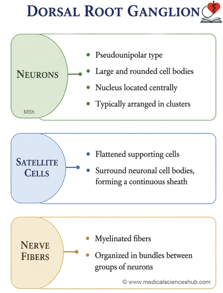
Structure of Sensory Ganglia
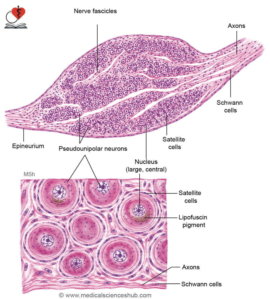
- They contain mainly pseudounipolar neurons (except vestibulocochlear ganglia, which are bipolar).
- Each neuron has:
- A peripheral process carrying input from receptors.
- A central process transmitting impulses to the CNS.
- Neuronal cell bodies are large, arranged in groups, often at the periphery.
- Nuclei are large, vesicular, and centrally placed.
- Groups of neurons are separated by bundles of myelinated nerve fibers.
Supporting Cells
- Each neuron is surrounded by satellite (capsular) cells, forming a complete capsule.
- These cells are flattened (low cuboidal).
- The ganglion is enclosed by a thin connective tissue capsule.
Autonomic Ganglia
- Autonomic ganglia supply smooth muscle and glands.
- The autonomic pathway involves two neurons:
- Preganglionic neuron: Located in the CNS; its axon reaches the ganglion.
- Postganglionic neuron: Located in the ganglion; its axon supplies target organs.
Examples
- Sympathetic: Sympathetic trunk, paravertebral ganglia (celiac, superior and inferior mesenteric, aorticorenal), and adrenal medulla.
- Parasympathetic: Ciliary, submandibular, otic, pterygopalatine, and terminal ganglia near organs.
Structure of Autonomic Ganglia
- They are smaller than sensory ganglia.
- Contain multipolar postganglionic neurons scattered throughout.
- Neurons have large, pale, eccentrically placed nuclei with prominent nucleoli.
- Cells are separated by bundles of mainly nonmyelinated fibers.
Supporting Cells
- Satellite cells are few and do not form a complete capsule due to multiple neuronal processes and synapses.
- The ganglion is enclosed by a connective tissue capsule.
- Nissl granules are prominent, and silver stains highlight neuronal processes.
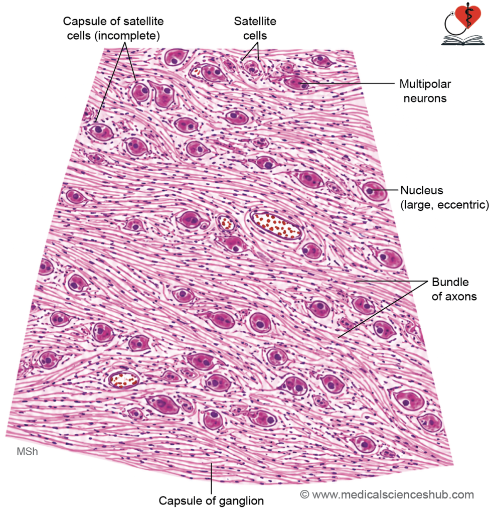
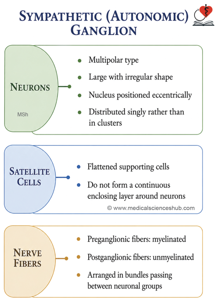
Table 9.5: Differences between sensory and autonomic ganglia
| Feature | Sensory (Dorsal Root) Ganglion | Autonomic (Sympathetic) Ganglion |
|---|---|---|
| Neuron type | Contains pseudounipolar neurons. | Contains multipolar neurons. |
| Cell body shape | Neurons are large and spherical. | Neurons are small and irregular. |
| Function | Composed of sensory neurons. | Contains postganglionic autonomic neurons. |
| Nucleus | Centrally placed, large and vesicular. | Eccentrically placed, large and vesicular. |
| Satellite cells | Form a complete sheath around neurons. | Incomplete covering by few satellite cells. |
| Arrangement | Neurons are arranged in clusters. | Neurons are scattered. |
| Nerve fibers | Fibers form bundles separating clusters. | Fibers run between scattered neurons. |
Important Questions
- Classify the neurons.
- List the difference between axon and dendrites.
- List the types of neuroglia.
- Write a short note on myelin sheath.
- Write a short note on histology of peripheral nerve (TS).
- Draw a well-labeled diagram for section of nerve stained by osmium tetroxide.
- Write a short note on histology of sensory ganglia.
- Write a short note on histology of autonomic or sympathetic ganglion.
- List the differences between dorsal root ganglion and sympathetic ganglion.
📝 Test Your Knowledge – Practice MCQs
Attempt the chapter MCQ quiz and assess your understanding of key concepts.
Click to see: Medical Disclaimer and Copyright Notice