
Competency
- AN72.1 Identify the skin and its appendages under the microscope and correlate the structure with function.
Introduction
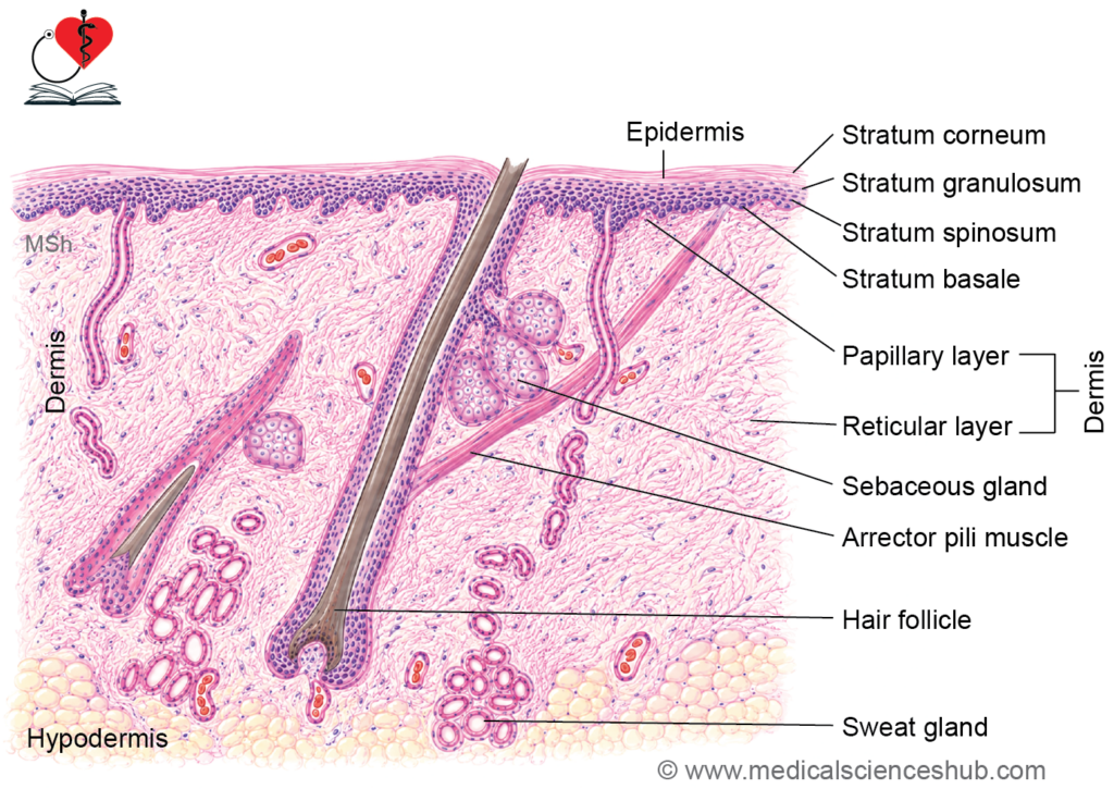
- The integumentary system consists of the skin and its associated appendages, including hair follicles, hairs, sweat glands, sebaceous glands, nails, and mammary glands.
- The skin is composed of two principal layers: the epidermis and the dermis.
- Beneath the dermis lies the hypodermis (subcutaneous tissue), which is rich in adipose tissue and helps anchor the skin to underlying structures.
- The skin forms the body’s protective outer covering and supports various accessory structures.
- It is the largest organ of the body, accounting for approximately 15–20% of total body weight.
Types of Skin
- Based on the thickness of the epidermis, skin is classified into thick skin and thin skin.
- Thick skin (glabrous skin) has a markedly thick epidermis with a prominent stratum corneum and lacks hair follicles. It is found on the palms and soles.
- Thin skin has a relatively thin epidermis and contains hair follicles and associated skin appendages.
- Thin skin covers almost the entire body surface except the palms and soles.
Structure of Skin
- Histologically, skin consists of two layers:
- Epidermis: It is a superficial layer that consists of keratinized stratified squamous epithelium.
- Dermis: It is a deeper layer that is made up of connective tissue.
Epidermis
- Epidermis consists of keratinized stratified squamous pithelium
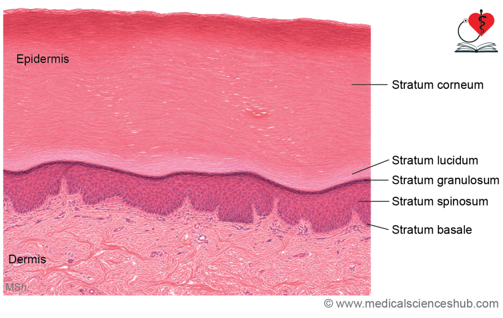
- Epidermis shows five layers of cells as follows:
- Stratum basale
- Stratum spinosum
- Stratum granulosum
- Stratum lucidum (only in thick skin)
- Stratum corneum
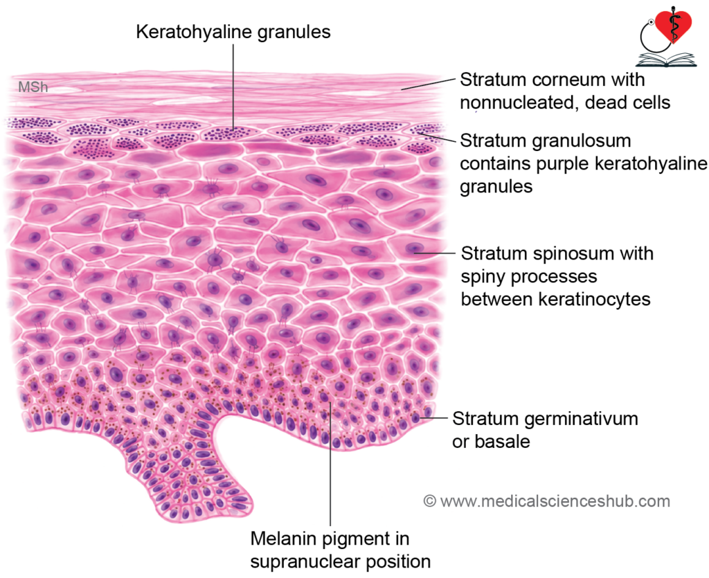
1. Stratum Basale (Stratum Germinativum)
- Stratum basale is the deepest layer of epidermis.
- Cells: It consists of a single layer of cuboidal (low columnar) cells that rest on basal lamina. It also contains melanocytes and mechanoreceptors.
- H&E staining: Cells show basophilic cytoplasm and melanin pigments.
- Functions: These cells divide mitotically to give rise to keratinocytes (cells of skin) that form superficial layers of skin. Hence, stratum basale is also called stratum germinativum
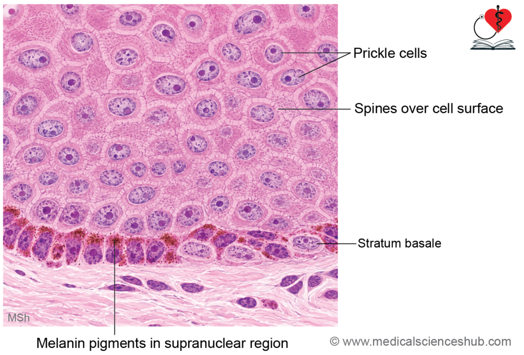
2. Stratum Spinosum (Malpighian Layer, Prickle Cell Layer)
- Above the stratum basale, several layers of thick stratum spinosum are present.
- Keratinocytes of stratum spinosum are polygonal cells that are connected with adjacent cells by desmosomes.
- Spines are artifact: During preparation of tissue for sectioning, cells often shrink. But these keratinocytes (prickle cells) remain adherent with each other at the site of desmosomes. It creates spines over the surface of these cells at the site of desmosomes. Thus, keratinocytes in this layer shows small spine-like projections on their external surface.
- Some cells of stratum spinosum may undergo mitosis; hence, stratum spinosum and stratum basale together can be considered as germinative zone.
- Node of Bizzozero: It is a site of thickened zone of desmosomes on light microscopy. [Giulio Bizzozero, Italian physician, 1846–1901].
3. Stratum Granulosum
- Stratum granulosum consists of 1–3 cells thick layer of keratinocytes.
- Cells of stratum granulosum contain granules of keratohyalin protein; hence, the name stratum granulosum.
- Keratohyaline is a precursor of filaggrin protein that on aggregation forms keratin filaments.
- H&E staining: Cells of stratum granulosum are intensely basophilic because of keratohyalin granules.
4. Stratum Lucidum (Lucid = Clear)
- Stratum lucidum is present only in thick skin.
- H&E staining: The cells of stratum lucidum are stained lightly with eosin and appears homogenous. Their cell boundaries are indistinct. The nuclei and cell organelles become disrupted and disappear toward superficial layer.
- Cells of stratum lucidum contain eleidin. It is a clear intracellular protein derived from keratohyaline and gets stained with hematoxylin. Later, eleidin gets converted to keratin in stratum corneum.
- Note: Some authors consider stratum lucidum as a part of stratum corneum.
5. Stratum Corneum
- This is the most superficial layer of epidermis.
- Cells of stratum corneum are nonnucleated, flat, scalelike (squamous) cells (dead cells) and they do not have cell organelles.
- Cells of stratum corneum are filled with keratin filaments.
- These cells are held together by extracellular lipid and carbohydrates that form a glue-like substance.
- Extracellular lipid makes skin impermeable to water.
- Thickness of stratum corneum depends on the amount of friction to which skin is exposed. Stratum corneum is very thick in palms and soles.
- Superficial cells of stratum corneum are being constantly shed off and are replaced by proliferation of stratum germinativum.
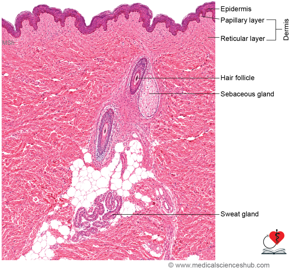
Dermis
- Dermis is made up of connective tissue containing collagen bundles, blood vessels, lymphatics, and nerve fibers.
- Dermis has two layers:
- Papillary layer
- Reticular layer.
1. Papillary Layer of Dermis
- It lies just deep to basal lamina of epidermis.
- It consists of loose connective tissue.
- It shows finger-like projections into the under surface of epidermis. These projections are called dermal papillae.
- Epidermis also protrude into dermis in the form of epidermal ridges, rete ridges, or rete pegs.
- Dermal papillae and epidermal ridges are longer in skin where mechanical stress is more. For example, skin of palm and dorsum of hand.
- Dermal papillae create superficial elevations on the surface of epidermis called dermal ridges. Pattern of dermal ridges is unique to each individual and genetically determined. Dermatoglyphics or fingerprint patterns form the basis for study of many genetic disorders.
- Transmission electron microscopy: Hemidesmosomes attach basal cells of epidermis with basal lamina.
- Immunohistochemistry: Papillary layer contains type I and type III collagen fibers.
- Blood vessels of papillary layer do not penetrate epidermis. Note: Epidermis is avascular and receives nutrition by diffusion.
- Nerves from papillary layer may terminate in the basal layer of epidermis.
2. Reticular Layer
- Reticular layer of dermis lies deep to papillary layer and it is a dense irregular connective tissue.
- Reticular layer is thicker and less cellular than papillary layer.
- It consists of thick, irregular bundles of type I collagen fibers and elastic fibers.
CLINICAL CORRELATION
• Basal cell carcinoma: It is the most common skin cancer. It arises from trichoblast or folliculosebaceous-apocrine germ.Neet It begins as painless skin elevations. It rarely metastasizes (spreads to distinct locations). Basal cell carcinoma occurs in hairy skin (mostly on face).
• Squamous cell carcinoma (epidermoid carcinoma): It arises from squamous cells of skin, part of respiratory tract, and gut. Squamous cell carcinoma of skin usually arises in sun-exposed parts (UV light exposed) in older people and mostly on face, ears, back of hands, scalp, and lips.
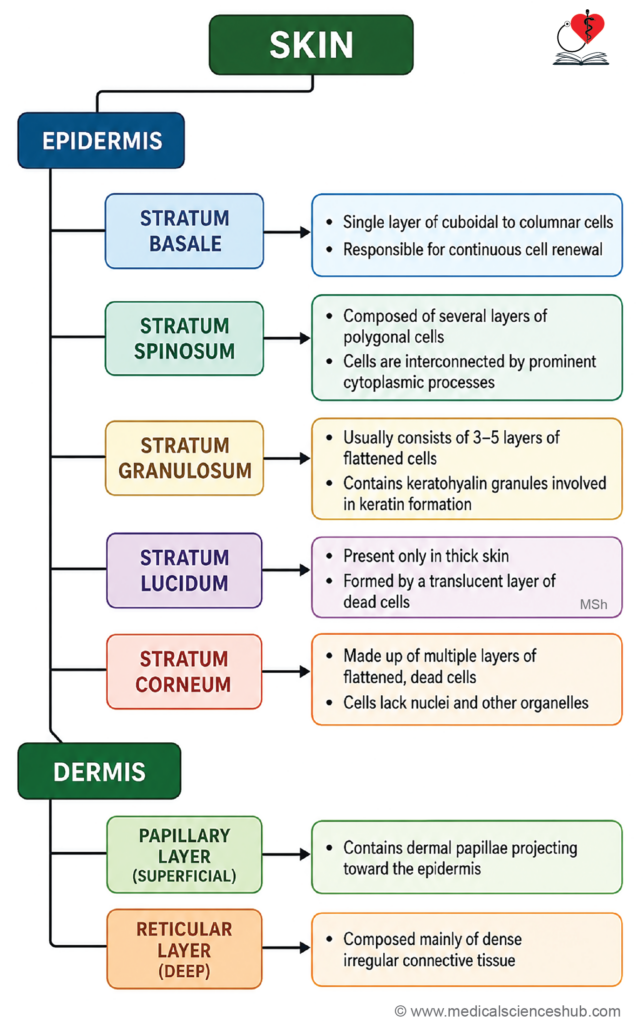
Table 11.1: Differences between thin and thick skin
| Feature | Thick Skin | Thin Skin |
|---|---|---|
| Stratum lucidum | Well-developed and present | Usually absent |
| Epidermal thickness | Relatively thick (approximately 0.4–1.5 mm) | Relatively thin (approximately 0.075–0.15 mm) |
| Epidermal ridges (rete ridges) | Prominent and well-defined | Less prominent |
| Hair follicles | Absent | Present |
| Arrector pili muscles | Absent | Present |
| Sebaceous glands | Absent | Present |
| Sweat glands | Numerous, especially eccrine glands | Fewer in number |
| Merkel cells and sensory receptors | More abundant | Less abundant |
| Locations | Palms of the hands and soles of the feet | Covers most other regions of the body |
Cells of Epidermis
- Skin has four types of cells: keratinocytes, melanocytes, Langerhans cells, and Merkel’s cells.
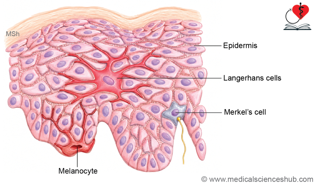
Keratinocytes
- Keratinocytes constitute approximately 85% of epidermal cells and are the predominant cell type of the skin.
- They originate from mitotically active stem cells in the stratum basale.
- Their primary functions are the production of keratin and the formation of the epidermal water barrier.
- Basal and deeper keratinocytes are rich in rough endoplasmic reticulum, which supports keratin protein synthesis.
- Keratin proteins assemble into keratin filaments (tonofilaments) that provide mechanical strength to the epidermis.
- In more superficial layers, these filaments aggregate into bundles known as tonofibrils.
- During differentiation, keratinocytes accumulate keratohyalin granules, which contain proteins such as filaggrin that promote aggregation of keratin filaments.
- The process of keratinization transforms living keratinocytes into flattened, anucleate cells of the outer epidermis.
- Cells of the stratum spinosum produce lamellar bodies, membrane-bound organelles containing lipids.
- These lipids are released into the extracellular space, where they contribute to the epidermal permeability barrier and help maintain cohesion between keratinocytes.
- The appearance of keratohyalin granules is an important indicator of terminal keratinocyte differentiation.
Melanocytes
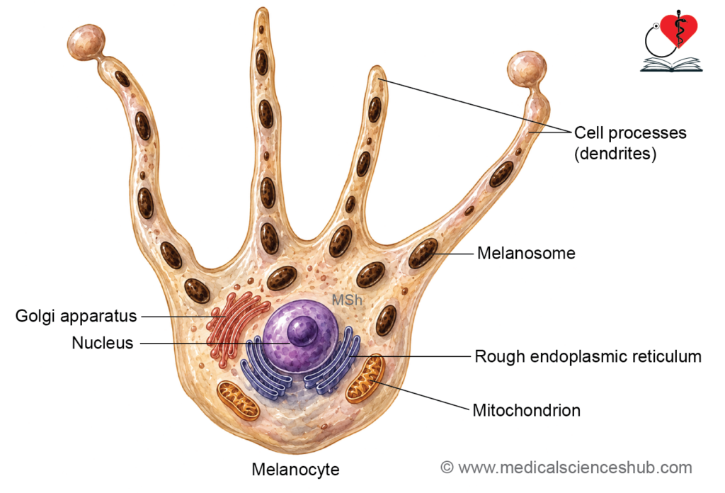
- Melanocytes are pigment-producing cells derived from neural crest cells and are located primarily in the stratum basale of the epidermis.
- They possess dendritic processes that extend between adjacent keratinocytes.
- Melanin is synthesized within specialized organelles called melanosomes and then transferred to keratinocytes through the dendritic processes.
- This transfer of pigment is an example of cytocrine secretion.
- Keratinocytes that receive and store melanin contribute to skin pigmentation and protection against ultraviolet radiation.
- Cells containing transferred melanin are sometimes referred to as melanophores.
Dendritic Cells of Langerhans
- Langerhans cells are specialized antigen-presenting cells found mainly in the stratum spinosum of the epidermis and are derived from bone marrow hematopoietic precursors.
- They are also present in the oral mucosa and other stratified squamous epithelia.
- These cells capture, process, and present antigens to T lymphocytes, thereby initiating immune responses.
- Under electron microscopy, they contain characteristic Birbeck granules (Birbeck bodies), which appear as tennis-racket-shaped cytoplasmic organelles.
- Langerhans cells participate in cell-mediated immunity and play an important role in delayed-type hypersensitivity reactions.
- Neoplastic proliferation of these cells results in Langerhans cell histiocytosis, a group of disorders characterized by abnormal accumulation of Langerhans cells.
- They can also serve as reservoirs for HIV infection in affected individuals.
Cells of Merkel
- Merkel cells are specialized mechanoreceptor cells involved in the perception of fine touch and tactile discrimination.
- They are located in the stratum basale of the epidermis and possess a lobulated nucleus with dense cytoplasm.
- These cells form functional complexes with adjacent sensory nerve endings, known as Merkel cell–neurite complexes.
- Merkel cells are particularly abundant in areas requiring high tactile sensitivity, such as the fingertips, oral mucosa, and regions associated with hair follicles.
- They play an important role in detecting sustained pressure and fine surface details, which is essential for tasks such as reading Braille.
- Merkel cell carcinoma is a rare but highly aggressive neuroendocrine malignancy of the skin.
Blood Supply and Nerve Supply of Skin
Blood Supply
- The epidermis is avascular and receives nutrients and oxygen by diffusion from blood vessels in the underlying dermis.
- The skin is supplied by three interconnected vascular plexuses:
- Subpapillary (papillary) plexus, located beneath the dermal papillae.
- Cutaneous (reticular) plexus, situated at the junction of the dermis and hypodermis.
- Deep vascular plexus, located above the deep fascia.
- Numerous arteriovenous anastomoses in the skin regulate blood flow.
- Reduced cutaneous blood flow helps conserve body heat, whereas increased blood flow promotes heat dissipation and assists in thermoregulation.
Nerve Supply
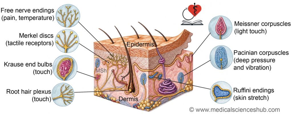
- The skin is supplied by cutaneous nerves that convey sensory information through free nerve endings, Meissner corpuscles, Pacinian corpuscles, and Ruffini endings.
- Cutaneous blood vessels receive autonomic sympathetic innervation, which causes vasoconstriction and reduces blood flow to the skin.
- Autonomic nerves also stimulate sweat gland secretion and induce contraction of arrector pili muscles, contributing to thermoregulation and piloerection.
CLINICAL CORRELATION
- Pemphigus is a group of autoimmune blistering disorders that affect the skin and mucous membranes.
- Pemphigus vulgaris results from autoantibodies directed against desmoglein 1 and desmoglein 3, key components of desmosomes, leading to loss of intercellular adhesion.
- In pemphigus foliaceus, blister formation occurs in the superficial layers of the epidermis.
- In pemphigus vulgaris, blisters typically develop in the lower epidermis, just above the basal layer.
- Bullous pemphigoid produces subepidermal blisters due to separation at the basement membrane zone, commonly within the lamina lucida.
Sensory Receptors of Skin
- The skin contains specialized sensory receptors that detect touch, pressure, vibration, temperature, pain, and skin deformation.
- Free nerve endings are the most widely distributed sensory receptors.
- They extend into the epidermis and around hair follicles.
- These unencapsulated endings detect pain, temperature, itch, crude touch, and hair movement.
- Pacinian corpuscles are large, encapsulated receptors located in the deep dermis and hypodermis.
- They consist of a central nerve fiber surrounded by concentric lamellae, giving an onion-like appearance in section.
- They respond to deep pressure and high-frequency vibration.
- Meissner corpuscles are found within the dermal papillae of hairless (glabrous) skin, particularly in the fingertips.
- They contain nerve endings surrounded by modified Schwann cells.
- They are responsible for light touch and tactile discrimination.
- Ruffini corpuscles are elongated, encapsulated receptors located in the deeper dermis.
- Their branching nerve endings detect skin stretch, sustained pressure, and tissue deformation.
- Krause end bulbs (bulboid corpuscles) are small encapsulated receptors associated with mucocutaneous regions such as the conjunctiva, lips, tongue, external genitalia, and certain joint capsules.
- They function primarily as cold-sensitive thermoreceptors.
- Merkel discs (Merkel cell–neurite complexes) are slowly adapting mechanoreceptors located in the stratum basale and around hair follicles.
- They consist of a sensory nerve ending closely associated with a Merkel cell.
- These receptors detect fine touch, texture, shape, and sustained pressure.
Appendages of Skin
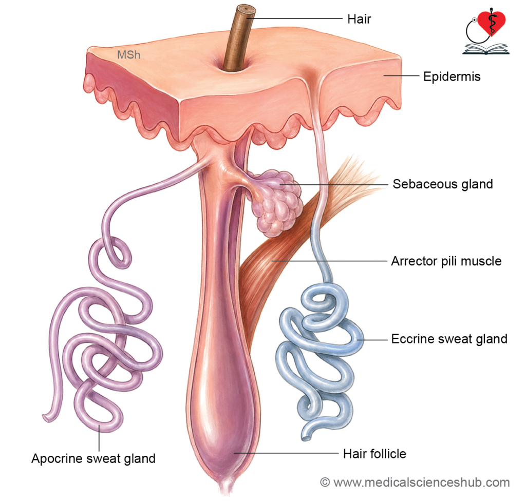
- The epidermis gives rise to the skin appendages, including hair follicles, hairs, nails, sebaceous glands, eccrine sweat glands, and apocrine sweat glands.
- These structures support protective and thermoregulatory functions of the skin.
Hairs
- Hair covers most of the body surface but is absent from the palms, soles, sides of the hands and feet, lips, parts of the external genitalia, and the ventral surfaces of the digits.
- Hair growth, distribution, and texture are influenced by hormonal factors, particularly androgens.
Parts of Hair
Hair has the following parts:
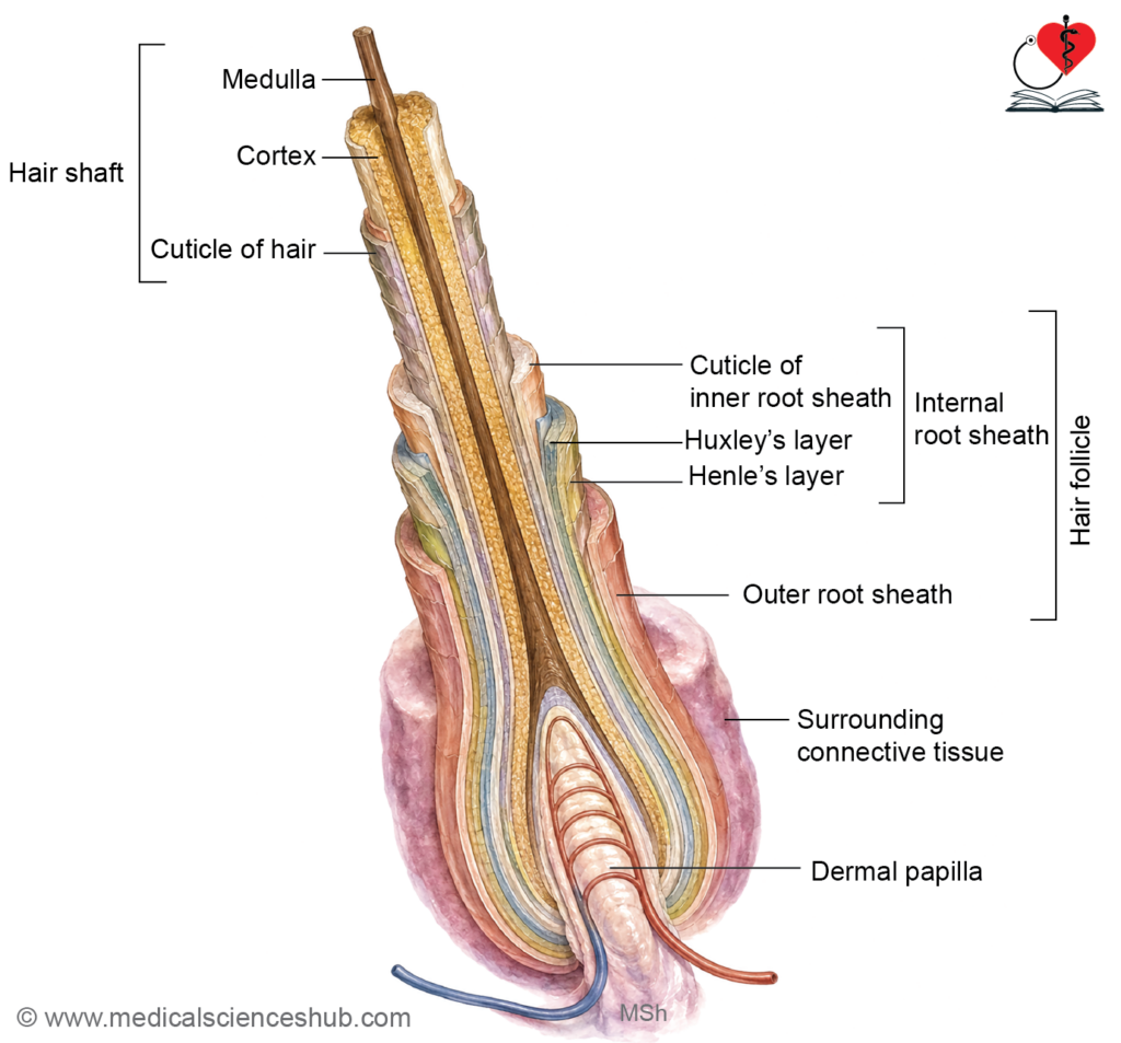
- Hair shaft: The visible portion that projects above the skin surface.
- Hair root: The portion embedded within the skin and enclosed by the hair follicle.
- Hair bulb: The enlarged terminal part of the root where active hair growth occurs.
- Hair papilla: A projection of vascular connective tissue from the dermis into the base of the hair bulb, providing nourishment.
- Hair follicle: A specialized epidermal invagination surrounded by dermal tissue that encloses and supports the hair root.
Hair Shaft and Root
- The hair shaft and hair root are composed of keratinized epithelial cells and represent specialized epidermal structures.
- The hair shaft consists of three concentric layers:
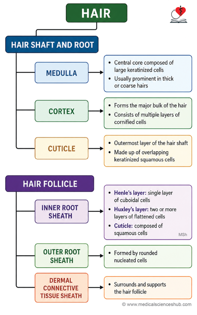
- Medulla: The central core of the hair, composed of large keratinized cells. It is typically present in thick hairs and may be absent in fine hairs.
- Cortex: The largest component of the hair shaft, forming most of its bulk. It contains hard keratin filaments and melanin pigments, which contribute to hair strength and color.
- Cuticle: The outermost layer, formed by overlapping keratinized squamous cells arranged like scales. It protects the underlying hair structure.
Hair Follicle
- The hair follicle is a specialized epidermal invagination surrounded by dermal connective tissue that encloses the hair root and produces hair.
- It is divided into several regions:
- Infundibulum: Extends from the skin surface to the opening of the sebaceous gland.
- Isthmus: Extends from the sebaceous gland opening to the attachment of the arrector pili muscle.
- Follicular bulge: Located near the insertion of the arrector pili muscle and contains epidermal stem cells important for hair regeneration.
- Hair bulb: The enlarged lower end of the follicle where active hair formation occurs.
- Dermal papilla: A vascular connective tissue projection that extends into the base of the hair bulb and provides nutritional support.
Layers of the Hair Follicle
- The follicular wall consists of:
- Inner root sheath
- Outer root sheath
- Dermal connective tissue sheath
Inner Root Sheath
- Surrounds the deeper portion of the growing hair and consists of:
- Henle’s layer: A single layer of flattened cells.
- Huxley’s layer: Usually one to two layers of cells containing trichohyalin granules.
- Cuticle of the inner root sheath: A layer of flattened cells that interlocks with the hair cuticle.
Outer Root Sheath
- Is continuous with the epidermis and contains living nucleated cells.
- It is separated from the surrounding connective tissue by a thick basement membrane (glassy membrane).
Hair Matrix
- The hair matrix consists of rapidly dividing cells surrounding the hair bulb and dermal papilla.
- These cells generate the hair shaft and inner root sheath during hair growth.
- Melanocytes within the matrix transfer melanin to developing hair cells, determining hair pigmentation.
CLINICAL CORRELATION
- The cortex contains most of the hair’s pigment and largely determines hair color.
- Hair pigmentation depends on melanin produced by melanocytes located in the hair bulb matrix.
Hair Growth Cycle
- Anagen phase: Active growth stage during which new hair is produced.
- Catagen phase: Short transitional stage characterized by cessation of hair growth.
- Telogen phase: Resting stage that culminates in shedding of the hair shaft.
- More than 80% of scalp hairs are normally in the anagen phase, which is prolonged compared with the telogen phase.
Types of Hair
- Terminal hairs are thick, coarse, and usually pigmented.
- Vellus hairs are short, fine, and lightly pigmented or unpigmented.
Alopecia Areata
- Alopecia areata is an autoimmune disorder characterized by localized, non-scarring hair loss.
- The affected skin typically appears normal, without significant scaling, erythema, or scarring.
- Exclamation-mark hairs at the margins of the bald patch are a characteristic clinical feature.
Arrector Pili Muscle
- Arrector pili muscles are small bundles of smooth muscle that extend from the dermis to the connective tissue sheath of a hair follicle.
- They are attached to hair follicles at an oblique angle and receive sympathetic autonomic innervation.
Functions
- Contraction of the arrector pili muscle causes the hair shaft to become more upright, producing piloerection or the characteristic gooseflesh appearance.
- Muscle contraction also creates a slight depression in the skin at its dermal attachment site.
- Because sebaceous glands are closely associated with the hair follicle, contraction of the muscle compresses the gland and facilitates the release of sebum onto the skin surface.
Sebaceous Glands
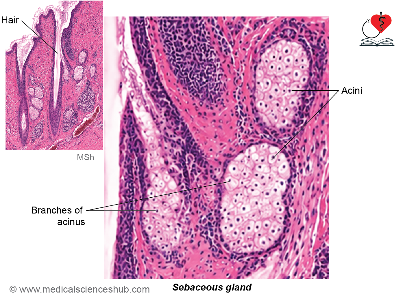
- Sebaceous glands are simple branched alveolar (acinar) holocrine glands located within the dermis and are usually associated with hair follicles.
- Most glands discharge their secretion into the pilosebaceous canal through a short duct that opens into the follicular infundibulum.
- Sebaceous glands independent of hair follicles are present in the lips, areola of the nipple (Montgomery glands), labia minora, and inner surface of the prepuce.
Structure
- Each gland consists of:
- A secretory portion (alveoli/acini).
- A short duct lined by stratified squamous epithelium.
- The secretory units are composed of:
- Basal cells, which rest on the basement membrane and undergo mitosis to replenish the gland.
- Sebocytes (secretory cells), which are large polygonal cells filled with lipid droplets.
- In routine histological preparations, lipids are dissolved during processing, giving sebocytes a characteristic foamy appearance.
Functions
- Sebocytes synthesize and accumulate sebum, an oily secretion that lubricates and waterproofs the skin and hair.
- During secretion, sebocytes undergo degeneration and disintegrate completely, releasing their contents into the duct. This mechanism is known as holocrine secretion.
- Sebaceous glands also produce antimicrobial peptides such as cathelicidins and β-defensins, which contribute to the skin’s innate defense system and support the epidermal barrier.
CLINICAL CORRELATION
- Seborrhea refers to excessive production of sebum due to increased activity of sebaceous glands, resulting in oily skin.
- Acne vulgaris is a common disorder of the pilosebaceous unit, particularly during adolescence.
- It develops when hair follicles become obstructed by sebum and keratinized cellular debris.
- Clinical manifestations include closed comedones (whiteheads), open comedones (blackheads), inflammatory papules, pustules, and potential scarring.
- Genetic predisposition and increased androgen levels during puberty are important contributing factors.
- A comedo is a plugged hair follicle containing accumulated sebum and keratin.
Sweat Glands
- Sweat glands are exocrine glands that produce sweat (perspiration) and contribute to thermoregulation.
- They are classified into two main types:
- Eccrine (merocrine) sweat glands, which are widely distributed throughout the body and are absent from the lips and parts of the external genitalia.
- Apocrine sweat glands, which are localized to the axilla, areola, perianal skin, and external genital region.
- Modified apocrine glands include the glands of Moll in the eyelids and ceruminous glands of the external acoustic canal, which produce earwax.
Eccrine Sweat Glands
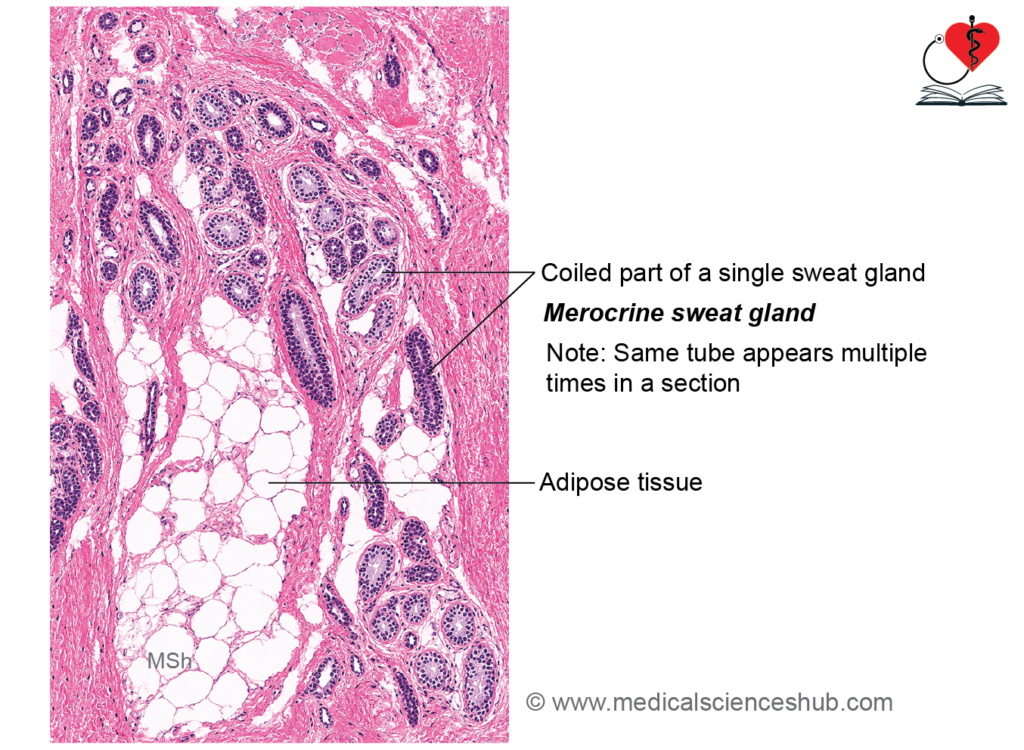
- Eccrine sweat glands are simple coiled tubular glands distributed over most of the body surface, except the lips and parts of the external genitalia.
- They are particularly abundant on the palms, soles, forehead, scalp, and axillae.
- Their primary function is thermoregulation through the production and evaporation of sweat.
Structure
- Each gland consists of two components:
- Secretory portion (body/fundus)
- Excretory duct
1. Duct
- The duct extends through the dermis and opens onto the skin surface via a sweat pore.
- It is lined by stratified cuboidal epithelium, typically composed of two layers of cuboidal cells.
- The duct has a narrower lumen than the secretory portion.
- Myoepithelial cells are absent from the duct.
- As the duct traverses the epidermis, it forms an intraepidermal spiral channel known as the acrosyringium.
2. Secretory part/body
- The secretory segment is a highly coiled tubular structure located in the deep dermis or superficial hypodermis.
- It is lined by a simple epithelium resting on a basal lamina and surrounded by connective tissue.
- Three cell types are present:
- Clear cells: Located adjacent to the basal lamina and connected by intercellular canaliculi. They secrete water and electrolytes, forming the watery component of sweat.
- Dark cells: Rich in rough endoplasmic reticulum and secretory granules. They produce glycoproteins and antimicrobial substances, including defensins and cathelicidins, through merocrine secretion.
- Myoepithelial cells: Contractile cells situated between the secretory epithelium and basal lamina. Their contraction assists in expelling sweat from the gland.
- Together, these cellular components produce sweat and contribute to temperature regulation, skin hydration, and innate antimicrobial defense.
Apocrine/Atypical Sweat Glands
- Apocrine sweat glands are specialized sweat glands located in the axilla, perianal region, and anogenital skin, including the mons pubis.
- Their ducts open into hair follicles, typically above the entry of sebaceous gland ducts.
- They are simple coiled tubular glands and consist of a secretory portion and an excretory duct.
- The secretory segment lies deep in the dermis or may extend into the hypodermis and has a characteristically wide lumen.
- It is lined by simple cuboidal to low columnar epithelial cells, supported by myoepithelial cells and a basal lamina.
- Electron microscopy demonstrates that secretion occurs primarily through merocrine (exocytotic) secretion.
- Modified apocrine glands include ceruminous glands of the external acoustic canal and glands of Moll in the eyelids.
- These glands produce protein-rich secretions, including pheromone-like substances.
CLINICAL CORRELATION
- Cystic fibrosis is an autosomal recessive disorder caused by mutations in the CFTR (Cystic Fibrosis Transmembrane Conductance Regulator) gene on chromosome 7.
- Defective CFTR channels impair chloride transport, resulting in reduced reabsorption of sodium chloride in sweat ducts and consequently elevated chloride levels in sweat.
- Apocrine sweat glands become functional at puberty and produce protein-rich secretions containing pheromone-like compounds.
- Eccrine sweat glands are primarily stimulated by increased body temperature and emotional stress.
- Apocrine glands respond mainly to emotional, sensory, and hormonal stimuli.
Nails
- Nails are specialized epidermal structures that support the fingertips and enhance precision during grasping and fine movements.
- They are composed of compact, keratinized, anucleate cells (corneocytes) containing hard α-keratin.
Structure of Nail
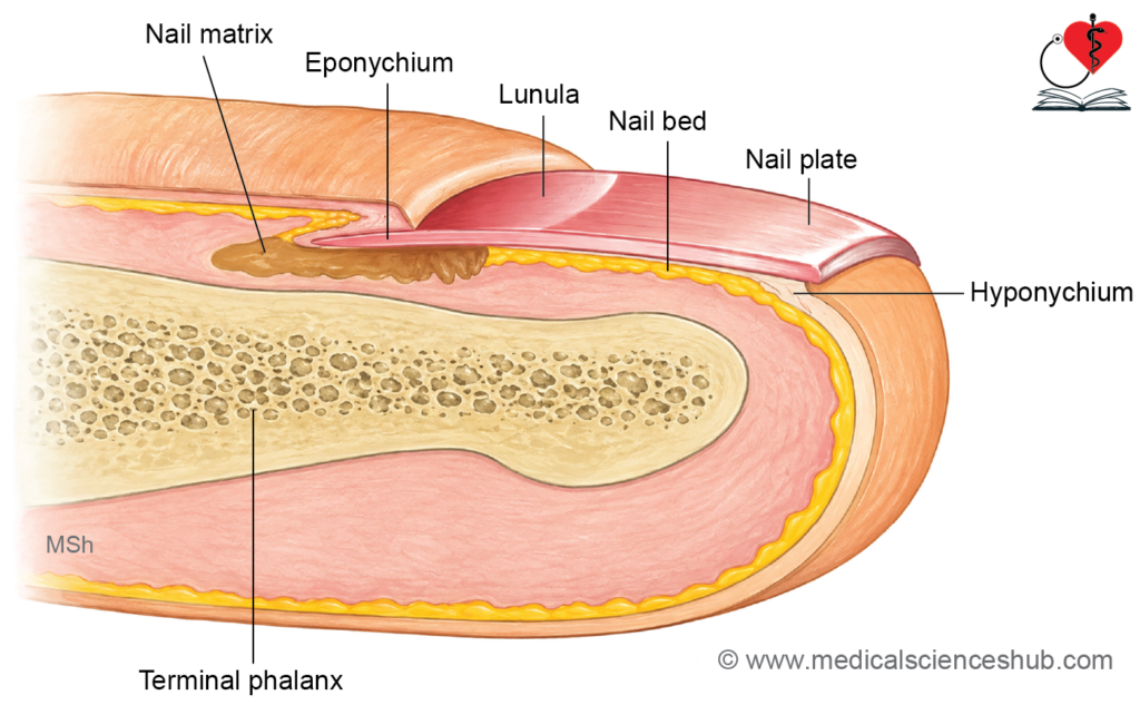
- Nail plate (body): The visible hard keratinized portion of the nail. It has a free distal edge and a proximal embedded root (radix).
- Nail bed: The highly vascular tissue beneath the nail plate. It supports the nail and is continuous with the epidermal stratum basale and stratum spinosum.
- Germinal matrix (nail matrix): The proliferative region at the root of the nail that produces most of the nail plate.
- Proximal nail fold: A fold of skin that covers and protects the nail root.
- Lunula: The pale, crescent-shaped area visible at the base of the nail, representing the distal part of the germinal matrix.
- Sterile matrix: The portion of the nail bed distal to the germinal matrix. It supports the nail plate but does not significantly contribute to nail production.
- Lateral nail folds (nail walls): Skin folds that overlap and protect the lateral margins of the nail plate.
- Lateral nail grooves: Furrows located between the nail plate and the lateral nail folds.
- Eponychium: A thickened extension of the stratum corneum beneath the proximal nail fold that protects the nail matrix.
- Hyponychium: The thickened epidermis beneath the free distal edge of the nail plate.
- Cuticle: A thin layer of keratinized tissue extending from the proximal nail fold onto the nail surface.
- Perionychium: The soft tissue surrounding the nail, including the nail folds and cuticle.
CLINICAL CORRELATION
- Onychia is inflammation of the nail folds, often associated with pus formation and possible shedding of the nail.
- Onycholysis refers to separation of the nail plate from the underlying nail bed.
- Onychocryptosis (ingrown toenail) occurs when the nail edge grows into the adjacent lateral nail fold, causing pain and inflammation.
- Onychodystrophy is a general term for abnormal nail growth or deformity.
- Onychogryphosis (ram’s horn nail) is marked thickening and excessive curvature of the nail plate.
- Paronychia is a bacterial or fungal infection involving the tissues surrounding the nail.
- Koilonychia (spoon-shaped nails) is characterized by a concave nail surface and is commonly associated with iron deficiency, although other causes may occur.
Nail Clubbing
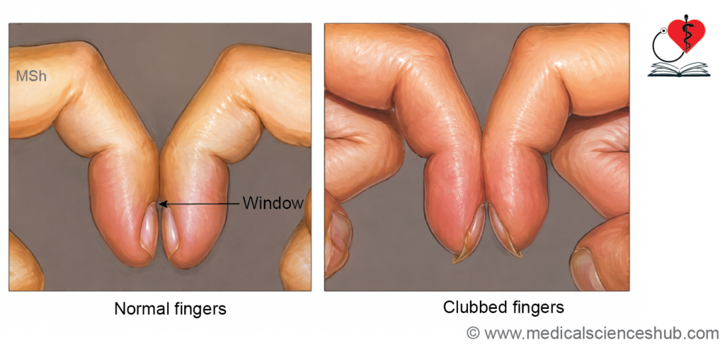
- Nail clubbing is characterized by enlargement of the distal digits, increased curvature of the nails, and thickening of the nail bed.
- It is commonly associated with chronic hypoxia and may occur in patients with long-standing pulmonary, cardiovascular, or other systemic diseases.
- Increased vascular connective tissue beneath the nail bed contributes to the characteristic appearance.
Clinical Assessment
- Schamroth’s window test: When corresponding fingernails are placed face-to-face, a small diamond-shaped gap is normally visible. Loss of this window suggests clubbing.
- Lovibond angle: The angle between the proximal nail fold and nail plate is normally about 160–180°. An angle greater than 180° is suggestive of clubbing.
Important Questions
- List the differences between thick and thin skin.
📝 Test Your Knowledge – Practice MCQs
Attempt the chapter MCQ quiz and assess your understanding of key concepts.
Click to see: Medical Disclaimer and Copyright Notice