
Competencies
- AN43.2 Identify, describe and draw the microanatomy of tongue, salivary glands.
- AN43.3 Identify, describe and draw microanatomy of …, lip,
Introduction
- The digestive system is responsible for ingestion, digestion, absorption of nutrients and water, and elimination of waste.
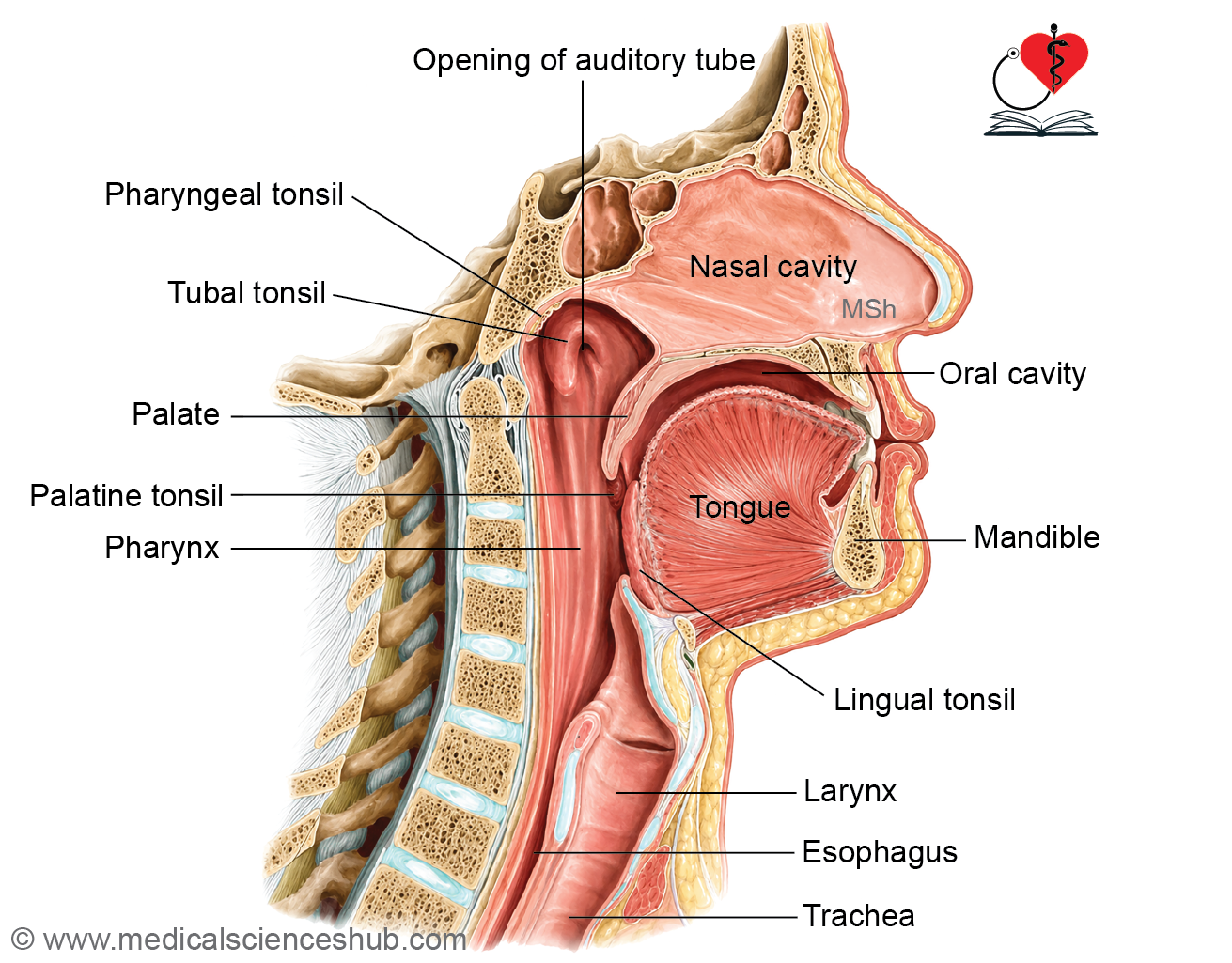
- It includes the oral cavity with the lips, teeth, tongue, and salivary glands.
- The gastrointestinal tract comprises the esophagus, stomach, small intestine, and large intestine.
- Associated digestive organs include the liver, gallbladder, and pancreas.
Teeth
- Teeth are embedded in the alveolar sockets of the maxilla and mandible.
- Children possess 20 deciduous (primary) teeth, with 10 teeth in each jaw: 4 incisors, 2 canines, and 4 molars.
- Adults normally have 32 permanent teeth, with 16 teeth in each jaw: 4 incisors, 2 canines, 4 premolars, and 6 molars.
- The gingiva (gum) covers the alveolar processes of the jaws and surrounds the cervical region of the teeth, providing support and protection.
Histology of Tooth
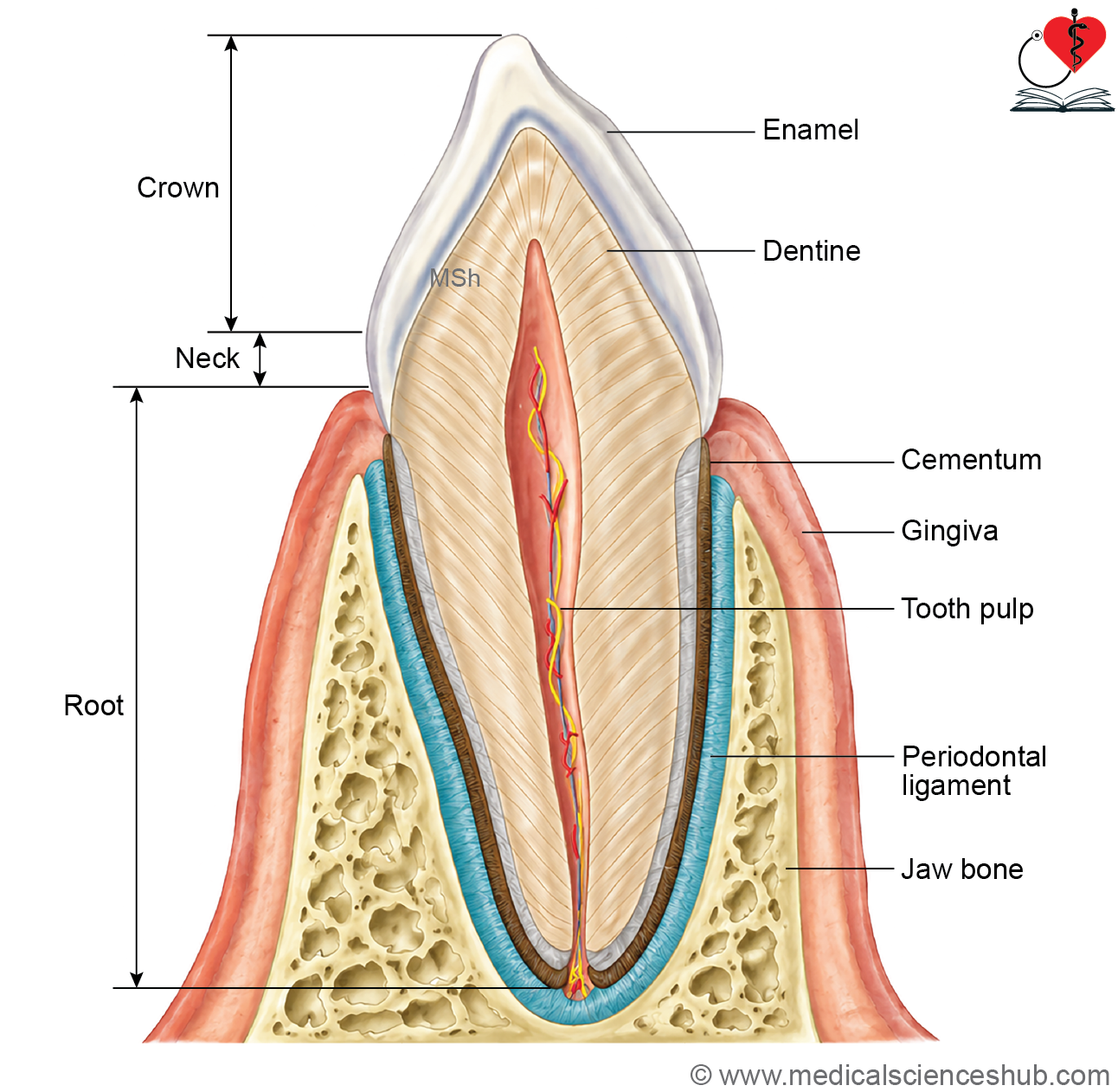
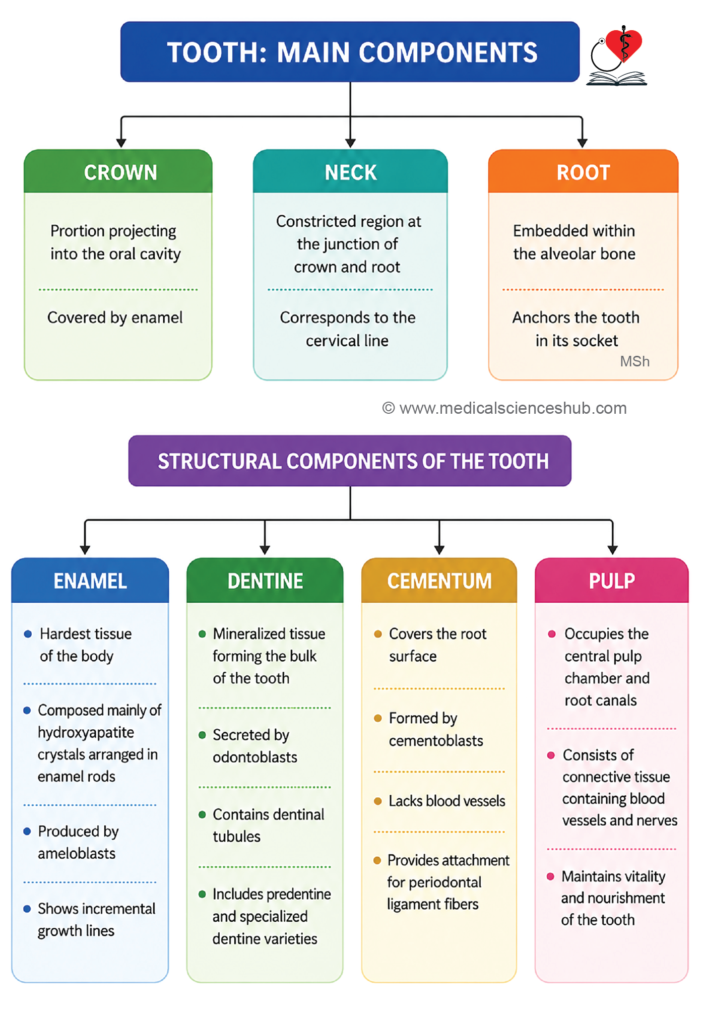
- A tooth consists of three anatomical parts: crown, neck, and root.
- The crown projects above the gingiva, the root is embedded within the alveolar bone, and the neck lies at their junction.
- Four specialized tissues form the tooth:
- Enamel: The hardest tissue in the human body, covering the crown.
- Dentine: Forms the bulk of the tooth and lies beneath enamel and cementum.
- Cementum: A calcified tissue covering the root surface.
- Pulp: Soft connective tissue containing blood vessels and nerves within the pulp cavity and root canal.
- The periodontal ligament anchors the cementum to the alveolar socket and supports the tooth during mastication.
Enamel
- Enamel is the hardest tissue in the human body and forms the outer covering of the tooth crown.
- It is highly mineralized, consisting of approximately 96–98% hydroxyapatite crystals by weight.
- The visible enamel-covered portion of a tooth is termed the clinical crown.
- Enamel is composed of tightly packed enamel rods (prisms) that extend from the dentinoenamel junction (DEJ) to the tooth surface.
- Each prism is surrounded by a thin organic layer known as the prism sheath.
Formation
- Enamel is produced by ameloblasts during tooth development.
- After tooth eruption, ameloblasts are lost; therefore, enamel cannot be regenerated or deposited further.
Microscopic Features
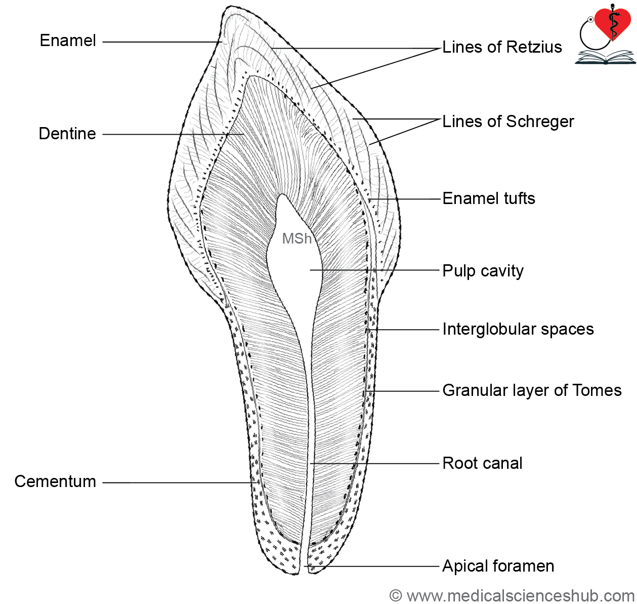
- Striae of Retzius are incremental growth lines produced by rhythmic enamel deposition during development.
- Neonatal lines are prominent growth lines that mark the transition between enamel formed before and after birth.
- Lines of Schreger appear as alternating light and dark bands caused by changes in the orientation of enamel rods.
Structural Variations
- Enamel tufts are hypomineralized extensions that arise from the dentinoenamel junction and project into enamel.
- Enamel lamellae are thin, leaf-like defects extending from the enamel surface toward the dentinoenamel junction.
- Enamel spindles are short extensions of odontoblastic processes or dentinal tubules that cross the dentinoenamel junction into enamel.
- These specialized structures reflect the developmental pattern and microscopic organization of enamel.
Dentine
- Dentine forms the bulk of the tooth and constitutes the wall of the pulp cavity.
- It is composed of approximately 70% mineral, 20% organic matrix, and 10% water, making it harder than bone but softer than enamel.
- The mineral component consists mainly of hydroxyapatite crystals, which provide rigidity and strength.
- Odontoblasts, located at the periphery of the pulp, are responsible for dentine formation throughout life.
- Numerous dentinal tubules extend from the pulp cavity toward the dentinoenamel or dentinocemental junction.
- These tubules contain odontoblastic processes known as Tomes’ fibers, which contribute to dentine sensitivity and nutrition.
- Newly formed, unmineralized dentine is called predentine; it later undergoes mineralization to become mature dentine.
- Incremental lines of von Ebner represent periodic deposition of dentine and reflect its rhythmic growth pattern.
- The granular layer of Tomes is a thin granular-appearing zone located beneath the cementum in the root dentine.
- Mantle dentine is the outermost, less mineralized layer adjacent to the dentinoenamel junction.
- Circumpulpal dentine forms the major portion of dentine and lies between the predentine and mantle dentine.
- Dentine provides structural support to enamel and protects the underlying pulp tissue.
Cementum
- Cementum is a thin, mineralized connective tissue that covers the root surface of the tooth and overlies dentine.
- It resembles bone in composition but is avascular and plays a key role in tooth attachment.
- Cementoblasts are cuboidal cells responsible for the formation and maintenance of cementum.
- Mature cementoblasts become cementocytes, which are enclosed within lacunae and communicate through canaliculi.
- The periodontal ligament anchors the tooth to the alveolar bone and transmits masticatory forces.
- Collagen fibers of the periodontal ligament become embedded within cementum as Sharpey’s fibers, providing strong attachment between the tooth root and surrounding alveolar socket.
Pulp
- The dental pulp occupies the pulp cavity and consists of connective tissue containing fibroblasts, macrophages, collagen fibers, mast cells, and extracellular matrix.
- It receives blood vessels and nerves through the apical foramen.
- The peripheral pulp is lined by odontoblasts, which produce dentine.
- A rich neurovascular plexus lies immediately beneath the odontoblastic layer, providing nutrition and sensory innervation to the tooth.
CLINICAL CORRELATION
- Dental caries is a microbial disease characterized by progressive destruction of the mineralized tissues of the tooth.
- Oral bacteria metabolize dietary carbohydrates, producing acids that cause demineralization of enamel and dentine.
- Persistent acid exposure leads to the formation and progression of carious lesions.
- Fluoride helps prevent dental caries by increasing the acid resistance of tooth structures.
- It promotes remineralization of early enamel lesions and also exhibits mild antimicrobial activity.
- Fluoride replaces hydroxyl ions in hydroxyapatite crystals to form fluorapatite, which is less soluble in acidic conditions.
- Regular exposure to fluoride through drinking water, toothpaste, and other sources contributes to improved dental health and reduced caries risk.
Tongue
- The tongue is a muscular organ composed of skeletal muscle covered by a mucous membrane.
- It plays essential roles in taste, speech, mastication, and swallowing (deglutition).
Gross Features of Tongue
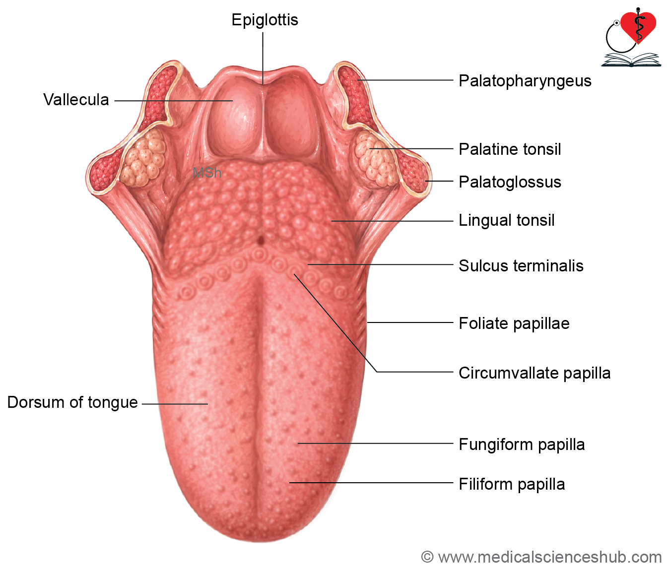
- Anatomically, the tongue consists of a root, body, and tip (apex).
- The root is attached to the mandible and hyoid bone, while the tip remains freely movable within the oral cavity.
- The body has dorsal and ventral surfaces.
- A V-shaped sulcus terminalis divides the dorsal surface into the anterior oral part (anterior two-thirds) and posterior pharyngeal part (posterior one-third).
- At the apex of the sulcus terminalis lies the foramen cecum, a remnant of the embryonic thyroglossal duct.
- The oral part appears rough due to numerous lingual papillae.
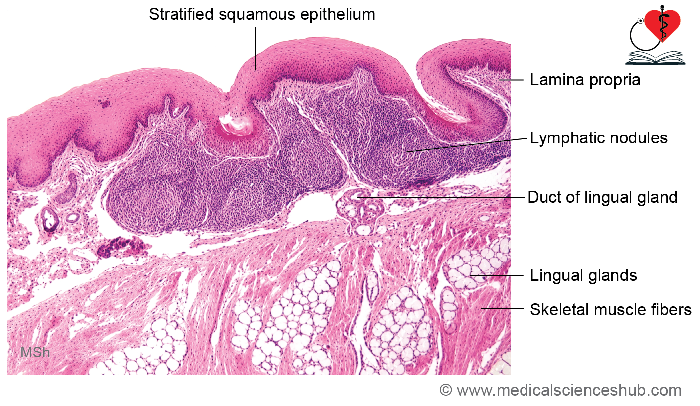
- The posterior pharyngeal part contains aggregates of lymphoid tissue known as the lingual tonsils, which contribute to immune defense.
Histology of tongue
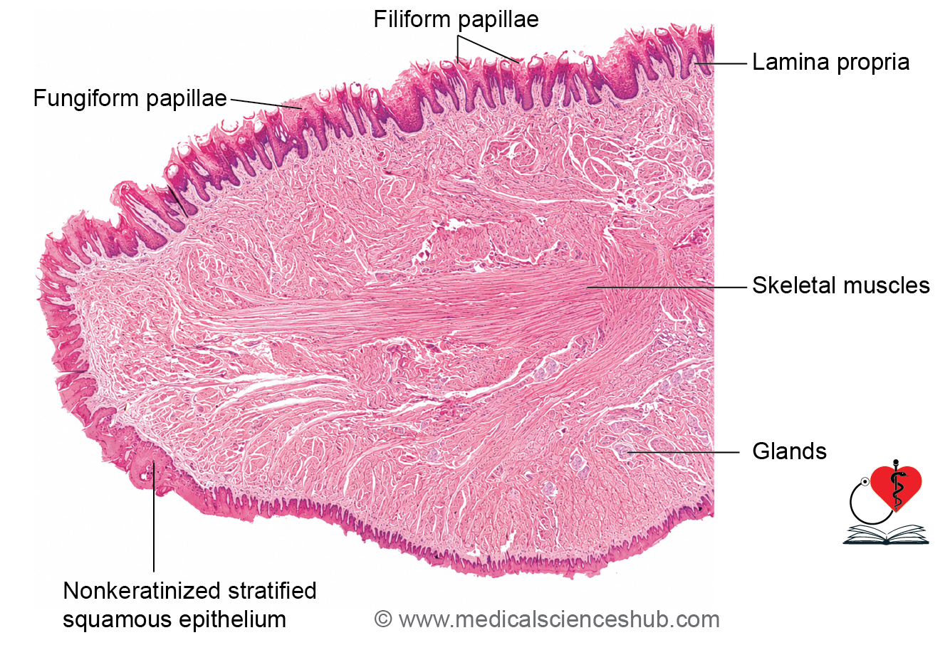
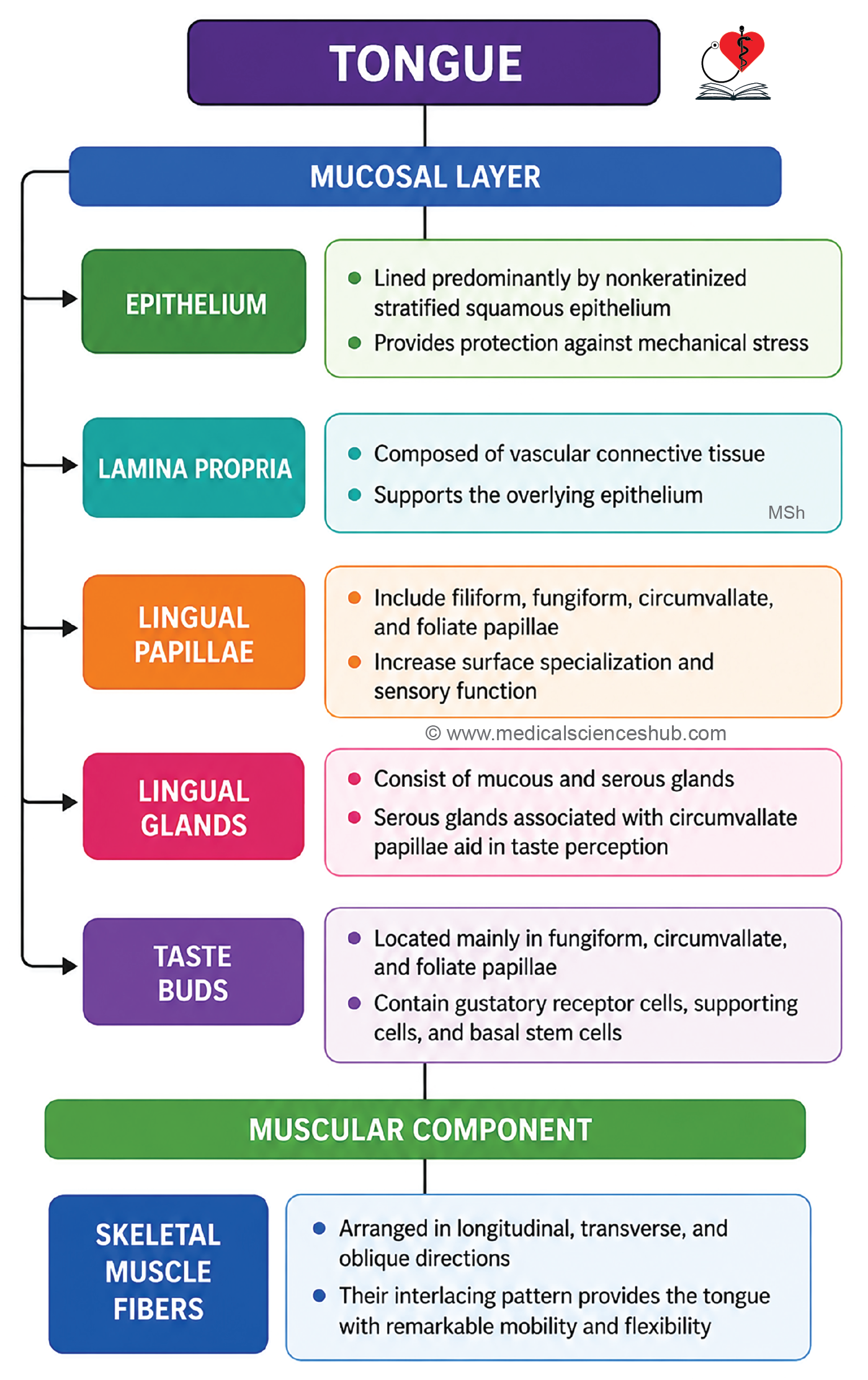
- The tongue is composed of a mucous membrane covering a core of skeletal muscle fibers arranged in three planes.
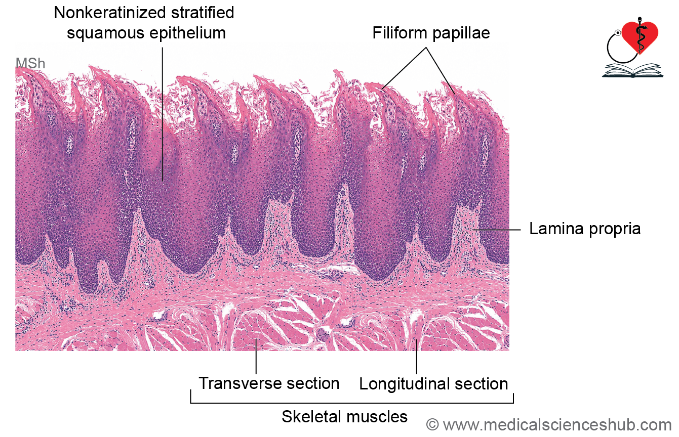
- The mucosa is lined by nonkeratinized stratified squamous epithelium supported by a loose connective tissue lamina propria.
- Four types of lingual papillae are present: filiform, fungiform, circumvallate, and foliate papillae.
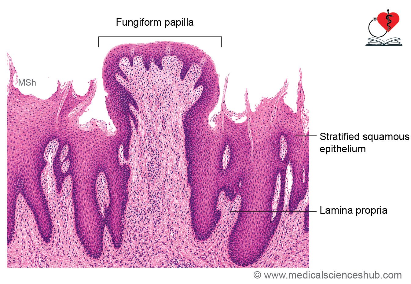
- Filiform papillae have keratinized conical tips, whereas fungiform papillae are mushroom-shaped.
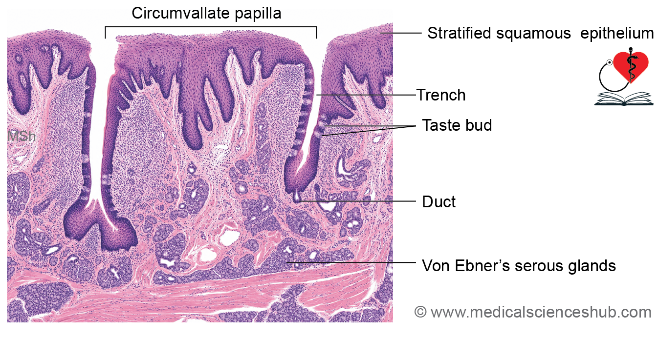
- Circumvallate papillae are surrounded by a circular trench, while foliate papillae are poorly developed in humans.
- Taste buds are present mainly in fungiform and circumvallate papillae and contain receptor, supporting, and basal cells.
- The tongue contains mucous lingual glands and von Ebner’s serous glands associated with the circumvallate papillae.
- Posterior one-third of tongue consists of mucous membrane (nonkeratinized stratified squamous epithelium and lamina propria) that covers the numerous mucous lingual salivary glands and their ducts, lymphatic follicles and a core of skeletal muscle fibers.
Muscles of Tongue
- The core of the tongue is composed of skeletal muscle arranged as intrinsic and extrinsic muscles.
- Intrinsic muscles are organized into longitudinal, transverse, and vertical bundles, enabling precise changes in tongue shape.
- Extrinsic muscles enter the tongue from surrounding structures and control its position and movement.
- Muscle bundles are separated and supported by connective tissue containing lingual glands, von Ebner’s serous glands, blood vessels, nerves, lymphatics, and small amounts of adipose tissue.
Papillae of Tongue
- The dorsal surface of the anterior tongue contains lingual papillae, which are projections of the lamina propria covered by epithelium.
- These papillae increase surface area and contribute to mechanical and sensory functions.
1. Filiform Papillae
- Filiform papillae are the most numerous and smallest papillae.
- They are conical projections covered by keratinized stratified squamous epithelium.
- Their tips are directed posteriorly and aid in food manipulation.
- They do not contain taste buds.
2. Fungiform Papillae
- Fungiform papillae are mushroom-shaped structures scattered among filiform papillae, especially near the tip of the tongue.
- They appear as small reddish or pink elevations due to their rich vascularity.
- Taste buds are present on their dorsal epithelial surface.
3. Circumvallate Papillae
- Circumvallate papillae are large dome-shaped papillae, usually numbering 8–12, located immediately anterior to the sulcus terminalis.
- Each papilla is surrounded by a circular trench or moat.
- Numerous taste buds are present along the lateral walls of the trench.
- Ducts of the von Ebner serous glands open into the trench and secrete fluid that washes away dissolved tastants.
- Their core contains connective tissue, blood vessels, and nerves.
4. Foliate Papillae
- Foliate papillae consist of parallel mucosal folds along the lateral margins of the tongue.
- They are relatively rudimentary in humans but are more prominent in certain mammals.
- A few taste buds may be present in their epithelium, particularly during early life.
Taste buds
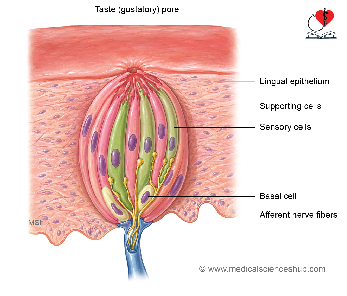
- Taste buds are specialized neuroepithelial sensory structures responsible for taste perception.
- They are found on fungiform and circumvallate papillae of the tongue, as well as in the soft palate, oropharynx, and epiglottis.
- Histologically, taste buds extend through the full thickness of the epithelium and appear as pale, oval, onion-shaped structures.
- Each taste bud opens onto the epithelial surface through a taste pore, where dissolved tastants interact with receptor cells.
Cell Types
- Gustatory (taste) cells are sensory receptor cells with apical microvilli projecting into the taste pore. Their basal ends synapse with afferent fibers of the facial, glossopharyngeal, and vagus nerves. These cells undergo continuous renewal.
- Supporting (sustentacular) cells provide structural and metabolic support and contribute secretions at the taste pore.
- Basal cells act as stem cells and replace both gustatory and supporting cells.
Mechanism of Taste Perception
- Dissolved chemicals (tastants) stimulate receptor cells and generate signals for the five primary tastes: sweet, salty, sour, bitter, and umami.
- Sweet, bitter, and umami tastes are mediated by G-protein-coupled receptors.
- Salty and sour tastes are detected primarily through ion channel-mediated mechanisms.
Functional Notes
- All regions of the tongue can detect all taste qualities, although certain areas may show relative sensitivity to specific tastes.
- Circumvallate papillae are particularly rich in taste buds responsive to bitter and umami stimuli.
Salivary Glands
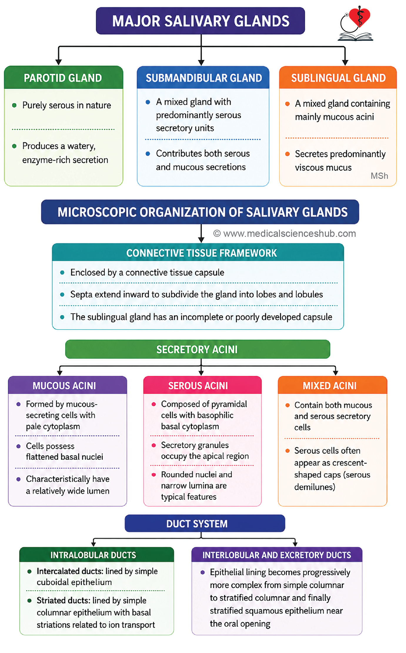
- Salivary glands produce and secrete saliva into the oral cavity, aiding lubrication, digestion, and oral health.
- They are classified into major salivary glands (parotid, submandibular, and sublingual) and minor salivary glands located within the mucosa of the lips, cheeks, palate, and tongue.
- Major glands discharge saliva through long excretory ducts, whereas minor glands open directly onto the mucosal surface.
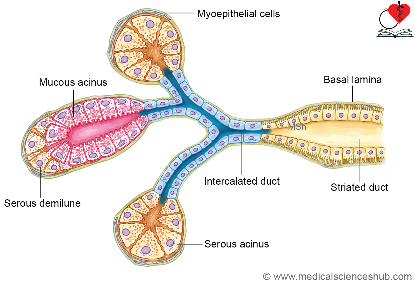
- Each gland consists of secretory acini and a branching duct system.
- The functional secretory unit, called a salivon, comprises an acinus, intercalated duct, and associated ductal components responsible for saliva modification and transport.
Histology of Salivary Glands
- Salivary glands are composed of a highly branched duct system and specialized secretory acini.
- Acini are formed by pyramidal secretory cells and are classified as serous, mucous, or mixed (seromucous) acini based on their secretions.
- Each gland is enclosed by a connective tissue capsule, which sends septa inward to divide the gland into lobes and lobules.
Salivary Ducts
The ductal system consists of intercalated ducts, striated ducts, and excretory ducts.
Intercalated duct
- These are lined by simple low cuboidal epithelium and may be associated with myoepithelial cells.
- They connect secretory acini to larger ducts and are especially prominent in serous glands.
- Their main functions are chloride ion absorption and bicarbonate ion secretion, contributing to saliva modification.
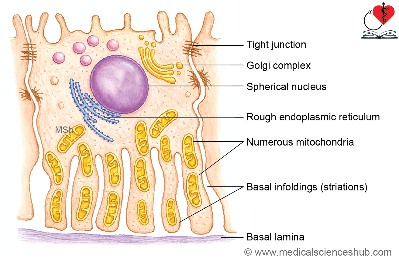
Striated ducts
- Striated ducts connect intercalated ducts to larger excretory ducts.
- They are lined by simple columnar epithelium.
- Basal plasma membrane infoldings containing numerous mitochondria produce the characteristic striated appearance.
- These ducts modify saliva by reabsorbing sodium ions and secreting potassium and bicarbonate ions.
- They are well developed in serous glands and are generally absent or poorly developed in predominantly mucous glands.
Excretory ducts
- Excretory ducts include interlobular, lobar, and main ducts.
- Their epithelial lining gradually changes from simple columnar to stratified squamous epithelium near the oral opening.
- The main excretory ducts ultimately discharge saliva into the oral cavity.
Secretory Acini
- Salivary glands are tubuloalveolar glands composed of serous, mucous, and mixed acini.
- These acini produce saliva with varying proportions of water, enzymes, and mucins.
Serous acinus
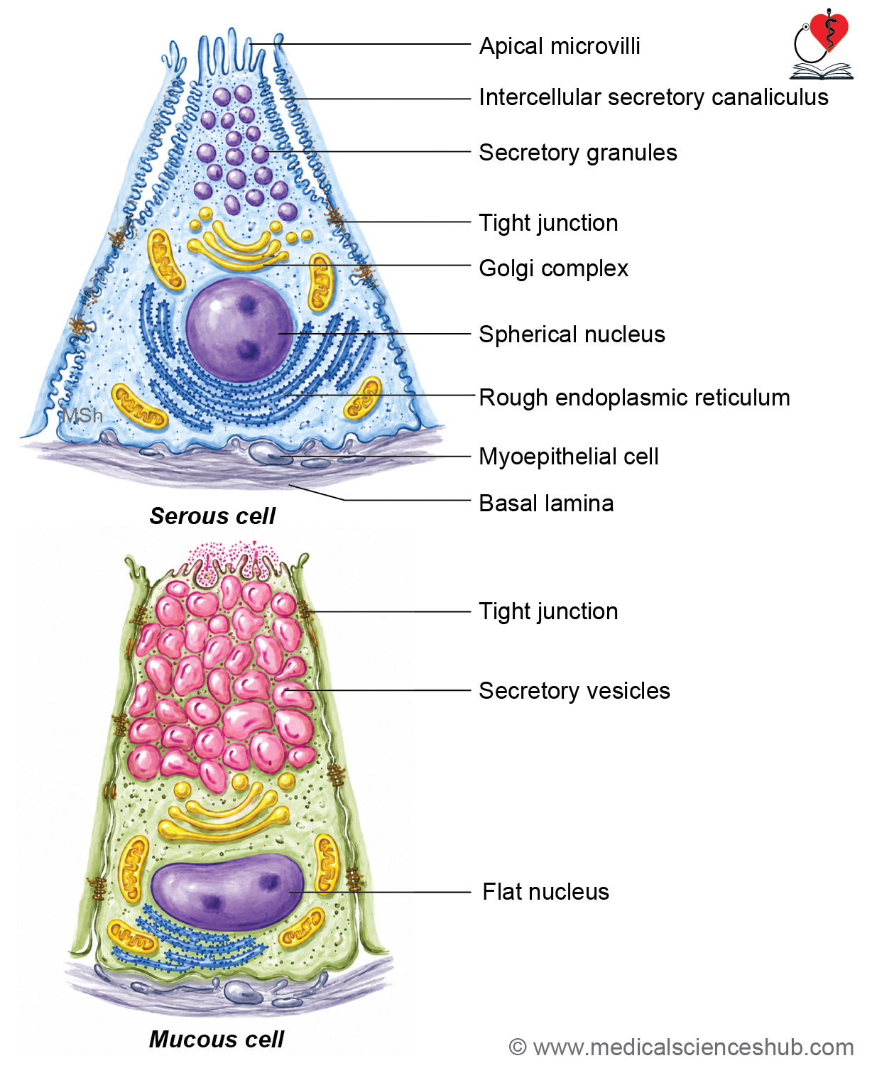
- Serous acini secrete a watery, protein-rich fluid containing digestive enzymes.
- They are lined by pyramidal secretory cells with a rounded basal nucleus.
- The basal cytoplasm is strongly basophilic due to abundant rough endoplasmic reticulum and ribosomes.
- The apical cytoplasm contains numerous secretory granules, producing an eosinophilic appearance.
- Their lumen is relatively narrow.
Mucous acinus
- Mucous acini produce a viscous secretion rich in mucins.
- Secretory cells are pyramidal with flattened, basally located nuclei.
- The cytoplasm appears pale or foamy because mucin is removed during tissue processing.
- These acini possess a wider lumen than serous acini.
Mixed acini
- Mixed acini secrete both mucous and serous components.
- They consist mainly of mucous cells with associated serous cells.
- In routine histological sections, serous cells may appear as crescent-shaped serous demilunes.
- Current evidence suggests that serous demilunes are largely a histological artifact produced during tissue processing when swollen mucous cells displace adjacent serous cells.
Myoepithelial cells
- Myoepithelial (basket) cells are flattened contractile cells located between the secretory epithelium and basal lamina.
- They contain actin filaments and assist in expelling glandular secretions from the acini into the duct system.
Parotid Gland
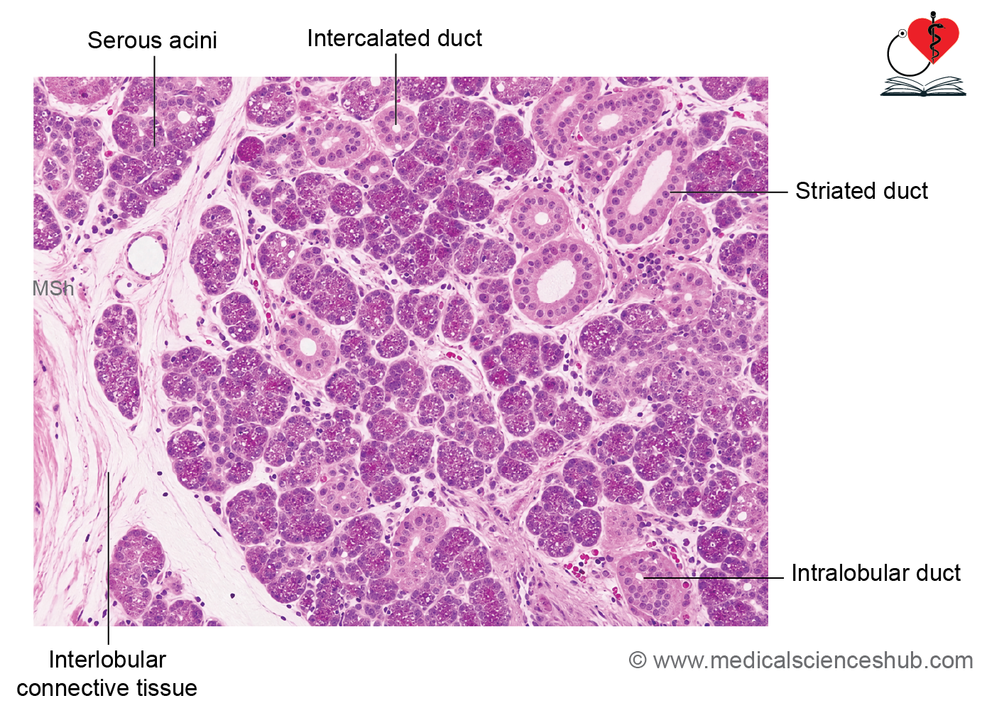
- The parotid gland is the largest of the major salivary glands and is a compound tubuloacinar serous gland.
- It is enclosed by a dense connective tissue capsule that divides the gland into lobes and lobules.
- Its main excretory duct, the Stensen duct, opens into the oral vestibule opposite the upper second molar tooth.
- The gland is composed almost entirely of serous acini, which produce a watery, enzyme-rich secretion.
- Acini are surrounded by myoepithelial cells that assist in expelling secretions.
- The duct system includes intercalated, striated, interlobular, and lobar ducts.
- Intercalated ducts are lined by simple cuboidal epithelium, whereas striated ducts are lined by simple columnar epithelium with characteristic basal infoldings.
- Well-developed striated ducts are a prominent histological feature of the parotid gland.
- Larger ducts show a gradual transition to stratified cuboidal or stratified squamous epithelium near their openings.
Submandibular Gland
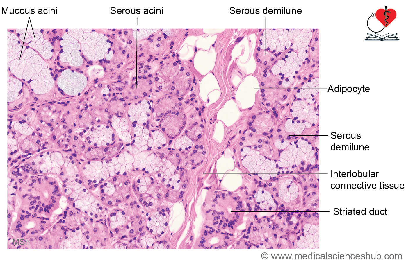
- The submandibular gland is a mixed compound tubuloacinar salivary gland, producing both serous and mucous secretions.
- Serous acini predominate, making the secretion mainly serous in nature.
- The gland is enclosed by a well-defined connective tissue capsule, which sends septa inward to divide it into lobes and lobules.
- Histologically, it contains numerous serous acini along with a smaller number of mucous acini, some of which are associated with serous demilunes.
- The duct system includes intercalated ducts, striated ducts, interlobular ducts, and larger lobar ducts.
- Compared with the parotid gland, intercalated ducts are fewer in number and shorter in length.
- In contrast, striated ducts are relatively longer and more prominent.
- The combination of predominantly serous acini and well-developed striated ducts is a characteristic feature of the submandibular gland.
Sublingual Gland
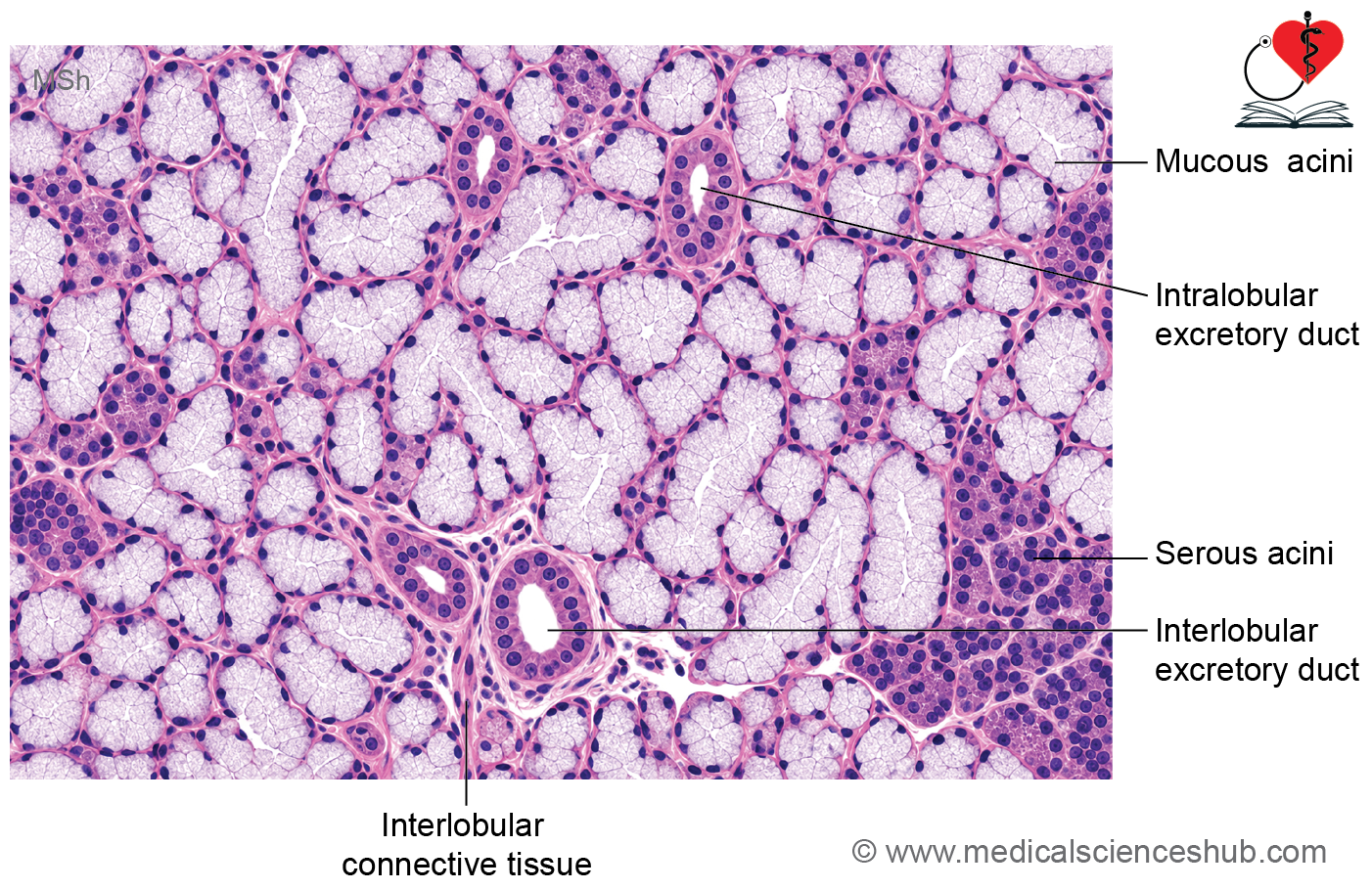
- The sublingual gland is the smallest of the major salivary glands and is a mixed compound tubuloacinar gland.
- Its secretion is predominantly mucous, with a smaller contribution from serous cells.
- Unlike the parotid and submandibular glands, it has a poorly developed or incomplete connective tissue capsule.
- The gland contains mainly mucous secretory units, which are often tubular in shape rather than purely acinar.
- A few serous and mixed (seromucous) acini may also be present.
- The duct system is relatively simple, with intercalated ducts being absent or very sparse and striated ducts poorly developed.
- Interlobular and larger excretory ducts are present and convey secretions to the oral cavity.
- The predominance of mucous acini is the key histological feature of the sublingual gland.
CLINICAL CORRELATION
- Pleomorphic adenoma is the most common benign salivary gland tumor.
- It arises from both ductal epithelial and myoepithelial cells, giving it a mixed histological appearance.
- The tumor most frequently involves the parotid gland and is therefore often termed a benign mixed tumor.
Important Questions
- Draw a well-labelled diagram of histology/ microanatomy of tongue.
- Draw a well-labelled diagram of histology/ microanatomy of circumvallate papilla.
📝 Test Your Knowledge – Practice MCQs
Attempt the chapter MCQ quiz and assess your understanding of key concepts.
Click to see: Medical Disclaimer and Copyright Notice