
Competency
- AN52.1 Describe and identify the microanatomical features of gastrointestinal system: Duodenum, jejunum, ileum, large intestine,appendix.
Introduction
- The small and large intestines form the major portion of the gastrointestinal tract. The small intestine is primarily responsible for digestion and nutrient absorption, while the large intestine absorbs water and electrolytes and forms and stores feces.
Small Intestine
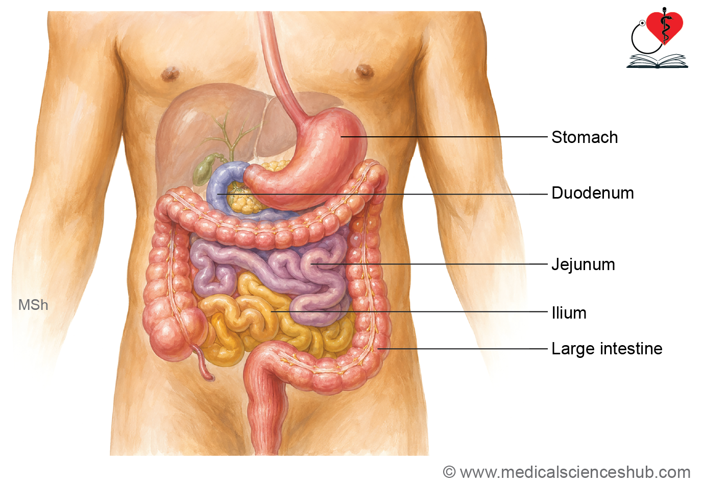
- The small intestine is a highly specialized digestive tube measuring approximately 5–6 m in length in a living adult and is the principal site of digestion and nutrient absorption.
- It is divided into three regions:
- Duodenum: The first segment, about 25 cm long, extending from the stomach to the duodenojejunal junction.
- Jejunum: The proximal two-fifths of the remaining small intestine, specialized for absorption.
- Ileum: The distal three-fifths, terminating at the ileocecal junction.
- Collectively, the jejunum and ileum are often referred to as the small bowel.
General Features of Small Intestine
- The small intestine is the primary site for digestion and absorption of nutrients.
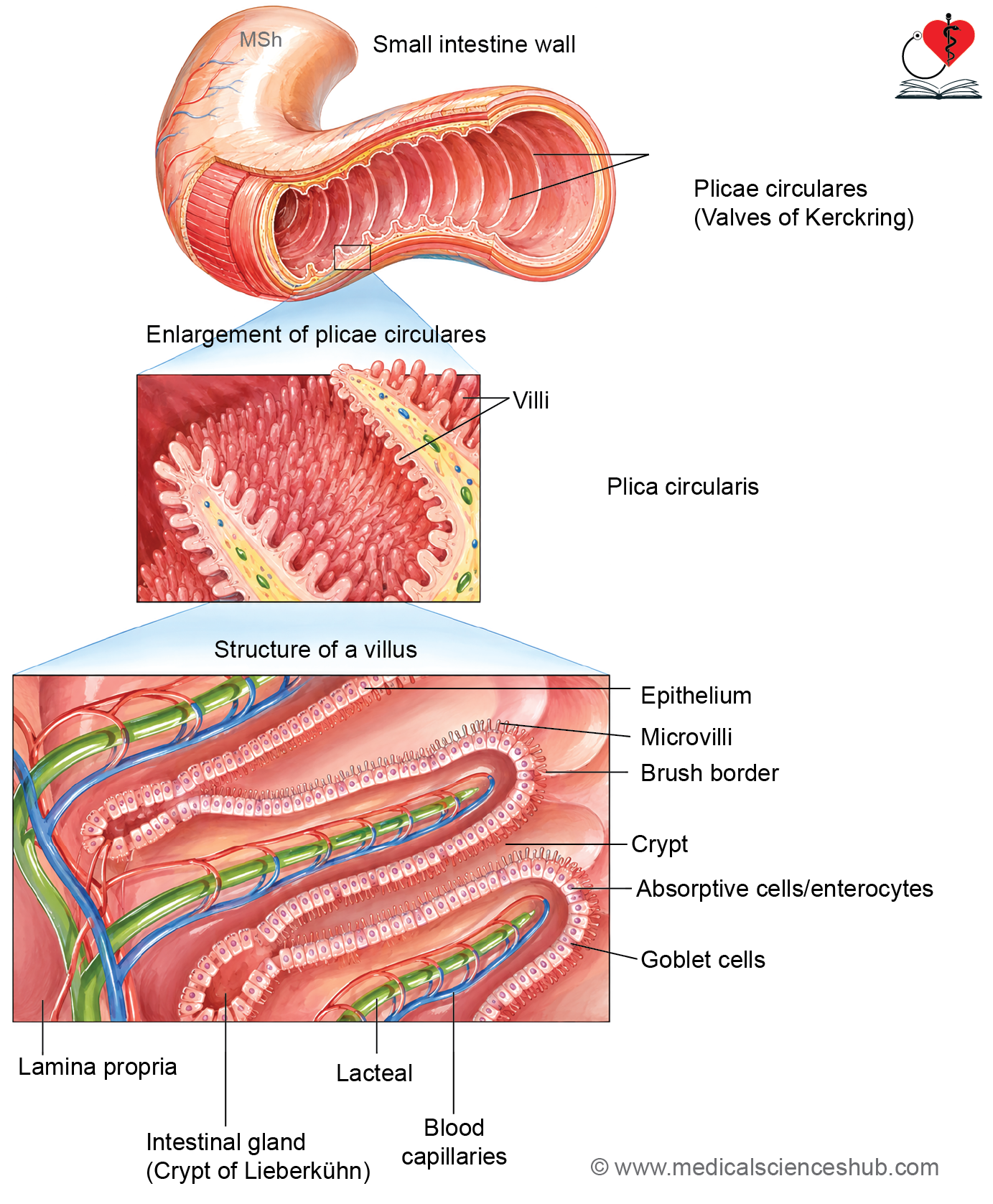
- To maximize absorptive efficiency, its mucosa exhibits several structural modifications that greatly increase the available surface area.
Surface Area Enhancements
- Plicae circulares (circular folds or valves of Kerckring) are permanent folds composed of mucosa and submucosa.
- They are most prominent in the duodenum (except the proximal few centimeters) and jejunum.
- They become less numerous distally and are largely absent in the terminal ileum.
- These folds slow the movement of intestinal contents and increase absorptive surface area.
- Villi are finger-like or leaf-shaped projections of the mucosa.
- Each villus contains a core of lamina propria with blood capillaries and a central lacteal (lymphatic vessel).
- Villi impart a velvety appearance to the intestinal lining.
- Their height gradually decreases from the duodenum toward the ileum.
- Microvilli are microscopic projections on the apical surface of absorptive epithelial cells.
- Together they form the brush border.
- They markedly increase the membrane surface available for digestion and absorption.
- Crypts of Lieberkühn are simple tubular intestinal glands located between adjacent villi.
- They extend through the lamina propria to the level of the muscularis mucosae.
- The glands open into the spaces between villi and are lined by epithelial cells continuous with the villous epithelium.
- The villus-to-crypt ratio is highest in the proximal small intestine and gradually decreases toward the distal ileum.
- The combined effects of plicae circulares, villi, microvilli, and intestinal crypts create an extensive absorptive surface, enabling efficient nutrient uptake.
Cells of Small Intestine
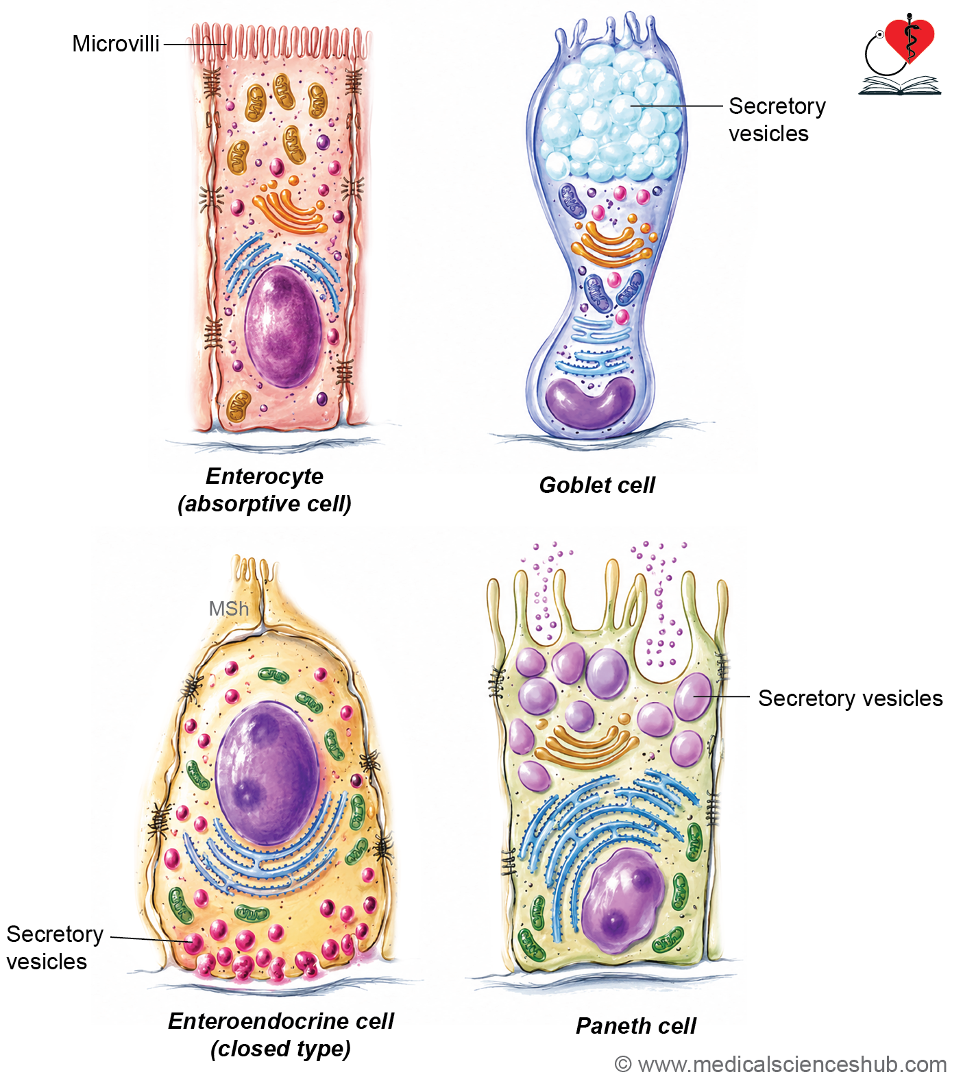
- The mucosa of the small intestine is lined by simple columnar epithelium composed of six principal cell types: enterocytes, goblet cells, Paneth cells, enteroendocrine cells, M cells, and stem (intermediate) cells.
Enterocytes/Absorptive Columnar Cells
- Enterocytes are the most abundant epithelial cells and are responsible for nutrient absorption.
- They are tall columnar cells with basally located oval nuclei.
- Their apical surface bears numerous microvilli, forming the brush border.
- Microvilli are covered by a glycocalyx containing digestive enzymes involved in the final breakdown of carbohydrates and peptides.
- Adjacent enterocytes are connected by tight junctions, which maintain the intestinal barrier.
- These cells have a short lifespan of about 4–6 days and are continuously replaced.
- Functions: Absorption of nutrients and secretion of enzymes required for terminal digestion.
Goblet Cells
- Goblet cells are unicellular mucus-secreting glands scattered among enterocytes.
- Their number increases progressively from the duodenum toward the ileum.
- They have a narrow basal portion and an expanded apical region filled with mucin granules.
- The nucleus is flattened and displaced toward the base.
- Function: Production of mucus that lubricates and protects the intestinal surface.
Paneth Cells (Zymogen Cells)
- Paneth cells are located at the base of the crypts of Lieberkühn.
- They contain prominent eosinophilic secretory granules and basally placed nuclei.
- Unlike most intestinal epithelial cells, they migrate toward the crypt base.
- Their lifespan is approximately one month.
- Functions:
- Secretion of lysozyme, α-defensins, and other antimicrobial substances.
- Regulation of intestinal microbial flora.
- Contribution to innate immunity and protection of intestinal stem cells.
Enteroendocrine/Argentaffin Cells
- These specialized cells are dispersed within the intestinal epithelium and crypts.
- They release regulatory peptides and hormones into the surrounding tissues and bloodstream.
- Open-type cells contact the lumen and respond to luminal contents.
- Closed-type cells do not reach the lumen.
- Functions: Secretion of hormones such as secretin, cholecystokinin (CCK), gastric inhibitory peptide (GIP), motilin, and other regulatory mediators that coordinate digestion.
M Cells
- M cells overlie Peyer’s patches and other lymphoid aggregates.
- Their apical surface has microfolds rather than microvilli.
- They contain basolateral pockets housing lymphocytes, macrophages, and dendritic cells.
- Function: Transport luminal antigens and microorganisms to immune cells, facilitating mucosal immune surveillance.
Intermediate Cells (Undifferentiated/Stem Cells)
- Stem cells are located primarily in the lower portions of the intestinal crypts.
- They continuously divide and replenish all epithelial cell types of the intestine.
- These cells are difficult to identify in routine histological preparations.
- Function: Maintenance and regeneration of the intestinal epithelium.
Table 15.1: Cells of small intestine
| Cell Type | Histological Features | Principal Functions |
|---|---|---|
| Enterocyte | Tall columnar absorptive cell with an oval nucleus near the base and a prominent brush border (striated border) | Absorption of nutrients and secretion of digestive enzymes associated with the brush border |
| Goblet Cell | Flask-shaped cell with a mucus-filled apical cytoplasm, flattened basal nucleus, and strong PAS-positive staining | Production and secretion of mucus for lubrication and protection of the intestinal lining |
| Paneth Cell | Located at the base of intestinal crypts; contains intensely eosinophilic cytoplasm, basal nucleus, and large secretory granules | Innate immune defense through secretion of lysozyme, α-defensins, and other antimicrobial substances; helps regulate normal intestinal microbiota |
| Enteroendocrine Cell | Scattered cells within the epithelium; identified by argentaffin or silver-staining properties | Secretion of gastrointestinal hormones and regulatory peptides that influence digestion and intestinal motility |
| M (Microfold) Cell | Specialized epithelial cell found overlying lymphoid follicles and Peyer’s patches; lacks a typical brush border | Uptake and transport of luminal antigens to underlying lymphocytes and antigen-presenting cells, facilitating immune surveillance |
| Stem (Undifferentiated) Cell | Columnar cell located in the lower part of intestinal crypts; possesses short, irregular microvilli | Continuous renewal of the intestinal epithelium by differentiating into various intestinal cell types |
Duodenum
- The duodenum is the first and shortest segment of the small intestine, measuring approximately 25 cm in length.
- It receives acidic chyme from the stomach and serves as the primary site for mixing with digestive secretions.
- Bile from the liver and pancreatic juice enter the second part of the duodenum through the major duodenal papilla.
- The alkaline bicarbonate-rich pancreatic secretion helps neutralize gastric acid and creates an optimal environment for digestive enzymes.
- The proximal few centimeters are intraperitoneal, whereas most of the duodenum is retroperitoneal.
Histology of Duodenum
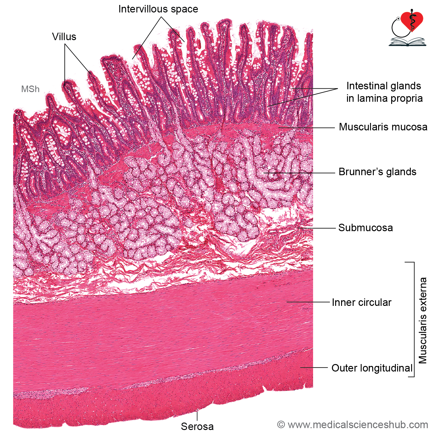
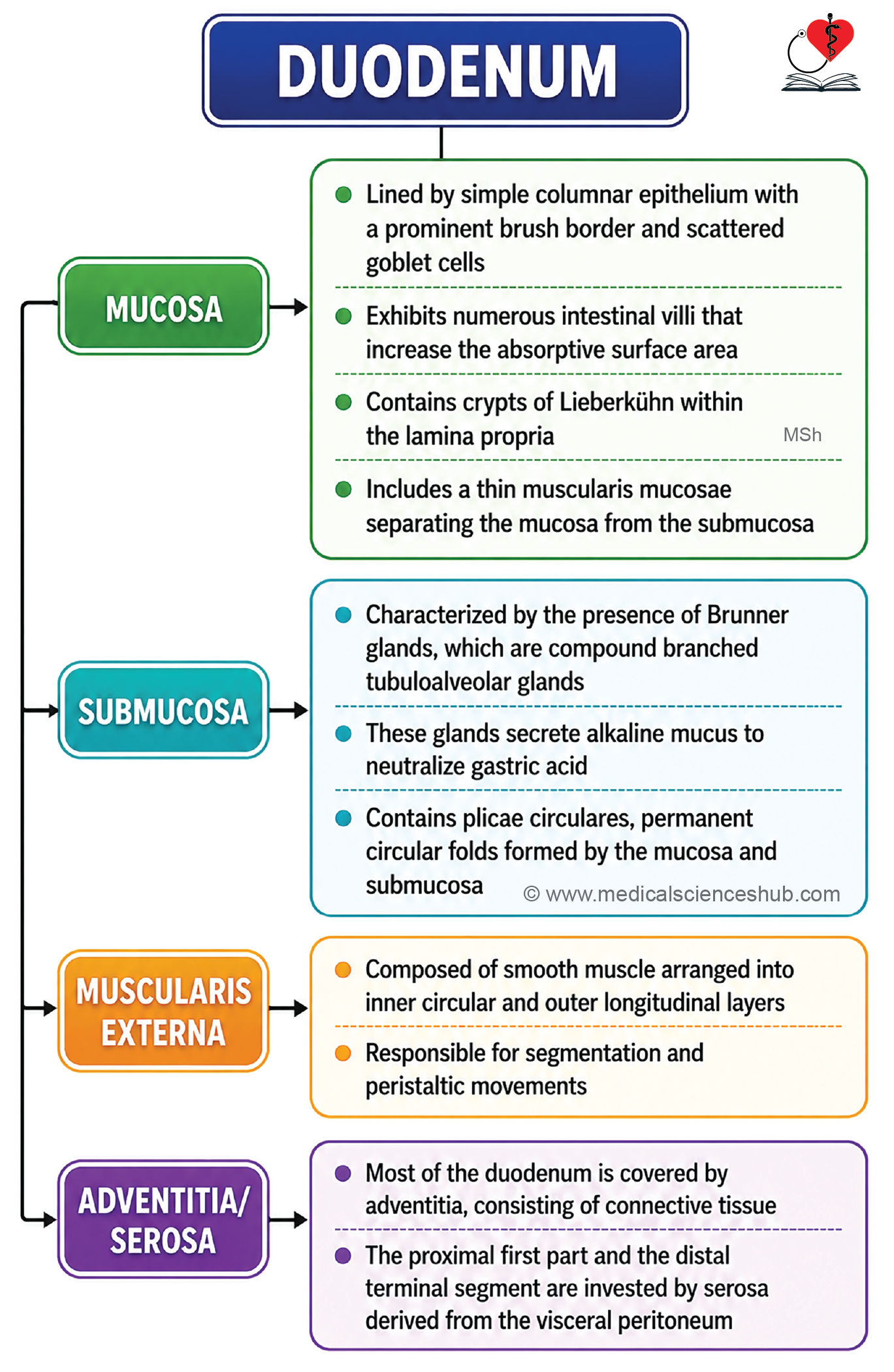
- The duodenal wall consists of mucosa, submucosa, muscularis externa, and serosa/adventitia.
Mucosa
- The mucosa exhibits several adaptations for absorption:
- Plicae circulares, permanent folds containing a core of submucosa.
- Villi, finger-like projections with a core of lamina propria.
- Each villus contains blood capillaries, a central lacteal (lymphatic vessel), and smooth muscle fibers.
- The surface is lined by simple columnar absorptive cells (enterocytes) with a brush border formed by microvilli.
- Goblet cells are present but relatively fewer than in the distal small intestine.
- Between villi are crypts of Lieberkühn, which contain enterocytes, goblet cells, Paneth cells, enteroendocrine cells, and stem cells.
- The muscularis mucosae consists of inner circular and outer longitudinal smooth muscle layers, with some fibers extending into the villi.
Submucosa
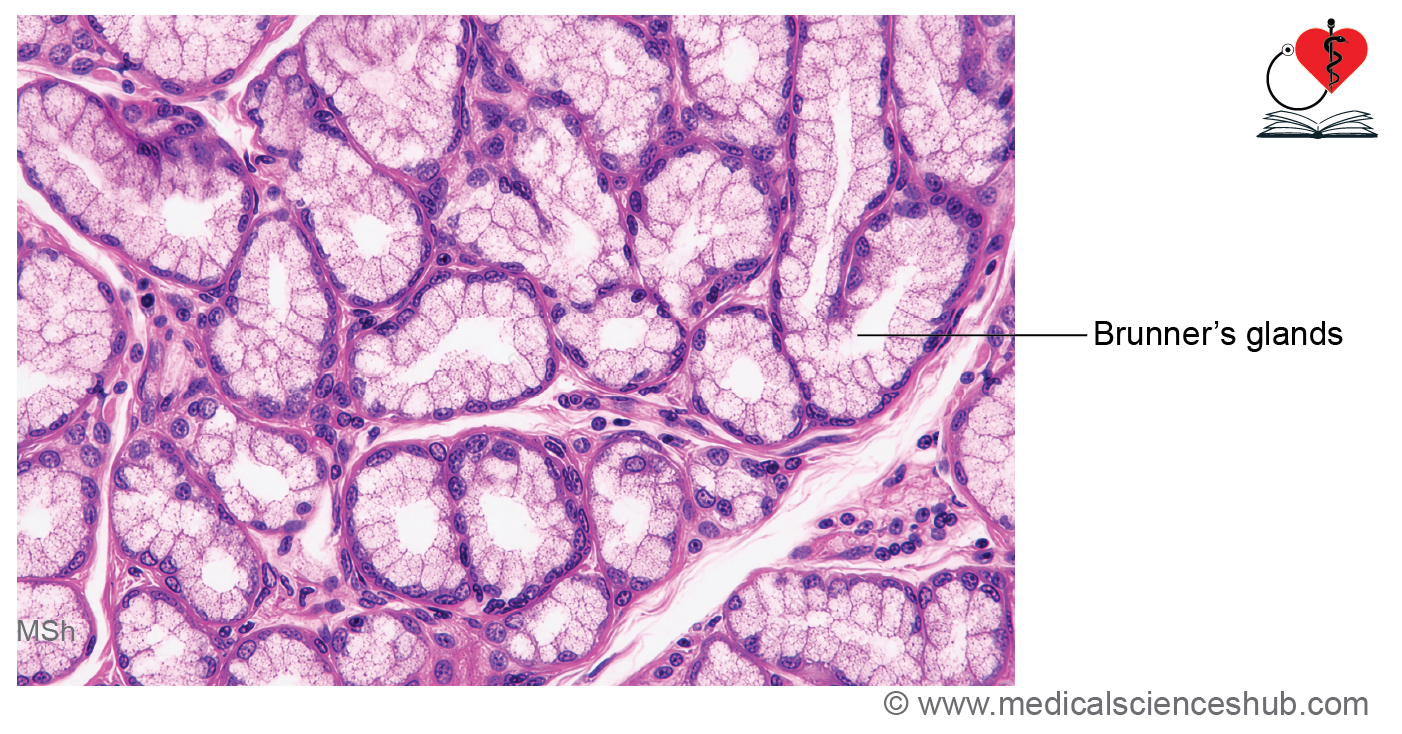
- The characteristic feature of the duodenum is the presence of Brunner’s glands.
- These are branched tubuloalveolar mucous glands located within the submucosa.
- Their ducts pass through the muscularis mucosae and open into the intestinal crypts.
- The glandular cells are mucous-secreting cells with pale cytoplasm and basally placed nuclei.
Functions of Brunner’s Glands:
- Secrete alkaline mucus that protects the duodenal mucosa from acidic gastric contents.
- Contribute bicarbonate-rich secretions that aid in neutralizing luminal acidity.
- Produce growth-promoting factors, including epidermal growth factor (EGF), which support mucosal repair and regeneration.
Muscle Layer
- The muscularis externa consists of inner circular and outer longitudinal smooth muscle layers responsible for intestinal motility.
Serosa/Adventitia
- Most of the duodenum is covered by adventitia, while a small proximal segment is covered by serosa.
Jejunum
- The jejunum is the middle segment of the small intestine, situated between the duodenum and ileum.
- It measures approximately 2 m in length and is a major site for nutrient absorption.
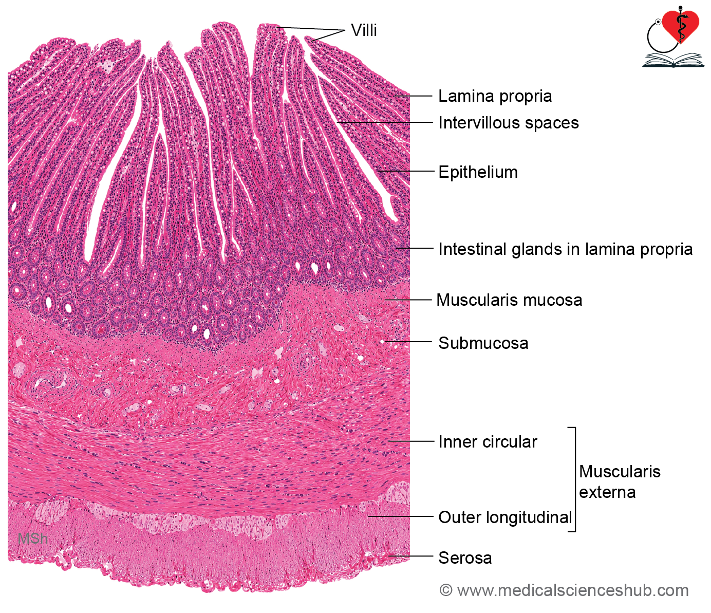
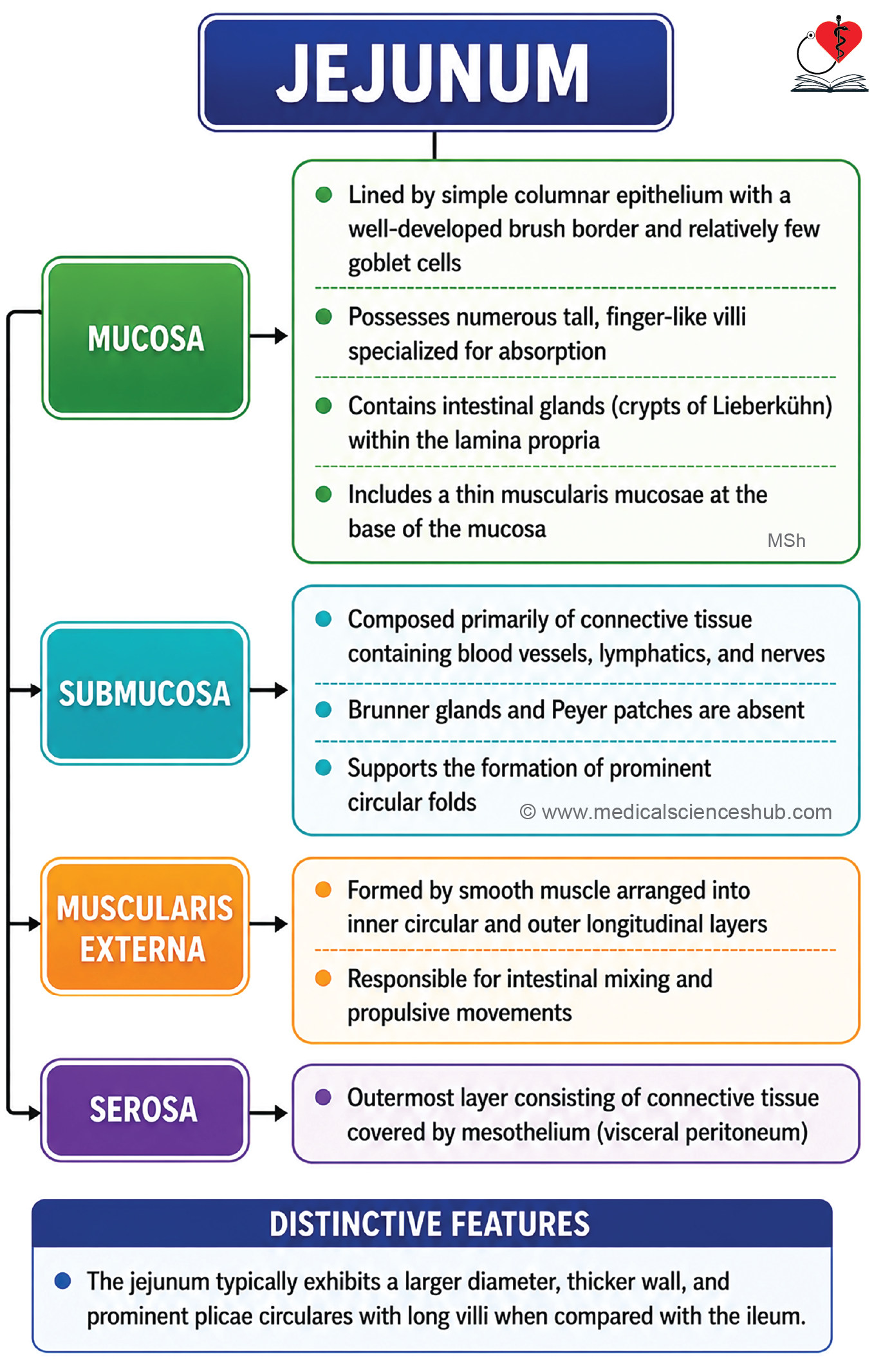
- Its wall consists of four layers: mucosa, submucosa, muscularis externa, and serosa.
Mucosa
- The mucosa contains prominent plicae circulares, permanent folds that increase the absorptive surface area.
- It also possesses numerous villi, which are typically tall and finger-like to tongue-shaped, giving the mucosa a characteristic appearance.
- Each villus contains a core of lamina propria with blood capillaries and a central lacteal.
- The lining epithelium consists of absorptive cells with microvilli and scattered goblet cells.
- Crypts of Lieberkühn and the muscularis mucosae are similar in structure to those of the duodenum.
Submucosa
- The submucosa is composed of connective tissue containing blood vessels, lymphatics, and the submucosal (Meissner’s) plexus.
- Unlike the duodenum and ileum, the jejunum lacks Brunner’s glands and Peyer’s patches, an important distinguishing feature.
Muscularis Externa
The muscularis externa consists of inner circular and outer longitudinal smooth muscle layers responsible for intestinal motility.
Serosa/Adventitia
The outer surface is covered by serosa (visceral peritoneum).
Ileum
- The ileum is the distal and longest segment of the small intestine, measuring approximately 3 m in length.
- It terminates at the ileocecal junction, where it opens into the cecum.
- Its wall consists of four layers: mucosa, submucosa, muscularis externa, and serosa.
Mucosa
- Plicae circulares are less prominent than in the jejunum and may be absent in the terminal ileum.
- The mucosa contains relatively short and sparse villi, which are typically finger-like in shape.
- The surface is lined by simple columnar epithelium with microvilli (brush border) and numerous goblet cells, which increase in number toward the distal intestine.
- The lamina propria contains intestinal glands (crypts of Lieberkühn) and abundant lymphoid tissue.
- The characteristic feature of the ileum is the presence of Peyer’s patches, which are aggregated lymphoid nodules involved in mucosal immunity.
- Peyer’s patches are primarily located in the lamina propria and frequently extend into the submucosa.
- The muscularis mucosae may become thin or interrupted over large Peyer’s patches.
Submucosa, Muscularis Externa, Serosa
- The submucosa contains connective tissue, blood vessels, lymphatics, and portions of Peyer’s patches.
- The muscularis externa consists of inner circular and outer longitudinal smooth muscle layers.
- The outer surface is covered by serosa (visceral peritoneum).
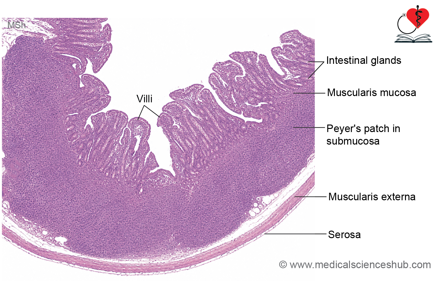
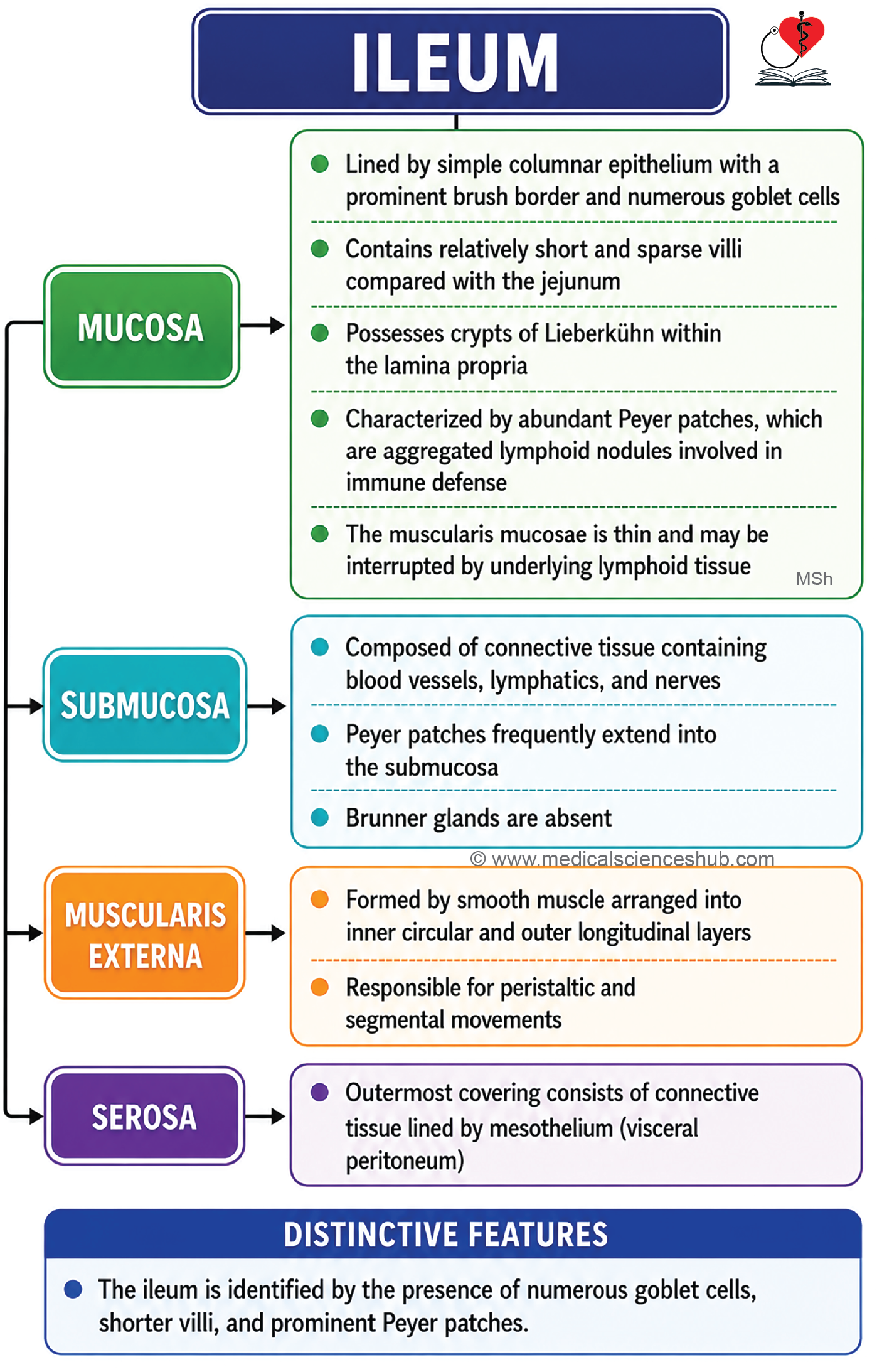
Table 15.2: Comparison between duodenum, jejunum, and ileum
| Feature | Duodenum | Jejunum | Ileum |
|---|---|---|---|
| Wall thickness | Relatively thick | Thick | Comparatively thin |
| Lumen | Broad | Broad | Narrower |
| Epithelium | Simple columnar epithelium with relatively few goblet cells | Simple columnar epithelium with relatively few goblet cells | Simple columnar epithelium with abundant goblet cells |
| Plicae circulares | Large and well developed | Most prominent and numerous | Small, sparse, or absent in distal part |
| Villi | Numerous, broad, and finger-shaped | Long, slender, and tongue-shaped | Shorter, fewer, and finger-shaped |
| Lamina propria | No aggregated lymphoid nodules | Scant lymphoid tissue | Contains prominent aggregated lymphoid nodules (Peyer’s patches) |
| Muscularis mucosae | Well developed | Well developed | Relatively thin |
| Submucosa | Contains characteristic Brunner’s glands | Brunner’s glands absent | Brunner’s glands absent; Peyer’s patches may extend into the submucosa |
| Special histological feature | Presence of Brunner’s glands producing alkaline mucus | Prominent plicae circulares and tall villi for maximal absorption | Abundant Peyer’s patches providing immune surveillance |
| Muscularis externa | Inner circular and outer longitudinal smooth muscle layers | Inner circular and outer longitudinal smooth muscle layers | Inner circular and outer longitudinal smooth muscle layers |
| Outer covering | Mostly serosa; proximal part may be covered by adventitia | Covered by serosa | Covered by serosa |
Peyer’s patches
- Peyer’s patches are aggregates of lymphoid nodules located primarily in the lamina propria of the terminal ileum.
- They appear as elongated areas of lymphoid tissue along the antimesenteric border of the ileum.
- Numerous Peyer’s patches are present and collectively form an important component of gut-associated lymphoid tissue (GALT).
- Large lymphoid aggregates may extend through the muscularis mucosae into the submucosa.
- The epithelium overlying Peyer’s patches contains specialized M cells (microfold cells).
- M cells possess surface folds rather than microvilli and transport luminal antigens to underlying lymphocytes, macrophages, and dendritic cells.
- Functions:
- Monitor intestinal microorganisms and foreign antigens.
- Initiate local immune responses.
- Help maintain normal intestinal microbial balance.
- Contribute to protection against enteric infections.
Large Intestine
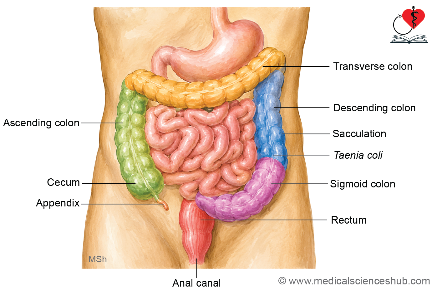
- The large intestine comprises the cecum, vermiform appendix, colon (ascending, transverse, descending, and sigmoid), rectum, and anal canal.
- Three characteristic gross anatomical features help identify the colon:
- Taeniae coli: Three longitudinal bands of smooth muscle formed from the outer longitudinal layer of the muscularis externa. They are absent in the appendix, rectum, and anal canal.
- Haustra: Sac-like dilatations of the colonic wall produced by the arrangement of the taeniae coli.
- Appendices epiploicae: Small peritoneal pouches filled with adipose tissue attached to the outer surface of the colon. These are absent in the rectum.
- These structural features contribute to the distinctive appearance and motility of the large intestine.
Colon
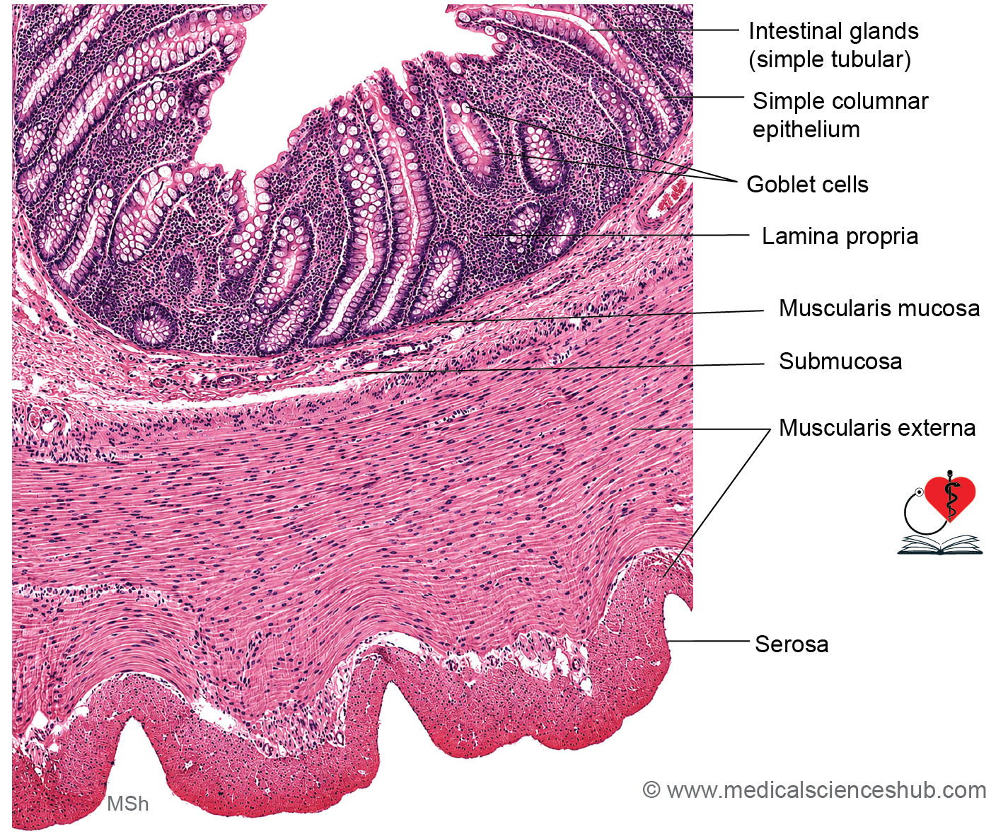
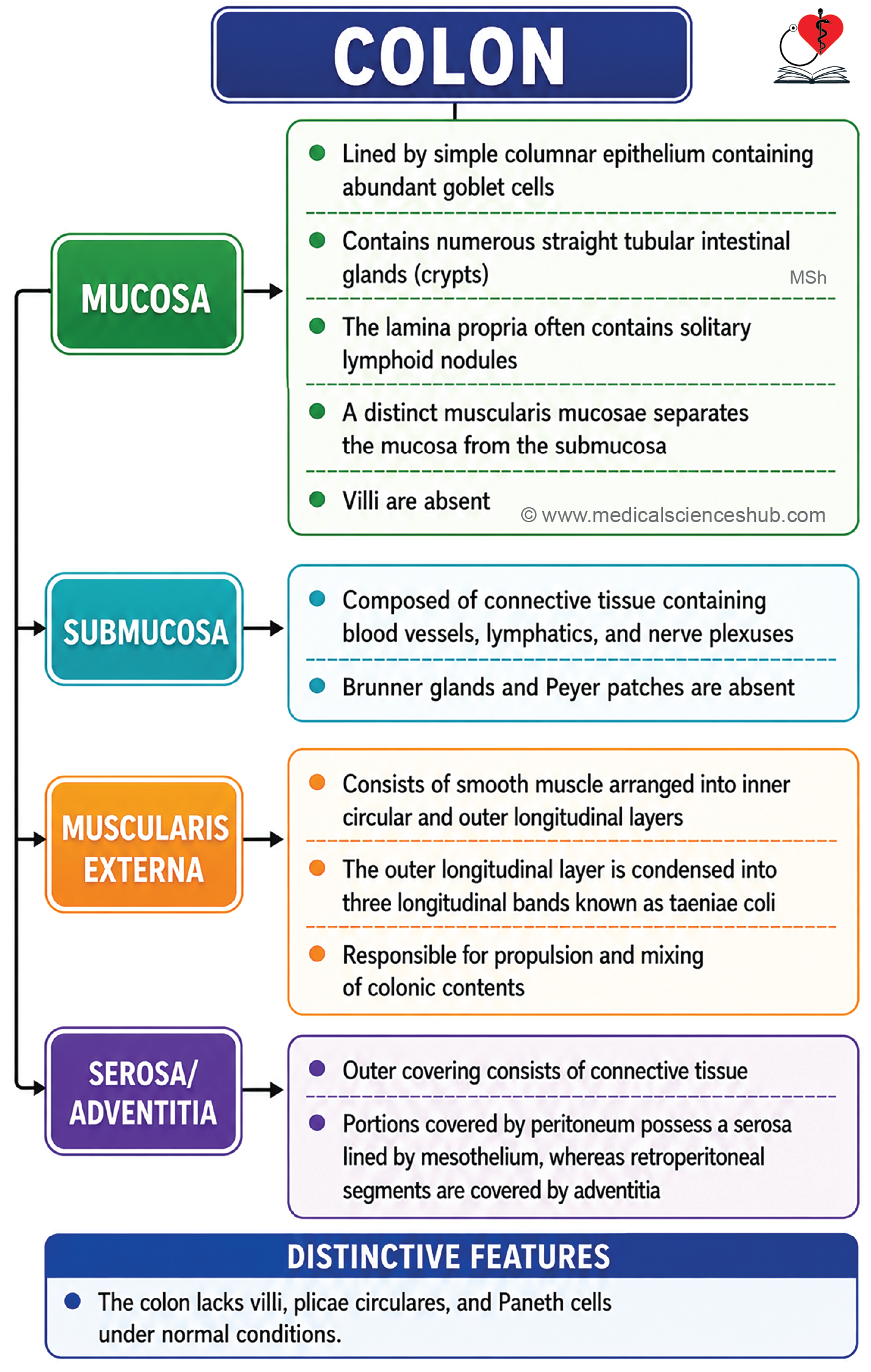
- The wall of the colon consists of four layers: mucosa, submucosa, muscularis externa, and serosa.
Mucosa
- The mucosa is composed of epithelium, lamina propria, and muscularis mucosae.
- The surface is lined by simple columnar epithelium containing numerous goblet cells and absorptive cells with short microvilli.
- Unlike the small intestine, the colon lacks villi and plicae circulares, resulting in a relatively smooth mucosal surface.
- The lamina propria contains numerous straight crypts of Lieberkühn, which extend deep into the mucosa.
- These glands are lined predominantly by absorptive cells and abundant goblet cells that secrete mucus for lubrication.
- Paneth cells are generally absent in the normal colon.
- Stem cells are located at the base of the crypts and continuously renew the epithelium.
- The colon is specialized for absorption of water and electrolytes and for mucus production.
- The lamina propria contains a dense collagen table beneath the epithelium, which helps regulate fluid and electrolyte transport.
- A pericryptal fibroblast sheath surrounds the glands and contributes to epithelial maintenance.
- Numerous lymphoid nodules and scattered immune cells provide local immune defense.
- Occasional lymphatic vessels are also present within the lamina propria.
Submucosa
The submucosa consists of dense irregular connective tissue containing blood vessels, lymphatics, and nerve fibers.
Muscularis Externa
- It is composed of an inner circular and an outer longitudinal smooth muscle layer.
- The outer longitudinal layer is condensed into three thick bands known as taeniae coli, a characteristic feature of the colon.
- Because the taeniae coli are shorter than the intestinal wall, the colon develops sac-like dilatations called haustra.
Serosa
- Most intraperitoneal portions of the colon are covered by serosa (visceral peritoneum), whereas retroperitoneal segments are covered by adventitia.
- The outer surface often shows fat-filled peritoneal projections called appendices epiploicae, which are characteristic of the colon.
Vermiform Appendix
- The vermiform appendix is a narrow, blind-ended tubular diverticulum arising from the cecum and is considered the narrowest part of the intestinal tract.
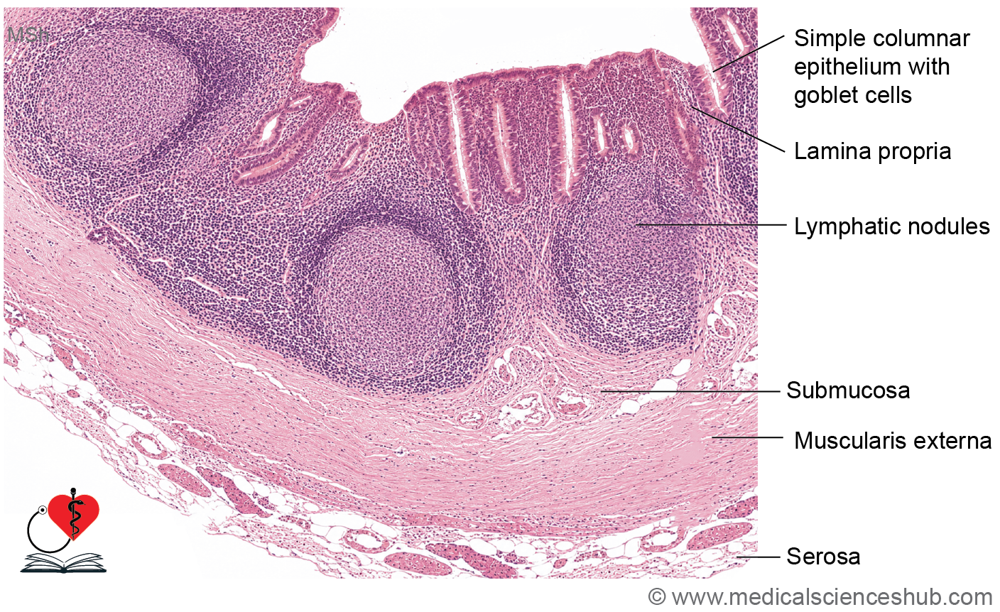
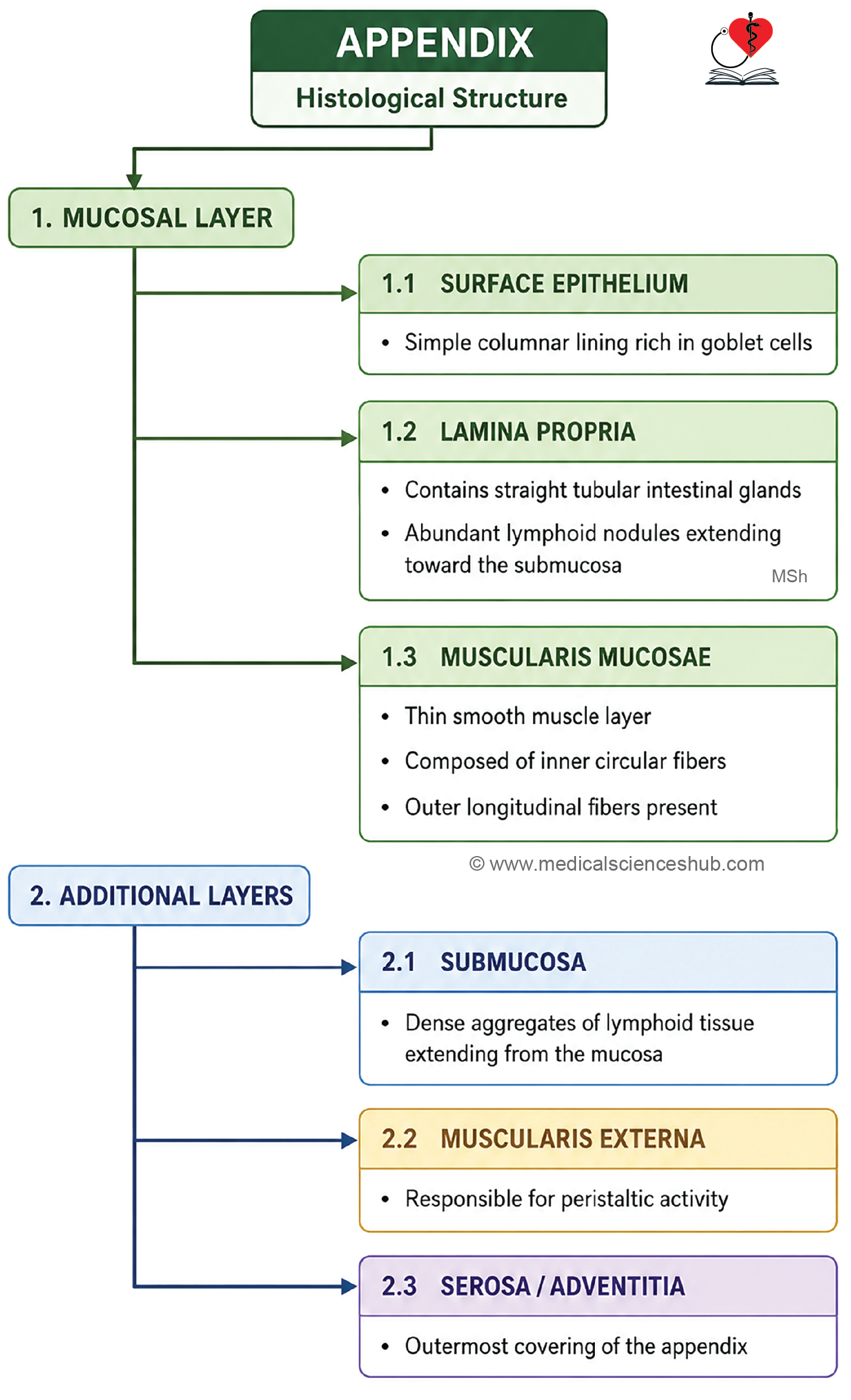
- Histologically, its wall consists of the four typical layers of the gastrointestinal tract: mucosa, submucosa, muscularis externa, and serosa.
Mucosa
- The mucosa is lined by simple columnar epithelium containing absorptive cells with microvilli, goblet cells, and occasional M cells.
- The lamina propria contains short crypts of Lieberkühn lined by columnar and goblet cells.
- A characteristic feature is the abundance of lymphoid follicles, which occupy much of the lamina propria and may extend through the muscularis mucosae.
Submucosa
The submucosa consists of connective tissue and contains extensions of the prominent lymphoid tissue from the mucosa.
Muscularis Externa
- It is composed of inner circular and outer longitudinal smooth muscle layers.
- Unlike the colon, the appendix lacks taeniae coli, an important distinguishing feature.
Serosa
The appendix is completely covered by serosa (visceral peritoneum), reflecting its intraperitoneal location.
Anal Canal
- The anal canal is the terminal segment of the gastrointestinal tract and measures approximately 4 cm in length.
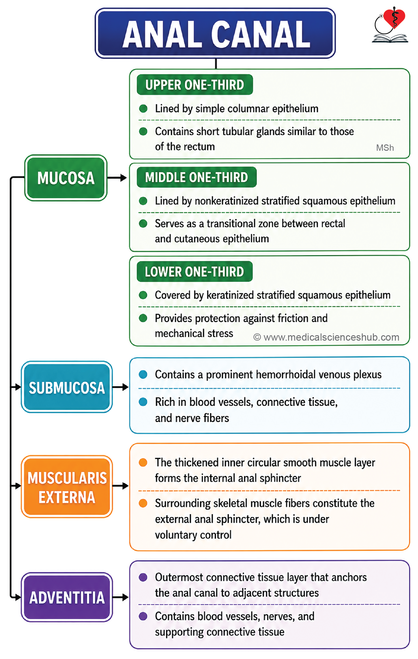
- Its wall consists of mucosa, submucosa, muscularis externa, and adventitia.
- The muscularis mucosae gradually disappears in the middle portion of the canal.
Mucosa
- The epithelial lining undergoes a characteristic transition:
- Upper one-third: Simple columnar epithelium.
- Middle one-third: Nonkeratinized stratified squamous epithelium.
- Lower one-third (below the pectinate line): Keratinized stratified squamous epithelium, continuous with the skin.
- The upper part contains 6–12 longitudinal mucosal folds known as anal columns.
- Adjacent anal columns are connected by transverse folds called anal valves.
- The line formed by the anal valves is the pectinate line, an important anatomical and embryological landmark.
- Small recesses above the anal valves form anal sinuses, which receive the openings of circumanal glands.
- The junction between the middle and lower thirds is marked by the white line of Hilton.
Submucosa
- The submucosa contains prominent venous networks called hemorrhoidal venous plexuses.
- These are divided into:
- Internal hemorrhoidal plexus above the pectinate line.
- External hemorrhoidal plexus below the pectinate line.
Muscularis Externa
- It consists of inner circular and outer longitudinal smooth muscle layers.
- Thickening of the inner circular layer forms the internal anal sphincter (involuntary smooth muscle).
- Outside the muscularis externa lies skeletal muscle forming the external anal sphincter, which is under voluntary control.
Adventitia
The outermost covering is adventitia, composed of connective tissue that anchors the anal canal to surrounding structures.
CLINICAL CORRELATION
- Anal fistula is an abnormal tract that creates a communication between the anal canal and the perianal skin.
- Internal hemorrhoids result from dilation of the internal hemorrhoidal venous plexus above the pectinate line. They may become more prominent in conditions associated with portal hypertension.
- External hemorrhoids arise from the external hemorrhoidal venous plexus below the pectinate line and are covered by skin.
- Colorectal adenocarcinoma is a common malignant tumor originating from the glandular epithelium of the large intestine.
- Ulcerative colitis is a chronic inflammatory disorder affecting the colon and rectum. It primarily involves the mucosa and submucosa and commonly presents with bloody diarrhea.
- Typhoid ulcers occur in the ileum due to infection by Salmonella Typhi. They are associated with inflammation of lymphoid tissue, particularly Peyer’s patches, followed by mucosal ulceration.
- Celiac disease is an immune-mediated disorder triggered by dietary gluten. It causes villous atrophy in the small intestine, leading to malabsorption.
- Crohn’s disease is a chronic inflammatory bowel disease that can involve any part of the gastrointestinal tract, most frequently the ileum and colon.
- Diverticulosis is characterized by herniation of the mucosa and submucosa through weak areas of the colonic wall, often adjacent to the taeniae coli.
Important Questions
- List the cells of small intestine with their functions.
- Write a short note on histology of duodenum.
- Write a short note on histology of jejunum.
- Write a short note on Peyer’s patches.
- Write a short note on histology of vermiform appendix.
📝 Test Your Knowledge – Practice MCQs
Attempt the chapter MCQ quiz and assess your understanding of key concepts.
Click to see: Medical Disclaimer and Copyright Notice