
Competency
- AN52.2 Describe and identify the microanatomical features of urinary system: Kidney, ureter and urinary bladder.
Introduction
- The urinary system consists of:
- A pair of kidneys
- A pair of ureters
- The urinary bladder
- The urethra
- The kidneys maintain internal homeostasis by filtering blood and producing urine.
Functions of the Kidneys
- Kidneys remove metabolic waste products and excess substances through ultrafiltration and urine formation.
- They regulate body fluid volume and maintain the balance of electrolytes, osmolarity, and acid-base status.
- The kidneys also function as important endocrine organs. It produces:
- Erythropoietin:
- Stimulates red blood cell production in the bone marrow.
- Its secretion increases in response to reduced blood oxygen levels.
- Renin:
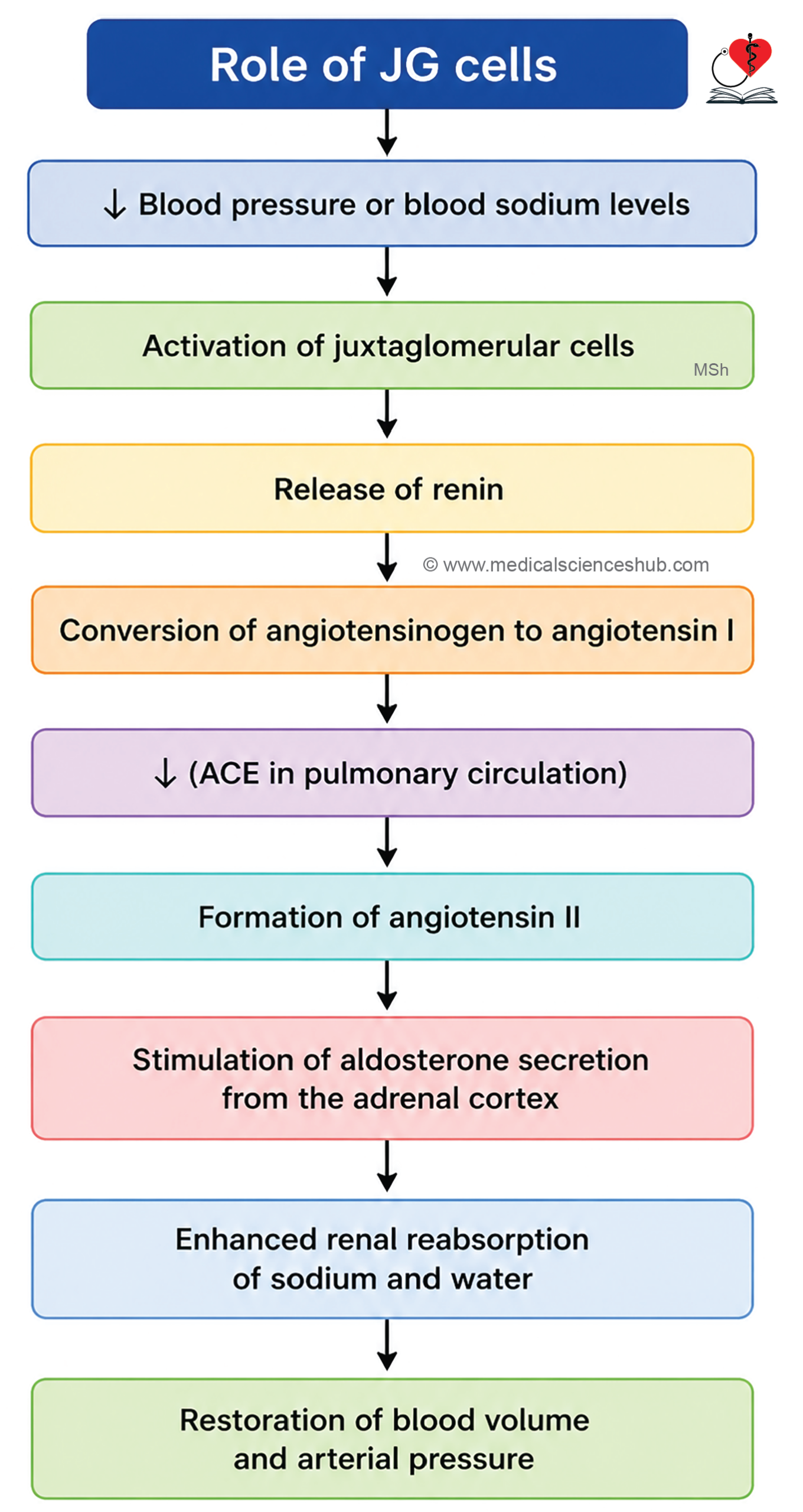
- Initiates the renin–angiotensin–aldosterone system (RAAS) by converting angiotensinogen to angiotensin I.
- Plays a key role in regulating blood pressure and fluid balance.
- Active Vitamin D (Calcitriol):
- Renal 1α-hydroxylase converts 25-hydroxyvitamin D into 1,25-dihydroxyvitamin D₃ (calcitriol).
- Calcitriol promotes intestinal absorption of calcium and phosphate and supports normal bone metabolism.
- Erythropoietin:
Kidney
Gross Structure of Kidney
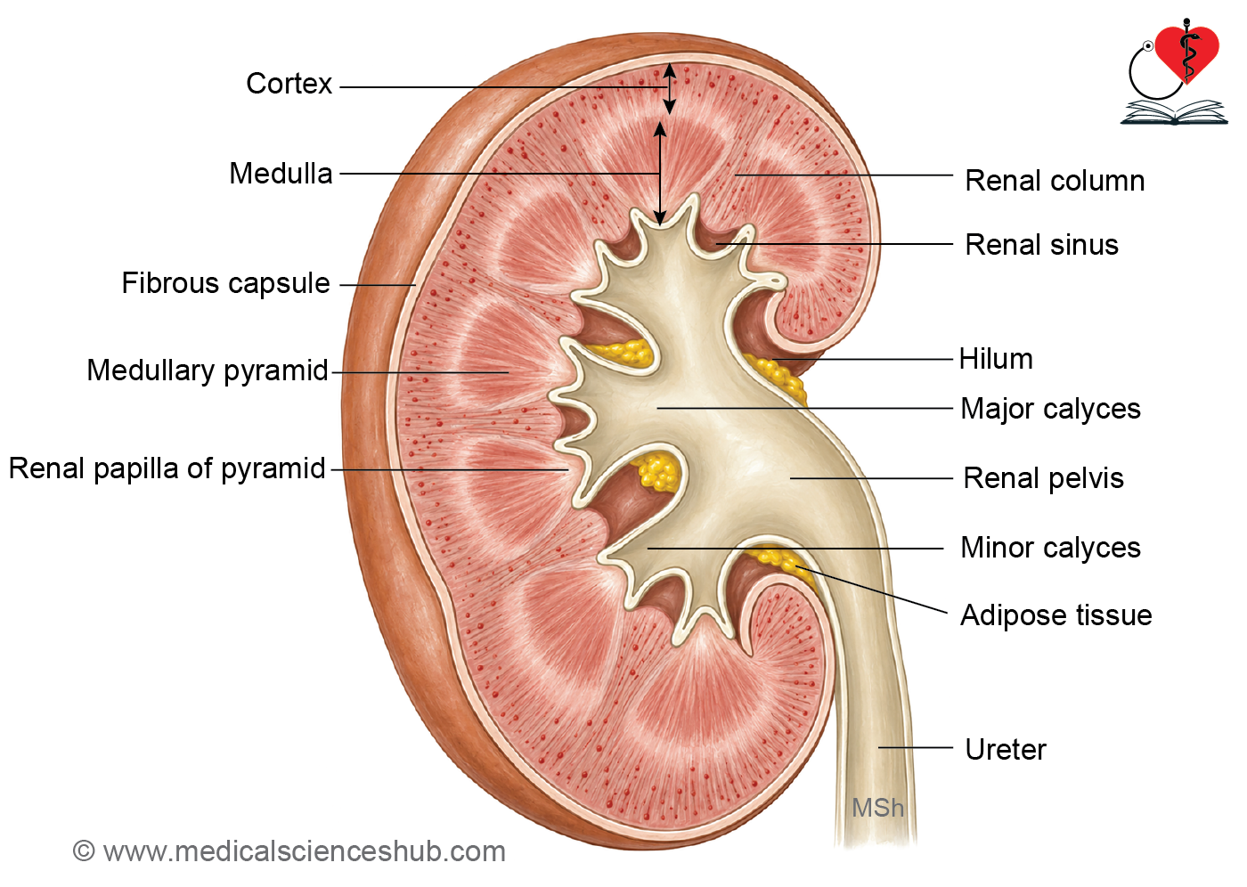
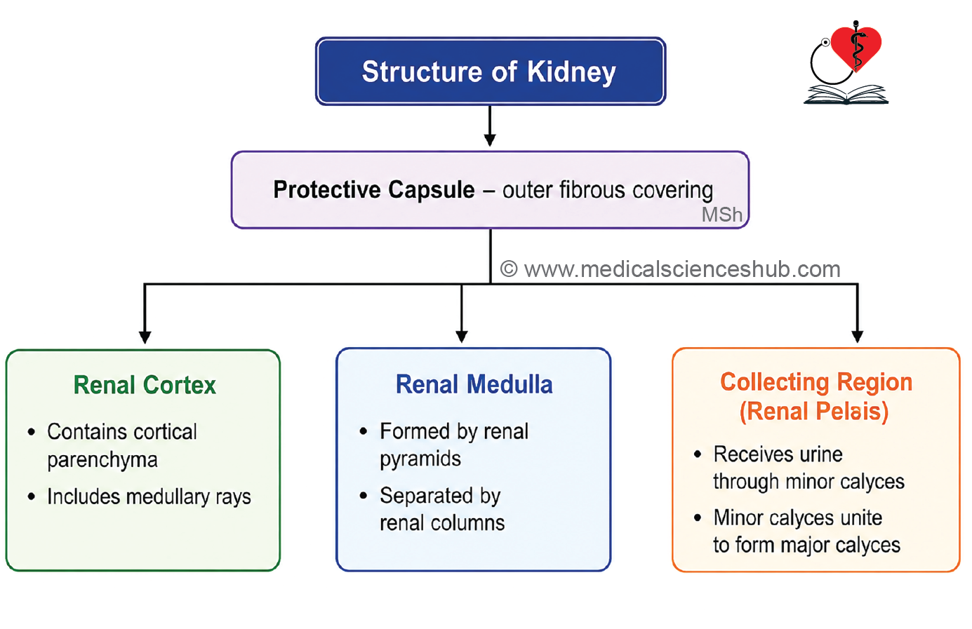
- Each kidney is a bean-shaped organ enclosed by a thin connective tissue capsule.
- The medial border contains the hilum, through which blood vessels, lymphatics, nerves, and the renal pelvis enter or leave the kidney.
- The cavity within the hilum that accommodates these structures is called the renal sinus.
- The renal pelvis collects urine and continues inferiorly as the ureter.
- It divides into major calyces, which further branch into several minor calyces that receive urine from the renal papillae.
Cortex and Medulla
- The kidney is divided into:
- An outer cortex, which appears reddish-brown because it receives most of the renal blood flow.
- An inner medulla, which is lighter in color.
- Approximately 90–95% of renal blood supply is directed to the cortex.
Renal Capsule
- The capsule consists of:
- An outer layer rich in collagen fibers and fibroblasts.
- An inner layer containing myofibroblasts.
Renal Pyramids and Papillae
- The medulla contains cone-shaped renal pyramids.
- The base of each pyramid faces the cortex, while the apex projects toward a minor calyx.
- The apex is called the renal papilla.
- The papillary tip contains multiple openings of collecting ducts, forming the area cribrosa.
Cortical Organization
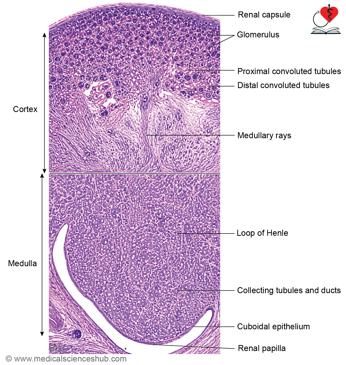
- Medullary rays (of Ferrein) are straight extensions of medullary tissue into the cortex and contain collecting ducts and straight nephron segments.
- The cortical tissue between adjacent medullary rays is termed the cortical labyrinth.
- Extensions of cortical tissue located between neighboring pyramids are known as renal columns (of Bertin).
Lobes and Lobules
- A renal lobe consists of one renal pyramid together with its overlying cortical tissue.
- Human kidneys typically contain 8–18 lobes, although lobulation is more distinct in the fetal kidney.
- A renal lobule is formed by a medullary ray and the surrounding cortical tissue drained by its associated collecting ducts.
Microscopic Structure of Kidney
Nephron
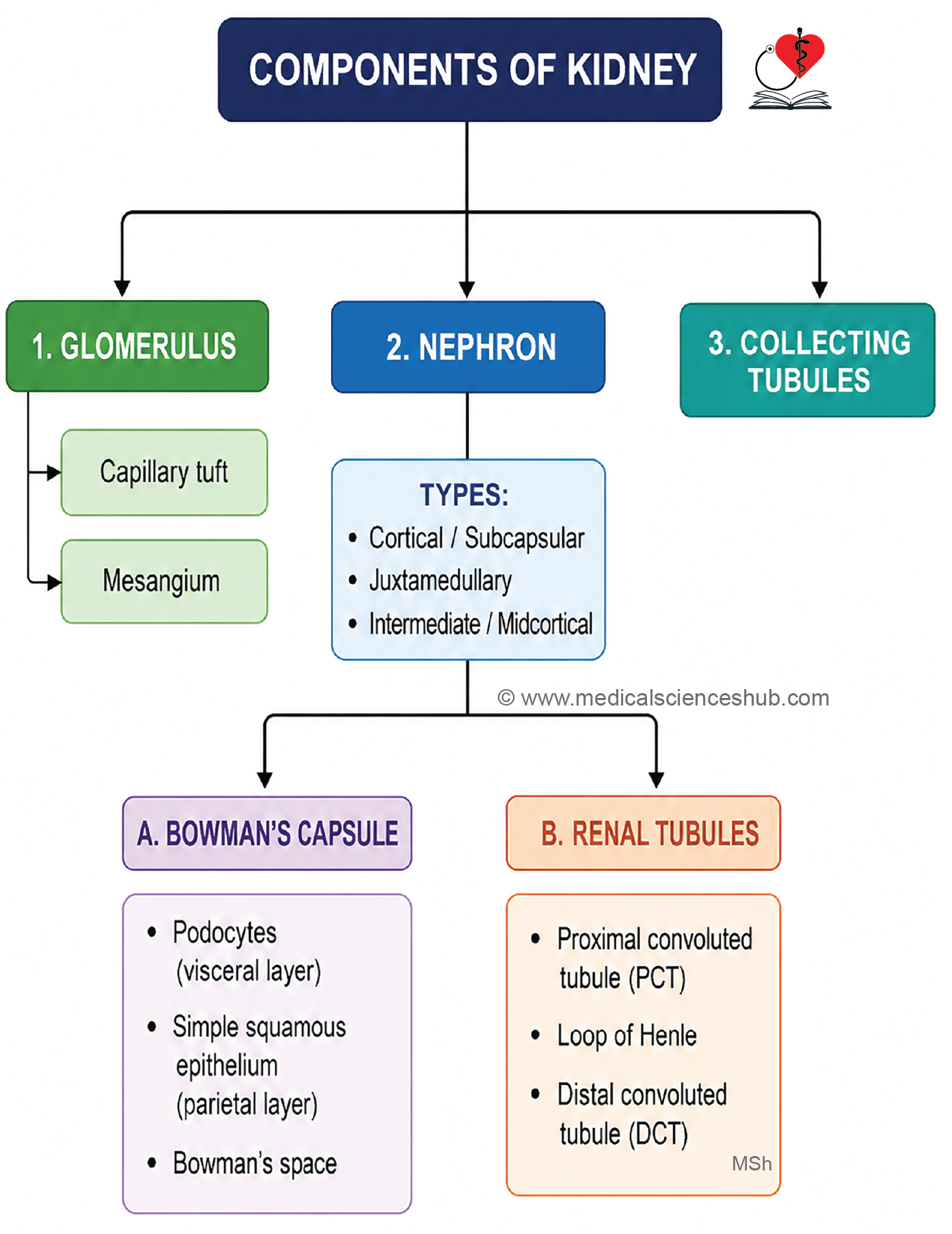
- The nephron is the structural and functional unit of the kidney responsible for urine formation.
- Each kidney contains approximately one million nephrons, giving a total of about two million nephrons in both kidneys.
- Every nephron consists of:
- A renal corpuscle, where blood filtration occurs.
- A renal tubule, which modifies the filtrate through reabsorption and secretion.
- The renal tubule is divided into:
- Proximal convoluted tubule (PCT)
- Loop of Henle
- Distal convoluted tubule (DCT)
- The distal convoluted tubule drains into a collecting tubule, which ultimately conveys urine toward the renal papilla and calyces.
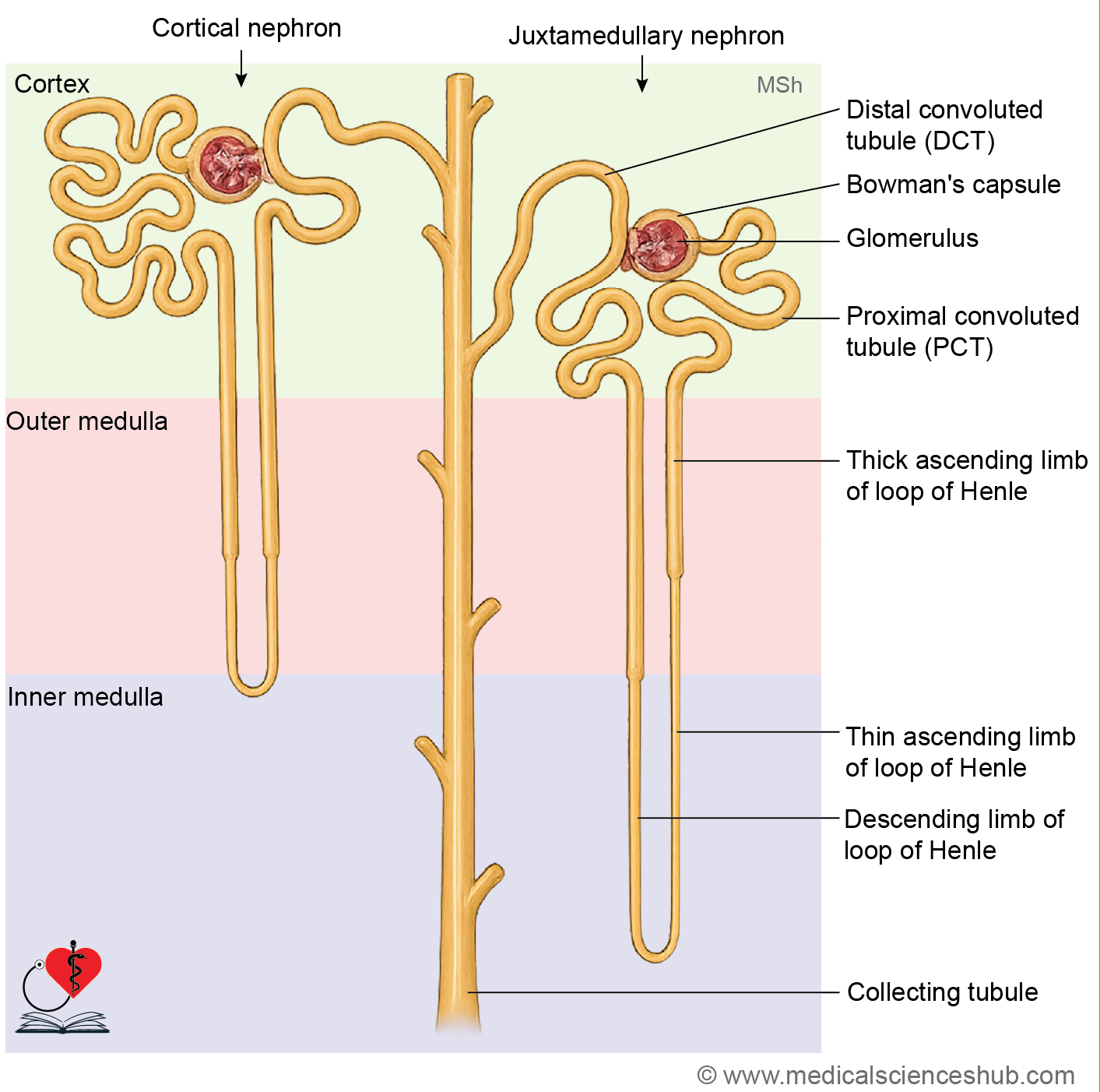
Types of nephrons
- Based on the location of their renal corpuscles, nephrons are classified into three types:
- Cortical (subcapsular) nephrons:
- Renal corpuscles are located in the outer cortex.
- They possess relatively short loops of Henle that extend only into the outer medulla.
- Juxtamedullary nephrons:
- Renal corpuscles are situated near the corticomedullary junction.
- They have long loops of Henle that extend deep into the medulla.
- These nephrons play a major role in generating concentrated urine and maintaining the renal osmotic gradient.
- Intermediate (mid-cortical) nephrons:
- Renal corpuscles are located in the middle region of the cortex.
- They possess loops of Henle of intermediate length.
- Cortical (subcapsular) nephrons:
- Together, these nephron types contribute to efficient filtration, reabsorption, and regulation of body fluid balance.
Renal Corpuscle/Malpighian Corpuscle
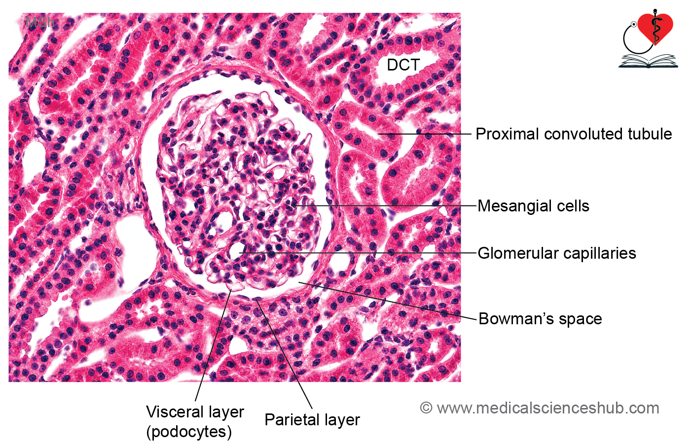
- The renal corpuscle is a spherical filtration unit located in the renal cortex and forms the initial part of the nephron.
- It consists of:
- The glomerulus, a tuft of specialized capillaries.
- Bowman’s capsule, a double-layered epithelial capsule surrounding the glomerulus.
- The urinary (Bowman’s) space, located between the two layers of Bowman’s capsule, where the filtrate is collected.
Glomerulus
- The glomerulus is a network of capillaries specialized for blood filtration.
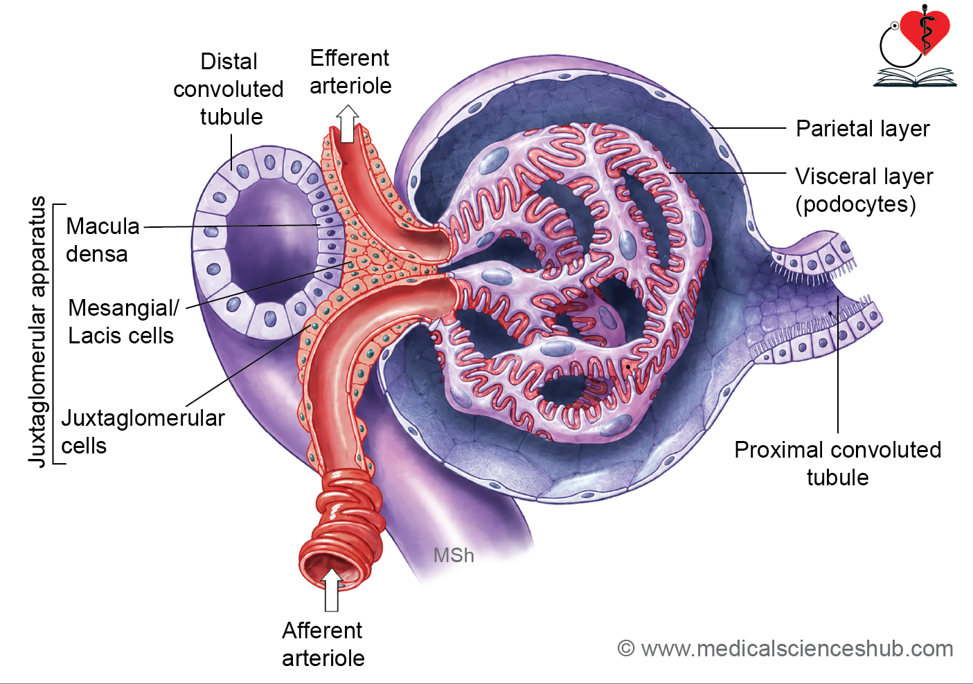
- Blood enters through the afferent arteriole and leaves via the efferent arteriole.
- The efferent arteriole is narrower than the afferent arteriole, helping maintain the pressure required for filtration.
Poles of the Renal Corpuscle
- The vascular pole is the site where the afferent arteriole enters and the efferent arteriole exits the glomerulus.
- The urinary pole lies opposite the vascular pole and marks the point where Bowman’s capsule continues as the proximal convoluted tubule (PCT).
Together, these structures initiate the process of glomerular filtration, the first step in urine formation.
Renal Tubule
- The renal tubule begins at Bowman’s capsule and extends to the collecting tubule.
- It consists of the:
- Proximal convoluted tubule (PCT)
- Proximal straight tubule
- Loop of Henle
- Distal straight tubule
- Distal convoluted tubule (DCT)
- The entire renal tubule is lined by simple epithelium, specialized for absorption and secretion.
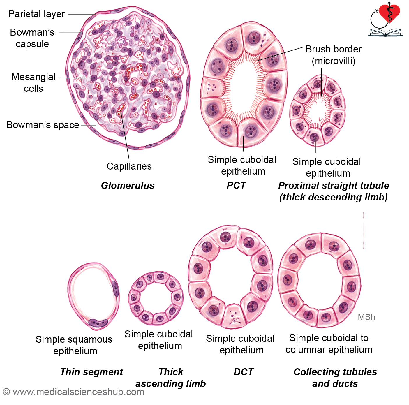
Proximal convoluted tubule (40–60 μm in diameter)
- The PCT is the first segment of the renal tubule and receives filtrate from the urinary pole of Bowman’s capsule.
- It measures approximately 40–60 μm in diameter.
- The lining consists of simple cuboidal epithelial cells with:
- A prominent brush border formed by densely packed microvilli.
- Centrally placed spherical nuclei.
- Eosinophilic cytoplasm due to abundant mitochondria.
Ultrastructural Features
- Long microvilli greatly increase the absorptive surface area.
- Tight junctions are present near the apical surface of adjacent cells.
- Numerous vertically arranged mitochondria produce basal striations, reflecting high metabolic activity.
Functions of the PCT
- The PCT is the major site of tubular reabsorption, reclaiming about 65–80% of the glomerular filtrate.
- It reabsorbs most filtered water through aquaporin-1 (AQP1) channels and transport mechanisms driven by the Na⁺/K⁺-ATPase pump.
- It also reabsorbs essential nutrients, including glucose, amino acids, small peptides, and electrolytes, returning them to the bloodstream.
Proximal Straight Tubule (Thick Descending Limb of Loop of Henle)
- The proximal straight tubule connects the proximal convoluted tubule to the loop of Henle.
- It has a relatively narrow lumen, and cell boundaries are often indistinct due to basolateral membrane infoldings.
- Some classifications consider it part of the PCT, whereas others include it in the thick descending limb of the loop of Henle.
- Its epithelial cells are shorter than those of the PCT and possess fewer microvilli, less prominent basal infoldings, and fewer mitochondria.
Thin Segment of Loop of Henle
- The length of the thin segment varies among nephron types and is longest in juxtamedullary nephrons.
- It is lined mainly by simple squamous epithelium with sparse microvilli and pale cytoplasm.
- Elongated nuclei often bulge into the lumen.
- In cross-section, it may resemble a small blood vessel, but it lacks blood cells within its lumen.
Distal straight tubule (thick ascending limb)
- This segment forms the thick ascending part of the loop of Henle.
- It is lined by cuboidal cells with eosinophilic cytoplasm and distinct cellular boundaries.
- The lumen appears wider than that of the proximal tubule.
- Brush border is absent, contributing to its clearer microscopic appearance.
Distal convoluted tubule (DCT)
- The DCT is shorter than the PCT and is lined by simple cuboidal epithelium.
- Cells contain centrally located round nuclei and lack a brush border, resulting in a wider and clearer lumen.
Functions of the DCT
- The DCT plays an important role in electrolyte and acid-base regulation.
- It reabsorbs sodium ions and bicarbonate while secreting potassium and hydrogen ions.
- Sodium reabsorption is enhanced by aldosterone, contributing to fluid and electrolyte balance.
Collecting Tubules and Ducts
- Collecting tubules arise in the renal cortex as continuations of the distal convoluted tubules (DCTs) and measure approximately 40–50 μm in diameter.
- As they pass through the medullary rays, multiple collecting tubules merge to form larger collecting ducts.
- Collecting ducts descend through the medulla toward the renal papilla and unite to form the papillary ducts (ducts of Bellini), which open at the tip of the renal papilla into the minor calyx.
Histological Features
- In cross-section, collecting tubules and ducts appear circular with distinct cell boundaries, helping differentiate them from convoluted tubules, which have irregular profiles and indistinct borders.
- Collecting tubules are lined by simple cuboidal epithelium without a brush border.
- The epithelium gradually becomes simple columnar as the ducts increase in size.
- Collecting tubules have narrower lumina than collecting ducts.
Cell Types
- Principal (light or clear) cells:
- The predominant cell type in collecting ducts.
- Possess a single primary cilium and a few short microvilli.
- Contain aquaporin-2 (AQP2) channels that mediate ADH-dependent water reabsorption.
- Show basal membrane infoldings.
- Mutations affecting AQP2 can lead to nephrogenic diabetes insipidus.
- Intercalated (dark) cells:
- Less numerous than principal cells.
- Lack prominent basal infoldings.
- Contribute to acid-base regulation by secreting hydrogen or bicarbonate ions.
- Together, collecting ducts play a key role in regulating water balance, electrolyte composition, and urine concentration.
Juxtaglomerular apparatus
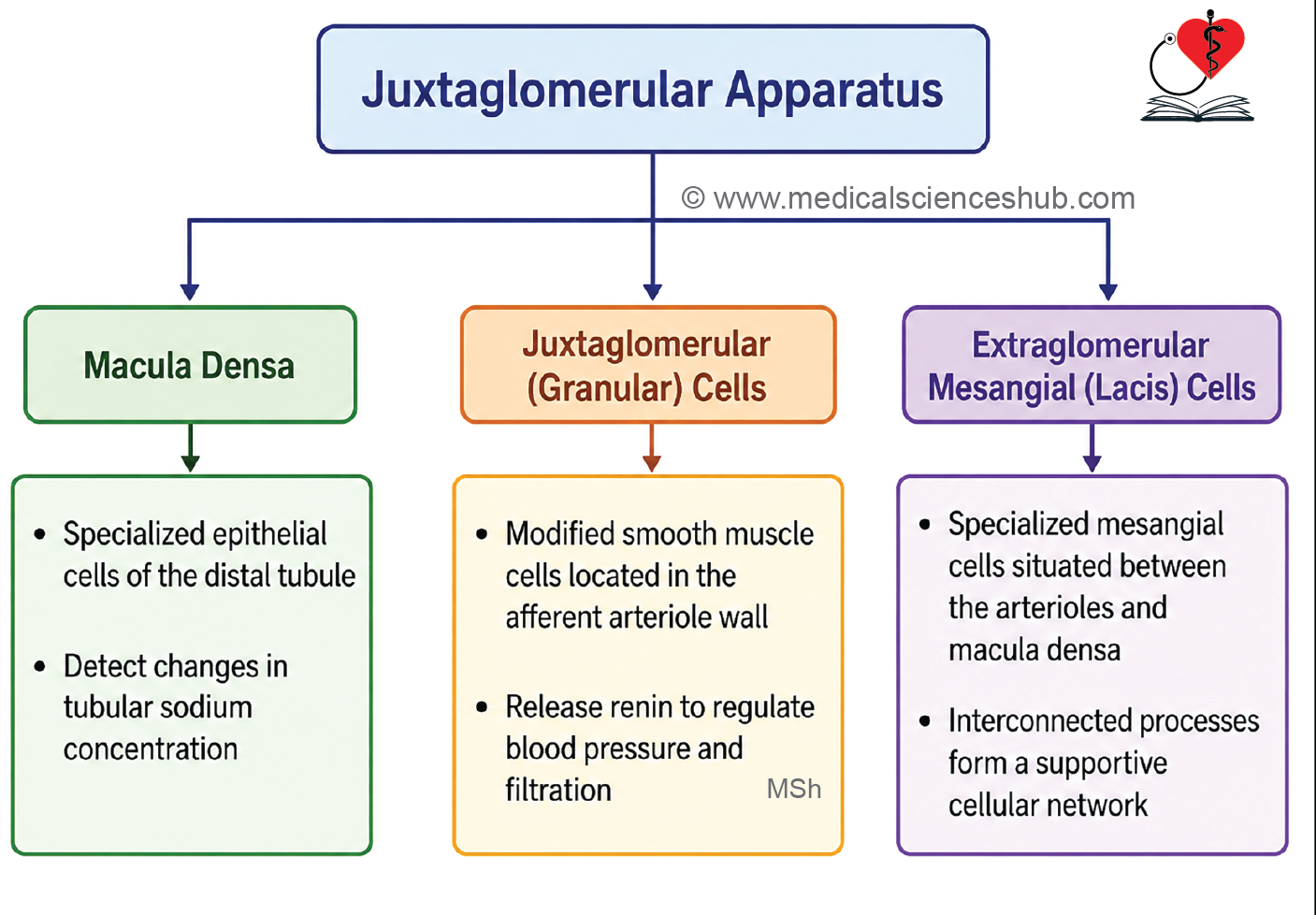
- The juxtaglomerular apparatus (JGA) is a specialized structure located near the vascular pole of the renal corpuscle.
- It plays a crucial role in regulating blood pressure, renal blood flow, and the glomerular filtration rate (GFR).
- The JGA consists of three components:
- Juxtaglomerular (JG) cells
- Macula densa
- Extraglomerular mesangial (Lacis) cells
- Juxtaglomerular Cells
- These are modified smooth muscle cells located in the wall of the afferent arteriole.
- Their primary function is the synthesis and secretion of renin, an enzyme involved in the renin–angiotensin–aldosterone system (RAAS).
- Macula Densa
- The macula densa is a specialized region of the distal convoluted tubule (DCT) adjacent to the afferent and efferent arterioles.
- Its cells are tall, closely packed, and columnar, unlike the typical cuboidal cells of the DCT.
- The densely arranged nuclei create the characteristic appearance known as the macula densa.
- These cells monitor tubular sodium chloride concentration and help regulate renin release.
- Extraglomerular Mesangial (Lacis) Cells
- These cells are located between the macula densa and juxtaglomerular cells.
- They possess branching, lace-like cytoplasmic processes.
- Lacis cells facilitate communication within the JGA and may contribute to regulation of the glomerular filtration rate through responses to angiotensin II and other signaling molecules.
Renal Microvasculature
- Each kidney receives blood through the renal artery, which branches sequentially into lobar, interlobar, arcuate, and interlobular arteries.
- Arcuate arteries arch along the corticomedullary junction at the base of the renal pyramids.
- Interlobular arteries extend through the cortex and give rise to afferent arterioles.
- Each afferent arteriole enters a renal corpuscle and forms a network of glomerular capillaries specialized for filtration.
- These capillaries reunite to form an efferent arteriole, creating a unique arterial portal system.
- Efferent arterioles of cortical nephrons form peritubular capillaries, which surround the renal tubules and facilitate reabsorption and secretion.
- Efferent arterioles of juxtamedullary nephrons give rise to the vasa recta, which participate in the renal countercurrent exchange system and help maintain the medullary osmotic gradient.
- Venous drainage follows the sequence:
- Peritubular capillaries → Interlobular veins → Arcuate veins → Interlobar veins → Renal vein.
- The renal capsule is drained by stellate veins, which ultimately empty into the interlobar venous system.
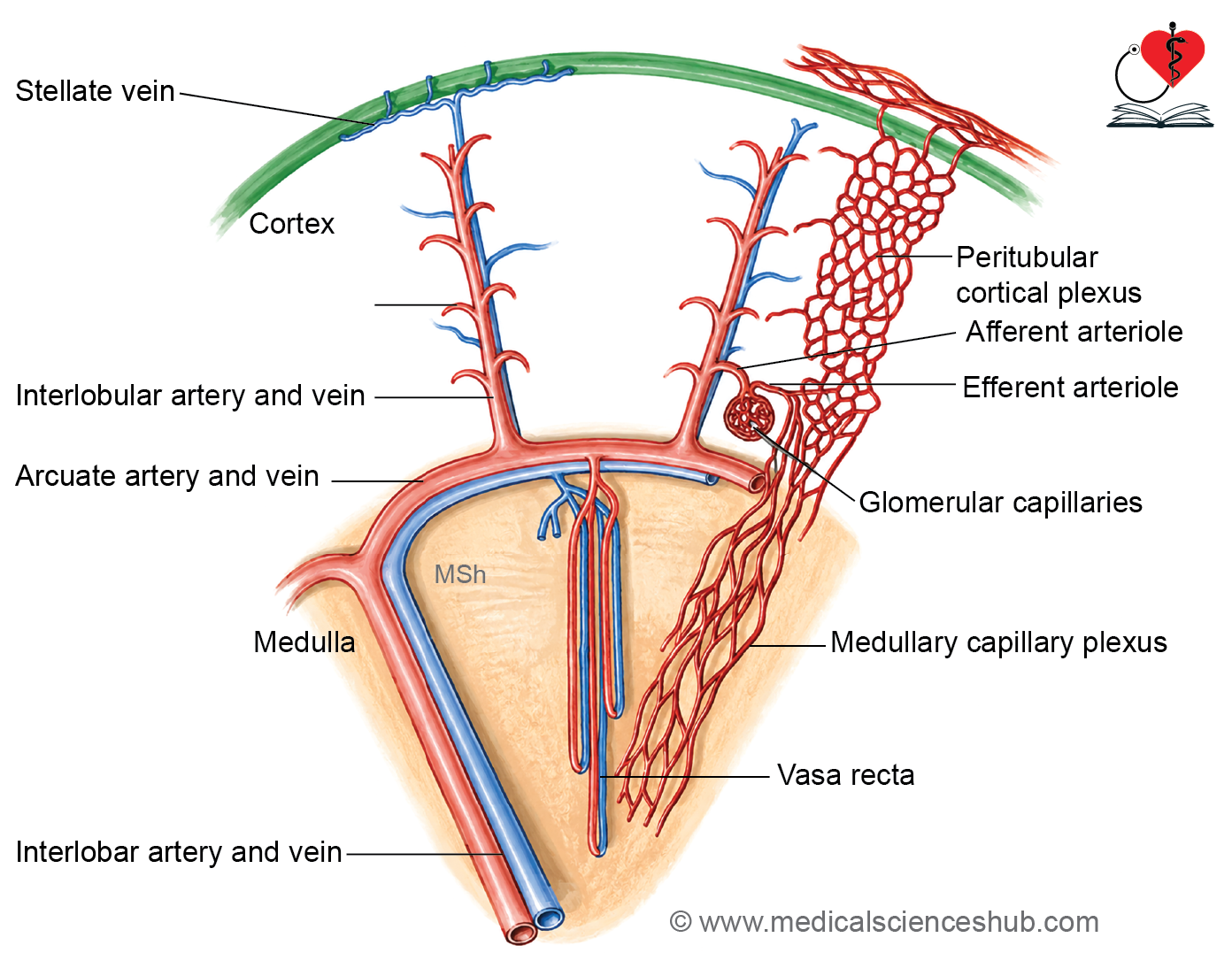
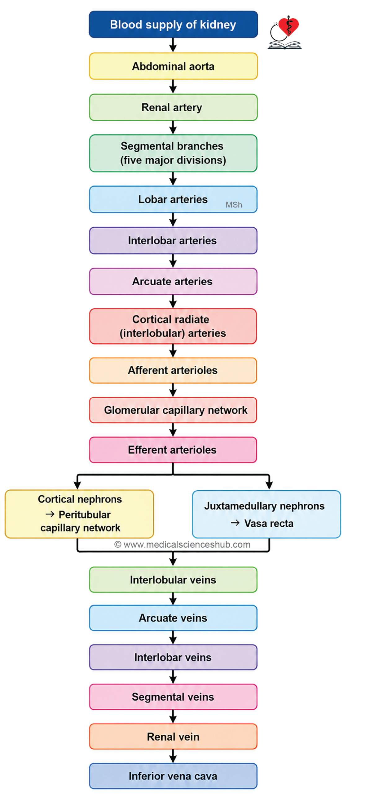
Table 17.1: Differences between proximal convoluted tubule and distal convoluted tubule
| Feature | Proximal Convoluted Tubule (PCT) | Distal Convoluted Tubule (DCT) |
|---|---|---|
| Length | Longer segment of the nephron | Shorter segment, approximately one-third the length of the PCT |
| Diameter | Larger diameter (about 60 μm) | Smaller diameter (about 15–30 μm) |
| Type of Epithelium | Simple cuboidal to low columnar epithelium | Simple cuboidal epithelium |
| Cytoplasmic Staining (H&E) | Cytoplasm appears more intensely eosinophilic (darker pink) | Cytoplasm appears less eosinophilic (lighter pink) |
| Nuclei in Cross Section | Fewer nuclei are visible due to larger cell size | More nuclei are visible and are evenly spaced |
| Luminal Appearance | Narrow, irregular, and often star-shaped lumen | Wider, smoother, and usually circular or oval lumen |
| Brush Border (Microvilli) | Well-developed brush border present | Brush border absent |
| Cell Boundaries | Indistinct because of extensive lateral interdigitations | More distinct and easier to identify |
| Primary Function | Reabsorption of water, electrolytes, glucose, amino acids, and other solutes | Selective reabsorption and secretion; involved in regulation of electrolyte and acid–base balance |
Ureters
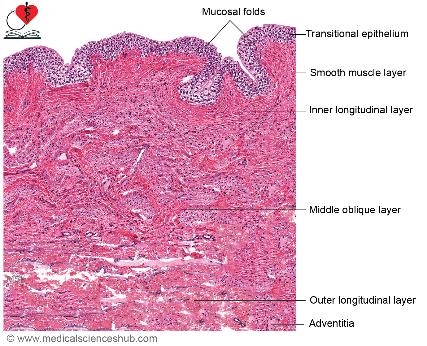
- The ureters are muscular tubes that transport urine from the renal pelvis to the urinary bladder.
- Each ureter measures approximately 25–35 cm in length.
- The lumen typically appears star-shaped in cross-section due to mucosal folding.
Mucous Membrane
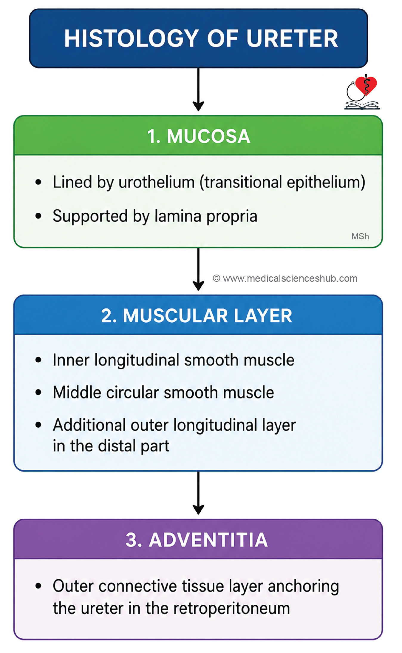
- The mucosa is lined by transitional epithelium (urothelium), usually 4–5 cell layers thick.
- Beneath the epithelium lies a lamina propria composed of connective tissue.
- Numerous longitudinal mucosal folds contribute to the characteristic star-shaped lumen.
Smooth Muscle Coat
- The wall contains smooth muscle arranged in:
- Inner longitudinal layer
- Middle circular layer
- Outer longitudinal layer
- The outer longitudinal layer is prominent mainly in the lower third of the ureter, becoming more distinct near the urinary bladder.
- Coordinated contractions of these muscle layers produce peristaltic movements that propel urine toward the bladder.
Adventitia
- The outermost layer is the adventitia, consisting of connective tissue that anchors the ureter to surrounding retroperitoneal structures.
- It contains blood vessels, lymphatic vessels, nerves, and adipose tissue.
- The specialized structure of the ureter ensures efficient and continuous transport of urine from the kidneys to the bladder.
Urinary Bladder
- The urinary bladder is a distensible muscular organ that temporarily stores urine before micturition.
- It receives urine through two ureteric openings and expels it via the urethra.
- The bladder wall consists of three principal layers:
- Mucosa
- Muscular coat (detrusor muscle)
- Serosa/Adventitia
Mucous Membrane
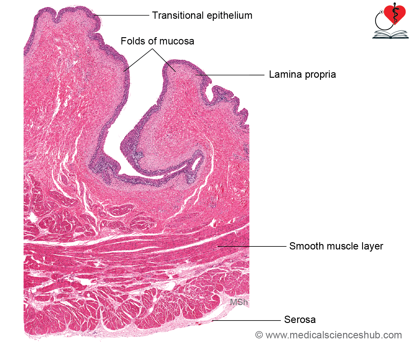
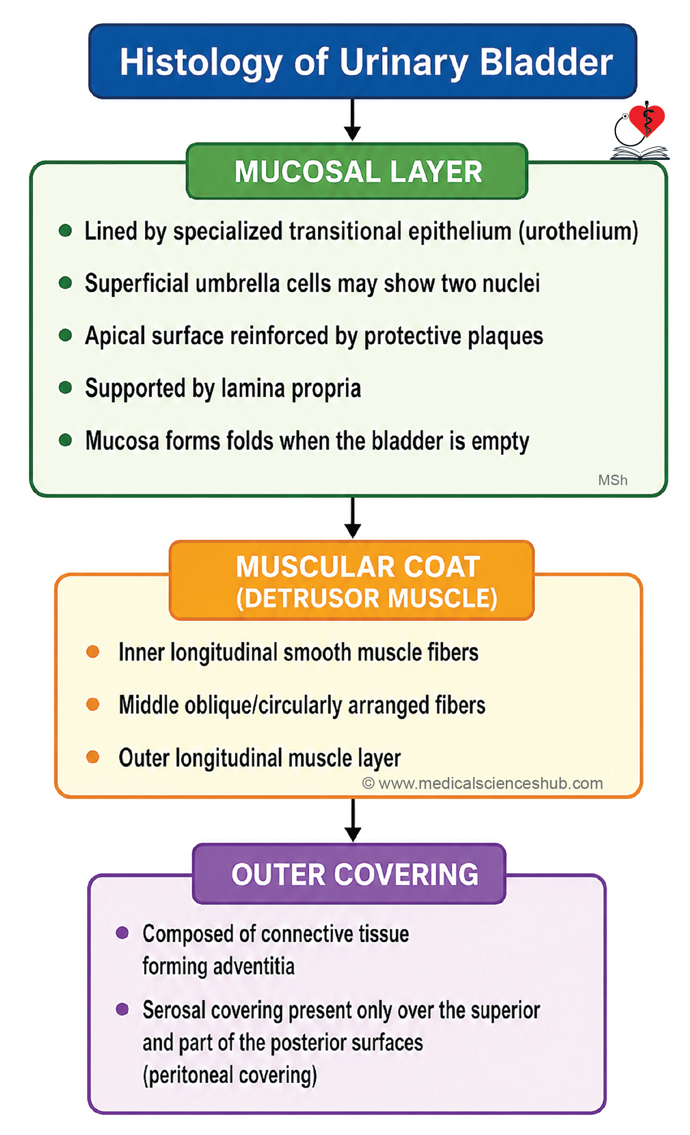
- The inner surface is lined by transitional epithelium (urothelium), specialized to accommodate repeated stretching.
- Superficial umbrella cells are dome-shaped and may be binucleated.
- In an empty bladder, epithelial cells appear cuboidal and project into the lumen; during distension, the epithelium becomes thinner and flatter.
- The epithelium typically decreases from about six cell layers in the empty state to three layers when stretched.
- Beneath the epithelium lies a fibroelastic lamina propria composed of loose connective tissue.
- Numerous mucosal folds are present in the empty bladder and disappear as the bladder fills.
- The trigone is a smooth triangular region where the mucosa is firmly attached to the underlying muscle and lacks prominent folds.
Muscle Coat/Detrusor Muscle
- The bladder wall contains a thick detrusor muscle formed by interlacing bundles of smooth muscle.
- Muscle fibers generally form inner and outer longitudinal layers with a less distinct middle circular or oblique layer.
- Near the bladder neck, circularly arranged smooth muscle contributes to the internal urethral sphincter (sphincter vesicae).
Serous Layer
- The superior surface and part of the posterior surface are covered by serosa (visceral peritoneum).
- The remaining external surfaces are covered by adventitia, consisting of connective tissue.
Functions of Plaques of Urinary Bladder
- The apical membrane of umbrella cells contains specialized urothelial plaques.
- These plaques and associated membrane vesicles allow the surface cells to change shape during bladder filling and emptying.
- This adaptation maintains a protective, impermeable barrier while permitting significant bladder distension.
CLINICAL CORRELATION
- Ectopia vesicae (bladder exstrophy) is a congenital defect in which the anterior wall of the urinary bladder and the overlying abdominal wall fail to develop properly, resulting in exposure of the bladder mucosa.
- Cystitis is inflammation of the urinary bladder, most commonly caused by bacterial infection, and may present with urinary frequency, urgency, and dysuria.
- Bladder stones develop due to crystallization and aggregation of urinary solutes within the bladder, often in the setting of urinary stasis or obstruction.
- Urothelial carcinoma (transitional cell carcinoma) arises from the urothelium lining the urinary tract. Important risk factors include cigarette smoking, chronic bladder irritation, urinary calculi, arsenic exposure, and occupational exposure to certain dyes and textile-related chemicals.
Urethra
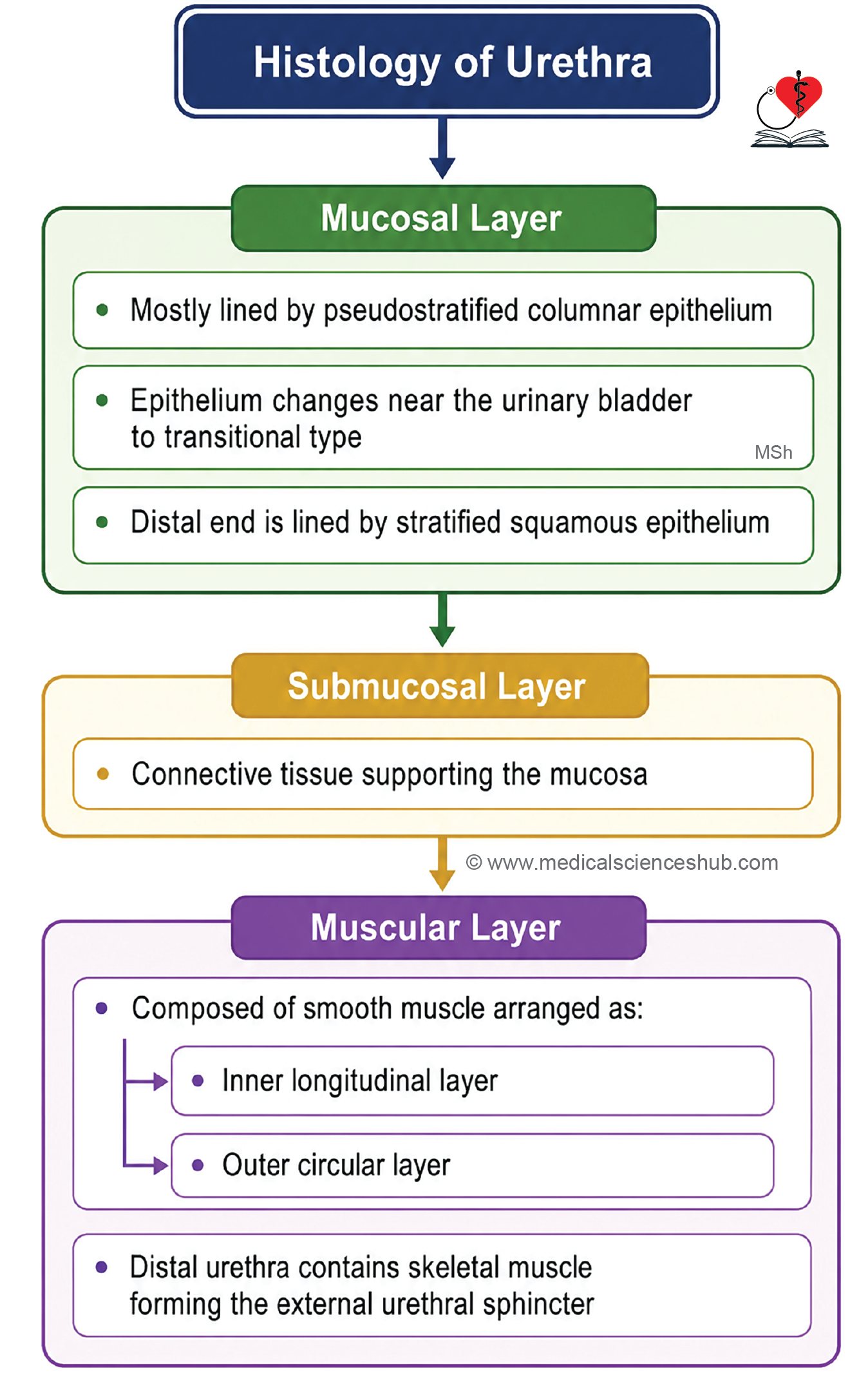
- The urethra is a fibromuscular tube that carries urine from the urinary bladder to the exterior of the body.
- The male urethra is approximately 20 cm long, whereas the female urethra is about 4 cm in length.
- Its wall is composed of three layers:
- Mucous membrane
- Submucosa
- Muscular coat
Mucous Membrane
- Most of the male urethra is lined by pseudostratified columnar epithelium.
- The prostatic urethra, near the urinary bladder, is lined by transitional epithelium (urothelium).
- Near the external urethral opening, the epithelium becomes stratified squamous.
- In females, the proximal urethra is lined by transitional epithelium, which gradually changes to stratified squamous epithelium distally.
Submucosa
- The submucosa consists of loose connective tissue containing blood vessels and nerves.
Muscle Coat
- The muscular layer contains inner longitudinal and outer circular smooth muscle fibers.
- The terminal part of the urethra is surrounded by skeletal muscle, forming the external urethral sphincter, which provides voluntary control of urination.
Important Questions
- Write a short note on glomerular filtration barrier.
- Write a short note on juxtaglomerular apparatus.
- List the differences PCT and DCT.
📝 Test Your Knowledge – Practice MCQs
Attempt the chapter MCQ quiz and assess your understanding of key concepts.
Click to see: Medical Disclaimer and Copyright Notice