
Competency
- AN52.2 Describe and identify the microanatomical features of male reproductive system: Testis, epididymis, vas deferens.
Introduction
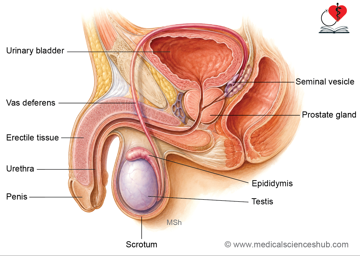
- The male reproductive system includes the paired testes, genital ducts (epididymis, ductus deferens, and ejaculatory ducts), accessory glands (seminal vesicles, prostate, and bulbourethral glands), urethra, scrotum, and penis.
- Its primary functions are:
- Production of spermatozoa through spermatogenesis.
- Synthesis and secretion of testosterone and other androgens.
- Storage, transport, and delivery of sperm into the female reproductive tract during reproduction.
Testis
- The testes are paired, ovoid male gonads located within the scrotum.
- They develop from the gonadal ridge (intermediate mesoderm) and primordial germ cells that migrate from the yolk sac. During development, the testes descend from the abdominal cavity into the scrotum.
Structure of Testis
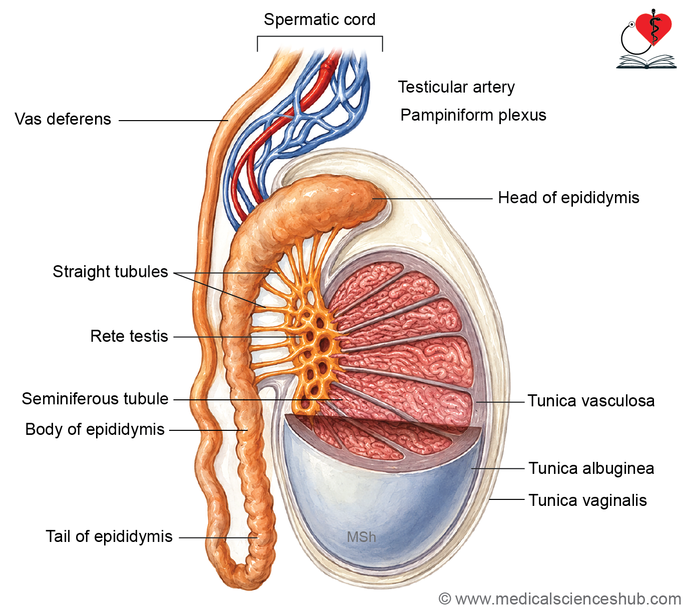
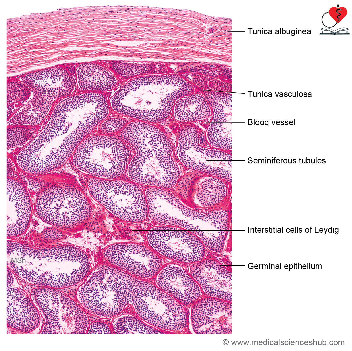
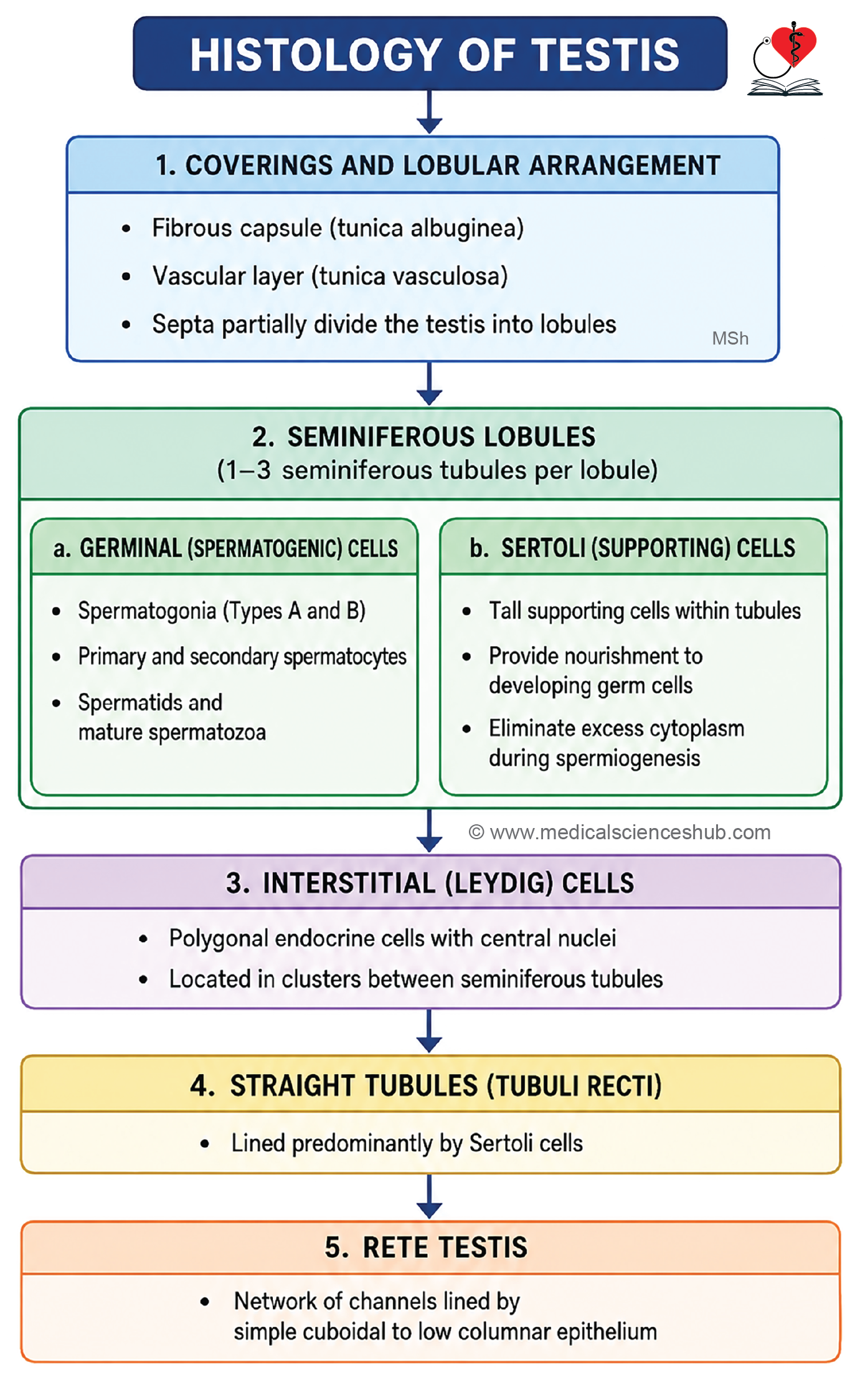
- The testis is enclosed by a dense fibrous capsule called the tunica albuginea.
- Deep to it lies the tunica vasculosa, a vascular connective tissue layer that supplies the testicular parenchyma.
- Connective tissue septa extend inward from the capsule and divide the testis into approximately 250–300 incomplete lobules.
- A thickened posterior region of the capsule forms the mediastinum testis, which contains blood vessels, lymphatics, and ductal structures.
Seminiferous Tubules and Duct System
- Each lobule contains 1–4 highly coiled seminiferous tubules, the sites of spermatogenesis.
- The tubules are supported by connective tissue stroma containing Leydig (interstitial) cells, which secrete testosterone.
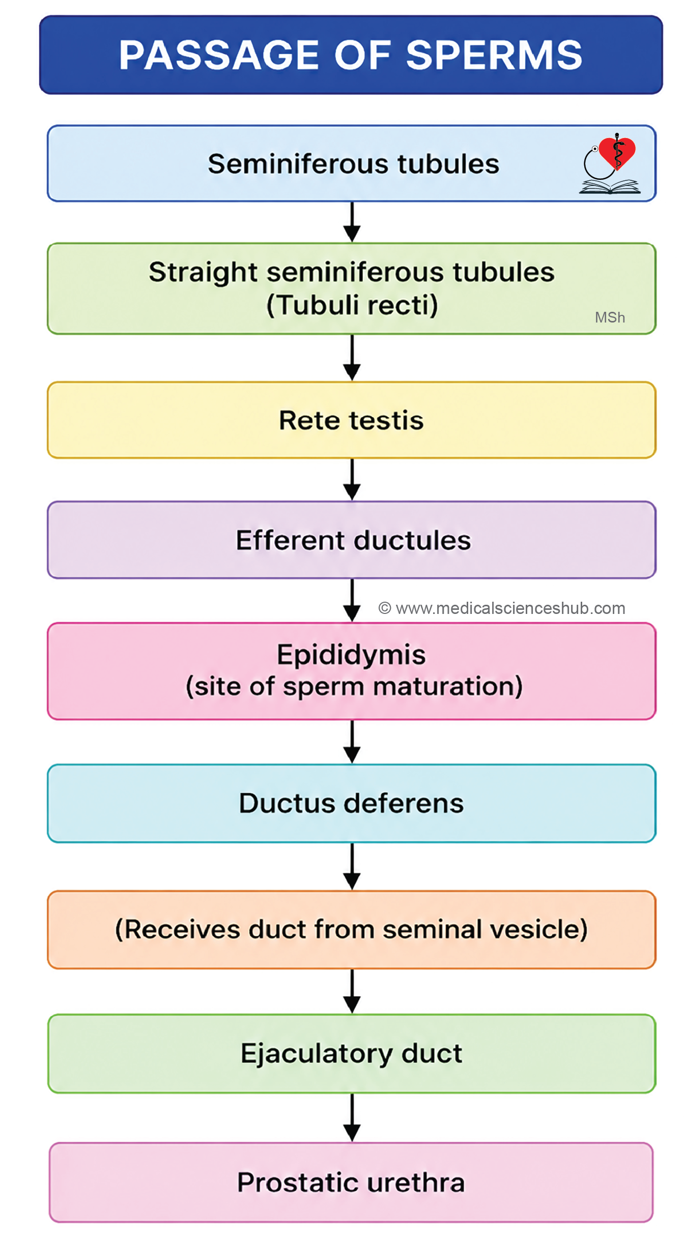
- Near the mediastinum, seminiferous tubules continue as straight segments called tubuli recti (straight tubules).
- The tubuli recti open into the rete testis, a network of interconnected channels within the mediastinum.
- The rete testis gives rise to 12–20 efferent ductules, which transport sperm to the epididymis.
- The epididymis continues as the ductus deferens, which carries sperm toward the ejaculatory ducts.
Microscopic Structure of Testis
Seminiferous Epithelium
- The seminiferous tubules are lined by a specialized stratified epithelium known as the seminiferous epithelium, which is responsible for sperm production.
- Each seminiferous tubule is approximately 50 cm long and 150–200 μm in diameter.
- The epithelium contains two principal cell types:
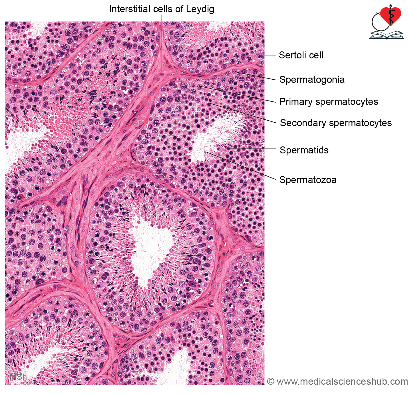
- Sertoli cells
- Spermatogenic (germ) cells
Sertoli cells (sustentacular cells)
- Sertoli cells are tall supporting cells that extend from the basal lamina to the lumen of the seminiferous tubule.
- They possess pale, euchromatic nuclei and form the true epithelial framework of the seminiferous tubules.
- Adjacent Sertoli cells are connected by tight junctions, creating the blood-testis barrier, which protects developing haploid germ cells from immune attack.
- Sertoli cells contain FSH receptors and respond to follicle-stimulating hormone.
Functions of Sertoli Cells
- Produce androgen-binding protein (ABP), which maintains a high local concentration of testosterone required for spermatogenesis.
- Secrete inhibin, which suppresses FSH secretion from the anterior pituitary.
- Produce anti-Müllerian hormone (AMH) during fetal life, promoting regression of Müllerian ducts.
- Synthesize proteins such as transferrin and plasminogen activator.
- Provide structural and metabolic support to developing germ cells.
- Phagocytose residual cytoplasm shed during spermiogenesis.
Spermatogenic (Germ) Cells
- These cells originate from primordial germ cells and undergo continuous differentiation to form mature spermatozoa.
- They are arranged in layers from the basement membrane toward the lumen in the following sequence:
- Spermatogonia
- Spermatocytes
- Spermatids
- Spermatozoa
- Not all developmental stages are visible in every cross-section of a seminiferous tubule.
Spermatogenic Cycle
- Spermatogenesis occurs in a coordinated wave along the length of the seminiferous tubule, known as the spermatogenic cycle.
- Because Leydig cell testosterone production is largely independent of testicular temperature, individuals with cryptorchidism may show impaired spermatogenesis but retain normal secondary sexual characteristics.
Lamina (Tunica) Propria
- The lamina propria is a thin, vascular connective tissue layer surrounding each seminiferous tubule.
- It contains specialized peritubular (myoid) cells located external to the basal lamina of the seminiferous epithelium.
- These contractile cells generate rhythmic contractions that facilitate the movement of spermatozoa and tubular secretions toward the duct system.
- Unlike typical connective tissue, this layer contains very few fibroblasts.
Leydig (Interstitial) Cells
- Leydig cells are located in the interstitial connective tissue between adjacent seminiferous tubules.
- They are large, polygonal cells with a central round nucleus and eosinophilic cytoplasm.
- Their cytoplasm contains abundant smooth endoplasmic reticulum, reflecting their role in steroid hormone synthesis.
- Leydig cells possess luteinizing hormone (LH) receptors; LH stimulation promotes testosterone production.
- Lipid droplets and lipofuscin pigment granules may be present within the cytoplasm.
Functions of Leydig Cells
- Synthesize and secrete testosterone, which is essential for spermatogenesis, development of male reproductive organs, and maintenance of secondary sexual characteristics.
Crystals of Reinke
- Some Leydig cells contain Reinke crystals, which are rod-shaped cytoplasmic inclusions of uncertain function.
- These structures are uncommon in normal testes but may be identified in certain Leydig cell tumors, where they can aid diagnosis.
Activity During Life
- Leydig cells are hormonally active during fetal development and contribute to male sexual differentiation.
- Their activity declines after birth and increases again at puberty under gonadotropin stimulation, remaining functional throughout adult life.
Spermatogenesis
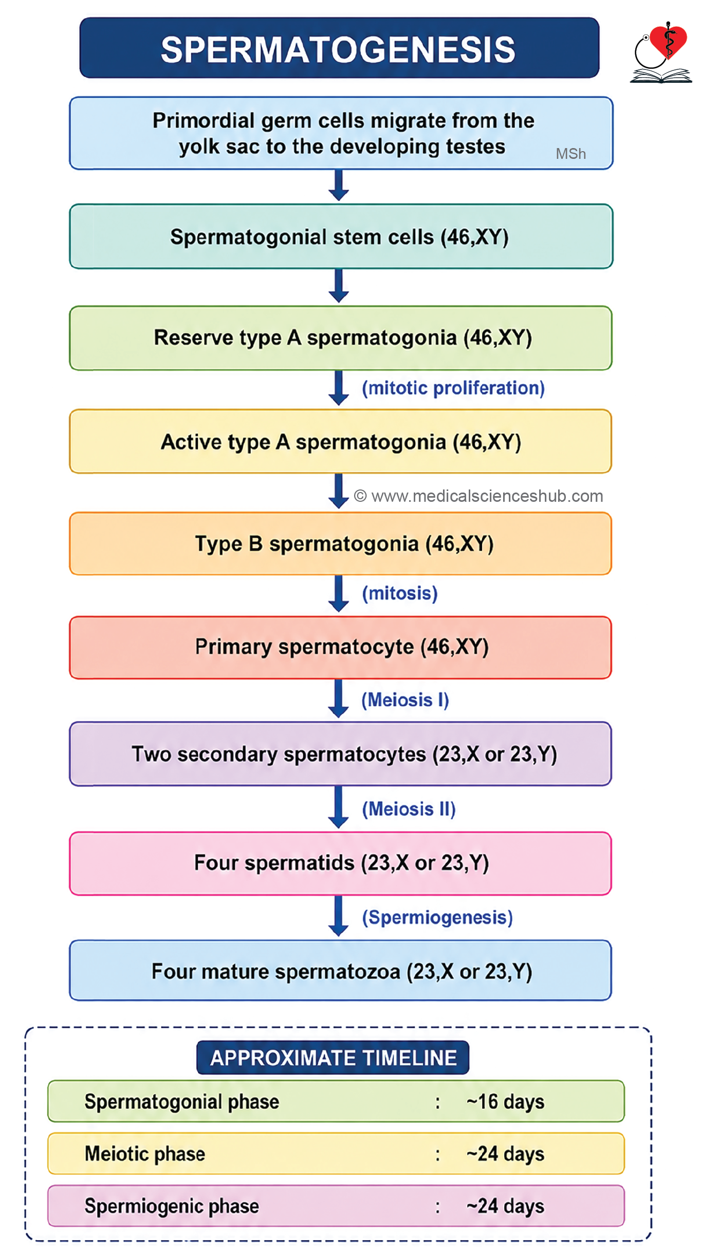
- Spermatogenesis is the process by which mature spermatozoa are produced from spermatogonial stem cells within the seminiferous tubules.
- It occurs in three sequential phases:
- Spermatocytosis
- Meiosis
- Spermiogenesis
Spermatocytosis
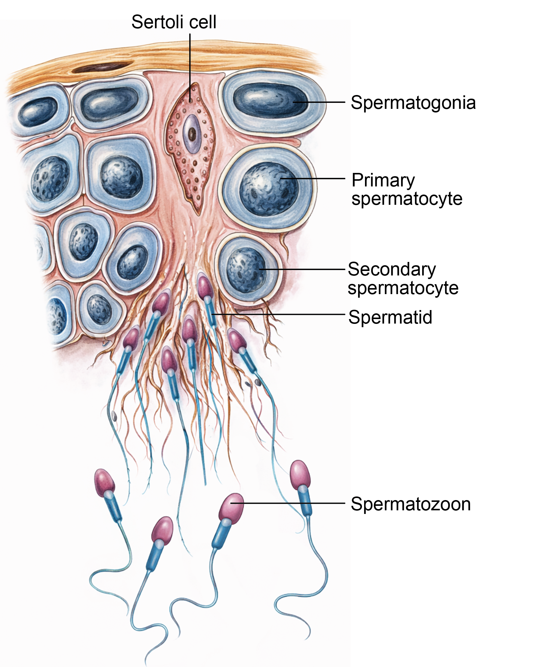
- This phase involves the transformation of spermatogonia into primary spermatocytes.
- Spermatogonia are classified according to nuclear appearance:
- Type A dark spermatogonia act as stem cells and possess densely stained nuclei.
- Type A pale spermatogonia arise from Type A dark cells and continue proliferative divisions.
- Type B spermatogonia differentiate and give rise to primary spermatocytes.
- Primary spermatocytes (46,XY) are the largest germ cells and occupy the middle region of the seminiferous epithelium.
Meiosis (Spermatocyte) Phase
- Primary spermatocytes undergo meiosis I to produce two secondary spermatocytes.
- Each secondary spermatocyte rapidly completes meiosis II, forming two spermatids.
- Thus, one primary spermatocyte produces four haploid spermatids carrying either an X or Y chromosome.
- Spermatids contain a haploid set of chromosomes and represent the first genetically mature germ cells.
Spermiogenesis (Spermatid Phase)
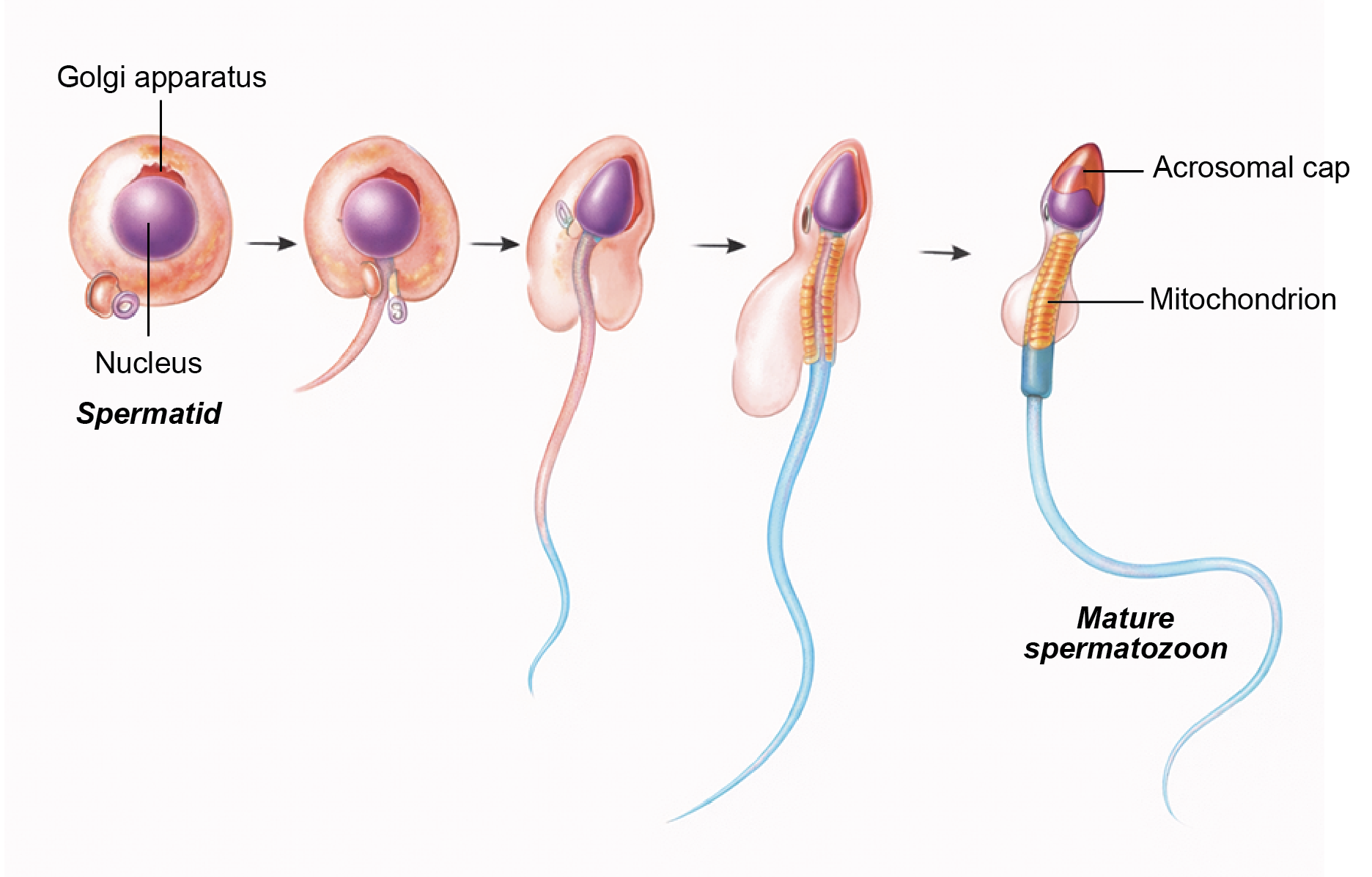
- Spermiogenesis is the transformation of spermatids into mature spermatozoa without further cell division.
Golgi Phase
- Golgi vesicles fuse to form the acrosomal vesicle, which establishes the anterior pole of the developing sperm.
- Centrioles migrate toward the posterior region.
Cap Phase
- The acrosomal vesicle spreads over the anterior portion of the nucleus to form the acrosomal cap.
Acrosomal Phase
- The nucleus elongates and becomes highly condensed.
- The flagellum develops from the distal centriole.
- Mitochondria accumulate around the proximal flagellum to form the middle piece.
- The tail differentiates into principal and end segments.
Maturation Phase
- Excess cytoplasm is shed as residual bodies, which are subsequently phagocytosed by Sertoli cells.
Spermiation
- The final release of mature spermatozoa from Sertoli cells into the lumen of the seminiferous tubule is called spermiation.
Structure of Mature Sperm
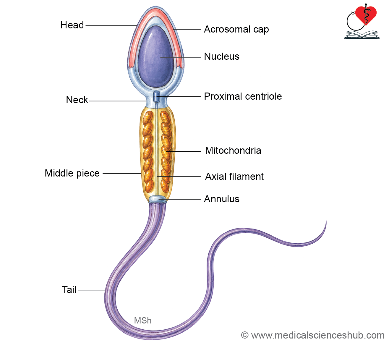
- Matured spermatozoa have a head, neck, middle piece and tail.
- Length: 50–60 μm including head: ~4 m, neck: ~0.3 m, middle piece: ~4 m, tail: ~40 m.
- Count: In a single ejaculation, 200–300 million sperms are emitted in a volume of 2–5 ml semen.
- Life: The usual period of viability after ejaculation is 48 hours, but may survive up to 4 days in the female genital tract.
Parts of Spermatozoon
A spermatozoon is divided into head, neck, middle piece, and tail.
Head
- The head is pyriform in shape. It contains a densely packed haploid nucleus with 23 chromosomes, either X or Y.
- The anterior two-thirds of the nucleus is covered by the acrosome, which is derived from the Golgi apparatus.
- The acrosome contains enzymes such as hyaluronidase and acrosin. These enzymes are released during the acrosome reaction and facilitate penetration of the corona radiata and zona pellucida of the oocyte.
Neck
- The neck connects the head to the middle piece.
- It contains the proximal centriole, which contributes to the connecting piece and plays a role after fertilization in forming the zygotic centrosome. The distal centriole gives rise to the axoneme of the flagellum.
Middle Piece
- The middle piece contains the axoneme, which extends from the neck into the tail.
- The axoneme has a characteristic arrangement of two central microtubules surrounded by nine peripheral doublets.
- Mitochondria are arranged helically around the proximal part of the axoneme, forming the mitochondrial sheath that provides energy for motility.
- An annulus marks the junction between the middle piece and the principal piece.
Tail
- The tail is divided into principal and end pieces.
- In the principal piece, the axoneme is surrounded by a fibrous sheath and plasma membrane.
- In the end piece, only the axoneme and plasma membrane are present.
Maturation and Capacitation of Spermatozoa
Definition: Capacitation is the functional maturation of spermatozoa that enables them to fertilize an oocyte. This phenomenon was first demonstrated by Chang and Austin in 1951.
Site: It occurs within the female reproductive tract, mainly in the uterus and uterine tubes.
Cellular changes during capacitation:
- Removal of seminal plasma proteins attached to the sperm head.
- Modification of the glycoprotein coat over the plasma membrane of the head region.
- Increased membrane fluidity and preparation of the acrosomal membrane for the acrosome reaction.
Effects of capacitation:
- Enhancement of sperm motility, including hyperactivated movement.
- Destabilization of the acrosomal membrane, which permits enzyme release during the acrosome reaction and facilitates penetration of the zona pellucida of the oocyte.
Clinical relevance:
- In in vitro fertilization procedures, spermatozoa are processed under laboratory conditions to achieve capacitation.
- In assisted reproductive techniques such as intracytoplasmic sperm injection, a single spermatozoon is introduced directly into the cytoplasm of the oocyte in cases of acrosomal or motility defects.
Duct System of Testis
- The duct system of the testis transports sperm through the following pathway: seminiferous tubules → straight tubules (tubuli recti) → rete testis → efferent ductules → epididymis.
Straight Tubule (Tubuli Recti)
- These are short terminal segments connecting seminiferous tubules to the rete testis.
- The proximal portion is lined by Sertoli cells and simple columnar epithelium.
- The distal portion is lined by simple cuboidal epithelium.
Rete Testis
- The rete testis is a network of interconnected channels located within the mediastinum testis.
- It is lined by low cuboidal to low columnar epithelial cells.
- Many lining cells possess a single primary cilium, which may assist in fluid movement and sensing luminal contents.
Efferent Ductules
- Efferent ductules convey sperm from the rete testis to the epididymis.
- Approximately 12–20 ductules are present.
- They are lined by simple ciliated columnar epithelium with absorptive cells and are supported by a thin lamina propria.
- A surrounding layer of circular smooth muscle helps propel sperm toward the epididymis.
Blood–testis barrier (Sertoli cell barrier)
- The blood–testis barrier is a specialized barrier that separates developing germ cells within the seminiferous tubules from the bloodstream.
Structure
- The barrier is formed by tight junctions and junctional complexes between adjacent Sertoli cells.
- These junctions divide the seminiferous epithelium into two compartments:
- Basal compartment: Located between the basement membrane and Sertoli cell junctions. It contains spermatogonia and early primary spermatocytes.
- Adluminal compartment: Located toward the lumen of the tubule. It contains advanced primary spermatocytes, secondary spermatocytes, spermatids, and spermatozoa.
- As germ cells mature, primary spermatocytes pass into the adluminal compartment through coordinated remodeling of Sertoli cell junctions without disrupting barrier integrity.
Functions
- The blood–testis barrier protects developing haploid germ cells from immune recognition.
- It prevents the formation of anti-sperm antibodies and maintains an optimal microenvironment for spermatogenesis.
- The barrier also regulates the selective movement of nutrients, hormones, and signaling molecules to developing germ cells.
Epididymis
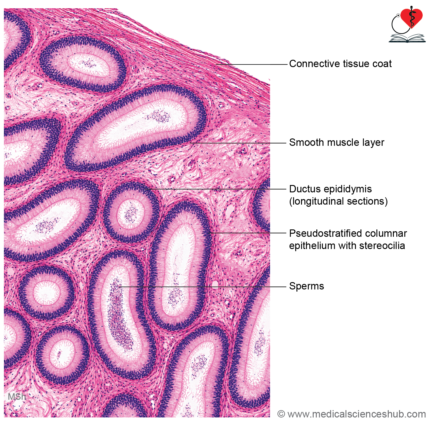
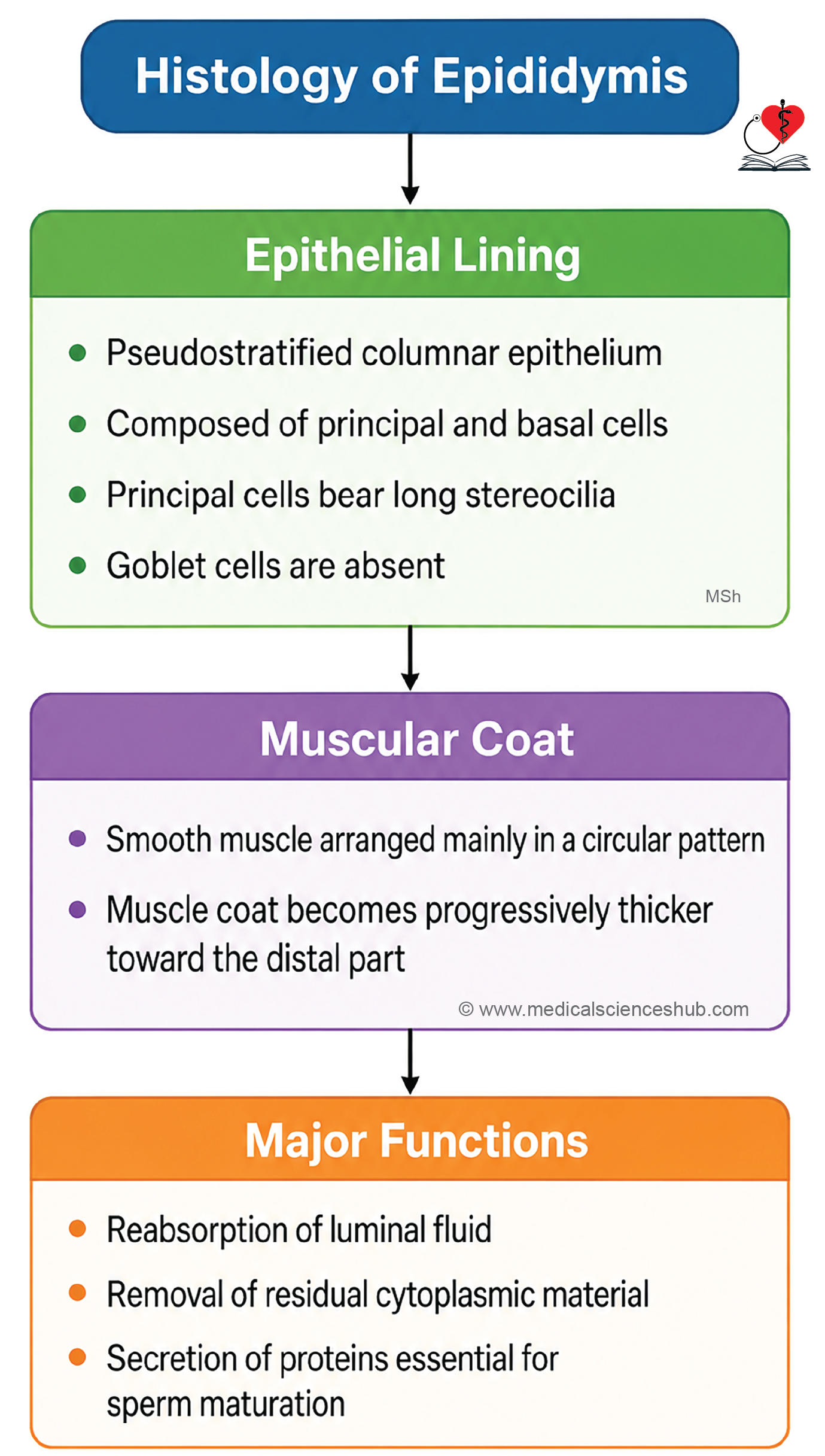
- The epididymis is a crescent-shaped structure located along the posterior border of the testis.
- It is approximately 7.5 cm long and serves as the site where spermatozoa undergo maturation and acquire motility.
- It is divided into three regions: head, body, and tail.
- The head contains the efferent ductules, while the body and tail contain the highly coiled duct of the epididymis.
Duct of the Epididymis
- The duct is a single, extensively coiled tube measuring about 4–6 m in length.
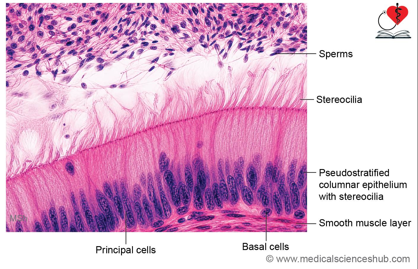
- It is lined by pseudostratified columnar epithelium composed of:
- Tall principal cells
- Small basal cells
- Principal cells possess long, non-motile stereocilia that increase the absorptive surface area.
- Occasional intraepithelial lymphocytes, known as halo cells, may also be present.
- The wall contains a thin layer of circular smooth muscle, which assists in sperm transport and ejaculation.
Functions
- Serves as a storage reservoir for mature spermatozoa.
- Promotes sperm maturation and acquisition of motility.
- Absorbs excess fluid arriving from the testis.
- Removes defective sperm and residual cytoplasmic material by phagocytosis.
- Secretes decapacitation factors, glycoproteins that coat sperm and are later removed during capacitation within the female reproductive tract.
CLINICAL CORRELATION
- Kartagener syndrome is an autosomal recessive disorder characterized by defective dynein arms, resulting in impaired ciliary and flagellar movement.
- Patients commonly develop chronic respiratory infections due to defective mucociliary clearance and may experience male infertility because of reduced sperm motility.
- Cryptorchidism refers to failure of one or both testes to descend into the scrotum.
- Normal spermatogenesis requires a temperature approximately 2–3°C below core body temperature.
- In an undescended testis, elevated temperature impairs germ cell development, often leading to azoospermia and infertility.
- Leydig cells usually continue producing testosterone; therefore, secondary sexual characteristics develop normally.
Factors Affecting Spermatogenesis
- Adequate nutrition and dietary status.
- Environmental influences and heat exposure.
- Congenital or developmental abnormalities.
- Infections involving the reproductive tract.
- Hormonal disturbances, including anabolic steroid use.
- Exposure to toxic chemicals and drugs.
- Ionizing radiation and other gonadotoxic agents.
- Young syndrome is a rare disorder associated with obstructive azoospermia and male infertility.
- It is often accompanied by chronic bronchiectasis and rhinosinusitis, reflecting abnormalities in mucociliary function.
Ductus Deferens/Vas Deferens
- The ductus deferens is a continuation of the epididymal duct and serves as the principal conduit for sperm transport.
- It is a thick-walled muscular tube extending from the epididymis to the ejaculatory duct.
- Within the spermatic cord, it ascends alongside the testicular artery, artery to the vas deferens, pampiniform venous plexus, lymphatics, nerves, and cremasteric structures.
- Its terminal expanded segment is known as the ampulla of the ductus deferens.
Histology of Ductus Deferens
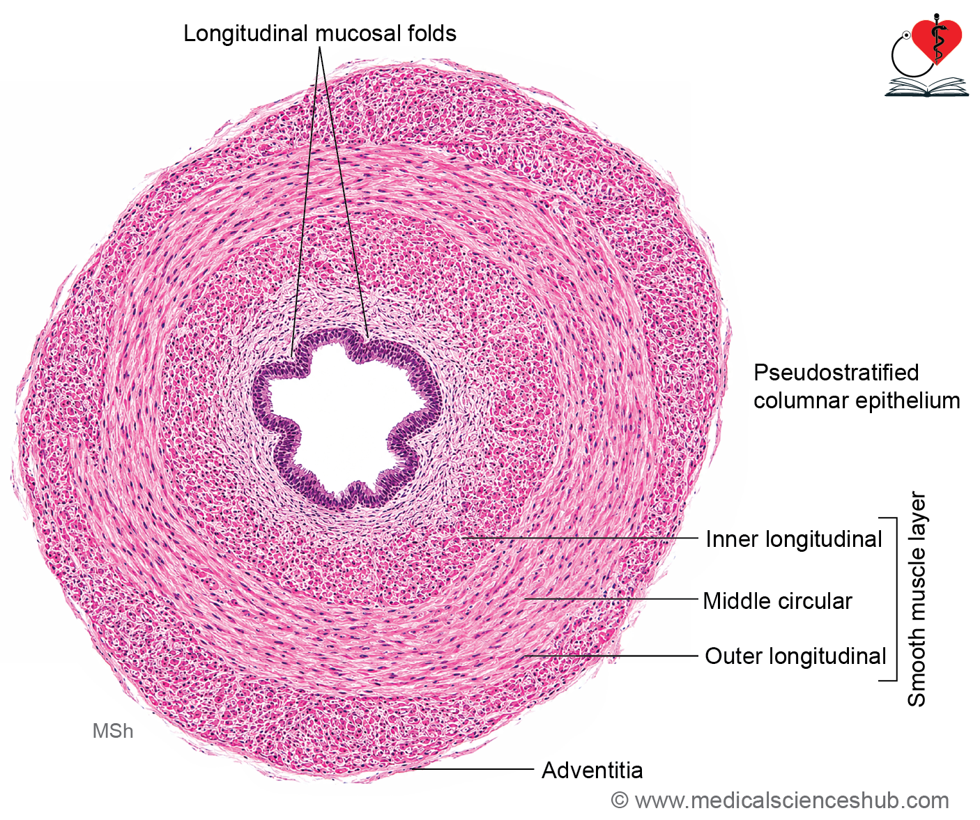
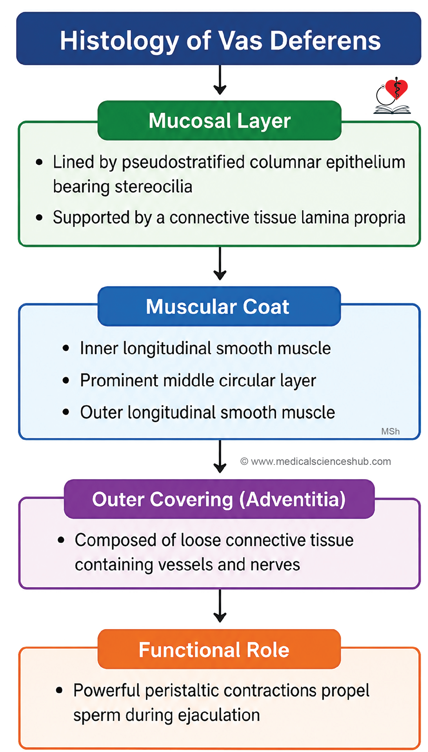
- Ductus deferens consists of three layers: Mucosa, muscle coat and adventitia.
Mucosa
- The lumen appears star-shaped due to prominent longitudinal mucosal folds.
- It is lined by pseudostratified columnar epithelium containing stereocilia.
- Two main cell types are present:
- Tall columnar cells with stereocilia extending to the lumen.
- Small basal cells, which function as progenitor cells.
- A thin lamina propria of connective tissue supports the epithelium.
Muscle Coat
- The wall contains a thick smooth muscle layer arranged as:
- Inner longitudinal layer
- Middle circular layer
- Outer longitudinal layer
- This well-developed muscular coat is the most prominent feature of the vas deferens.
- Coordinated peristaltic contractions propel sperm rapidly during ejaculation.
Adventitia
- The outer layer consists of loose connective tissue containing blood vessels, lymphatics, and nerves.
Functions of Vas Deferens
- Transports sperm from the epididymis to the ejaculatory duct during ejaculation.
- Powerful muscular contractions ensure rapid sperm propulsion.
- The vas deferens primarily serves as a conducting tube and is not a major site of sperm storage.
Important Questions
- Write a short note on spermiogenesis.
- Write a short note on structure of spermatozoa or sperm.
📝 Test Your Knowledge – Practice MCQs
Attempt the chapter MCQ quiz and assess your understanding of key concepts.
Click to see: Medical Disclaimer and Copyright Notice