
Competency
- AN52.2 Describe and identify the microanatomical features of prostate and penis.
Introduction
- The accessory male sex glands and penis play essential roles in male reproduction. The seminal vesicles, prostate, and bulbourethral glands contribute secretions to semen, while the penis functions as the organ of copulation and provides a passage for semen and urine.
Seminal Vesicles
- Seminal vesicles are paired, elongated accessory glands located posterior to the urinary bladder.
- Each gland consists of a highly coiled tubular structure that appears folded, producing an irregular lumen.
- The duct of each seminal vesicle joins the ampulla of the ductus deferens to form an ejaculatory duct.
Histology of Seminal Vesicle
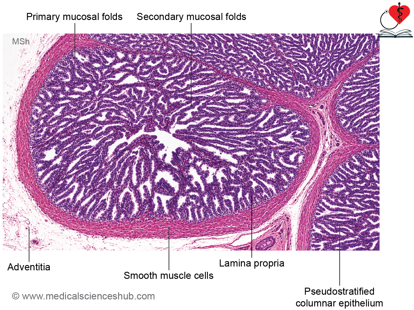
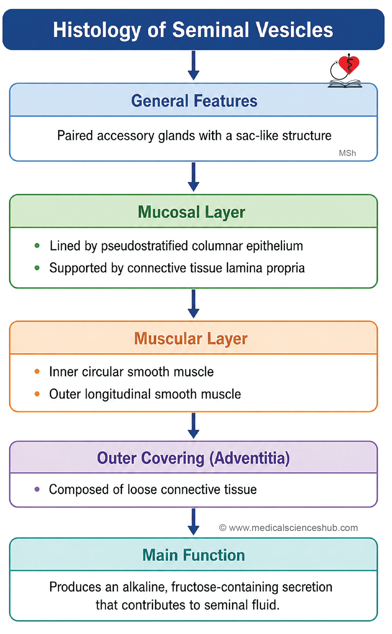
- Wall of seminal vesicle consists of three layers: Mucosa, muscle layer, and adventitia.
Mucosa
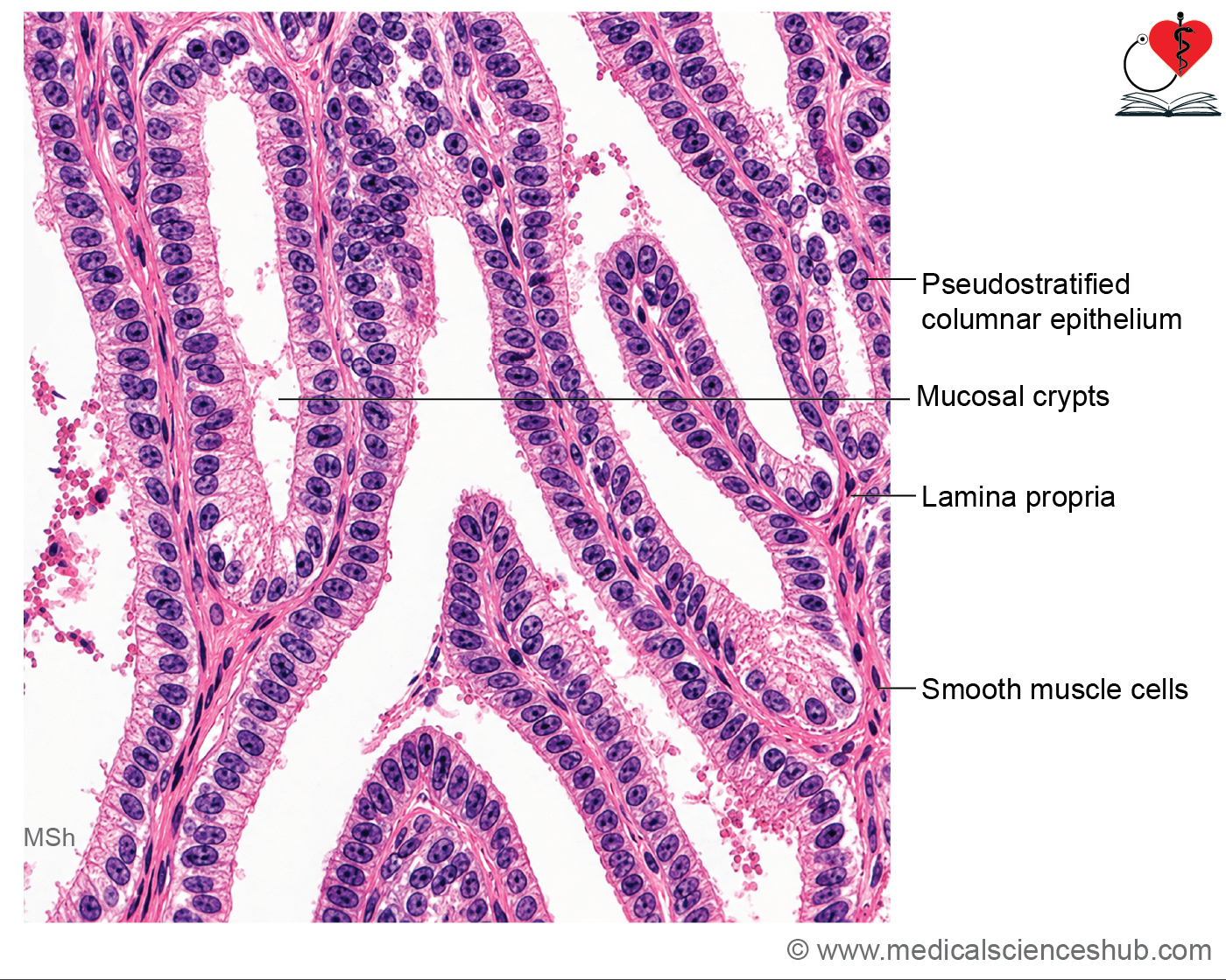
- The mucosa forms numerous complex folds, creating interconnected recesses and chambers that open into the main lumen.
- It is lined by pseudostratified columnar epithelium, composed of:
- Secretory principal cells (columnar to cuboidal)
- Small basal cells
- The underlying lamina propria contains loose connective tissue, elastic fibers, and scattered smooth muscle cells.
- Extensive mucosal folding is a characteristic histological feature of the seminal vesicle.
Muscle Layer
- The muscular wall consists of smooth muscle arranged in:
- Inner circular layer
- Outer longitudinal layer
- Contraction of these muscle layers helps expel seminal vesicle secretions into the ejaculatory ducts during ejaculation.
Adventitia
The outermost layer is a thin coat of loose connective tissue that supports the gland and contains blood vessels and nerves.
Functions of Seminal Vesicle
- Seminal vesicles contribute approximately 60–70% of semen volume.
- Their secretion, known as seminal fluid, is rich in fructose, which serves as a major energy source for spermatozoa.
- The secretion also contains prostaglandins, which may facilitate sperm transport within the female reproductive tract.
- Seminal fluid is mildly alkaline, helping to neutralize the acidic environment of the vagina.
- Lipochrome pigments present in the secretion contribute to the characteristic yellowish appearance of semen.
Prostate Gland
- The prostate gland is the largest accessory gland of the male reproductive system.
- It is approximately the size of a walnut and surrounds the proximal part of the urethra, known as the prostatic urethra.
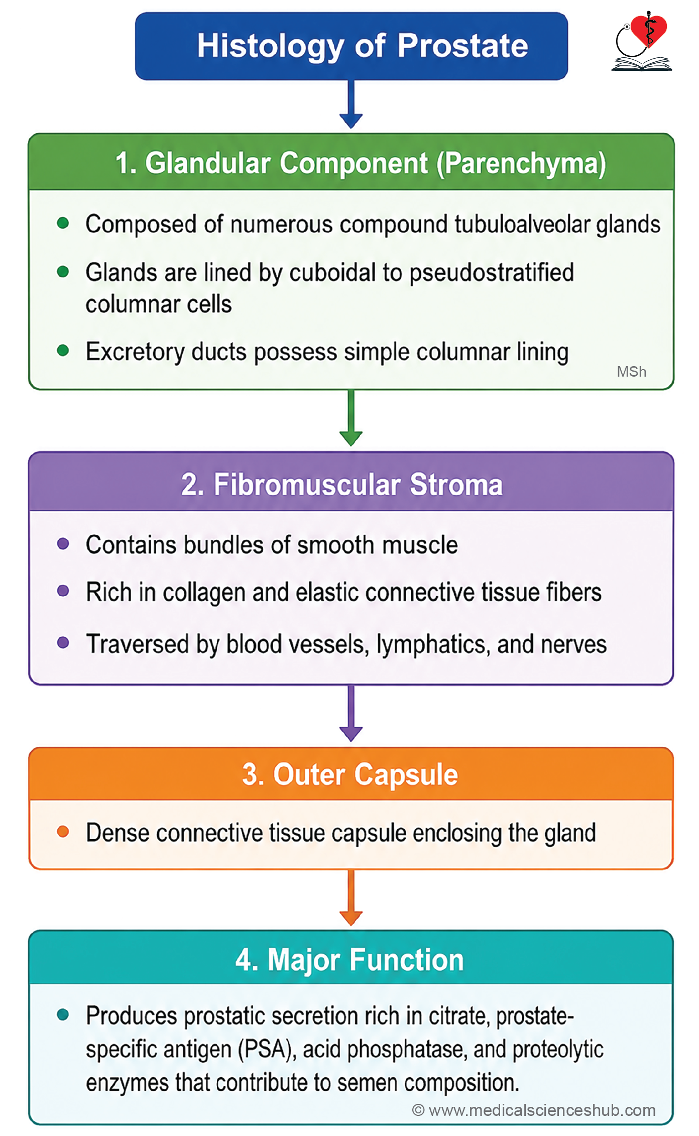
- The gland contains about 30–50 compound tubuloalveolar glands embedded within a fibromuscular framework.
Histology of Prostate Gland
Parenchyma
- The prostate is enclosed by a dense connective tissue capsule.
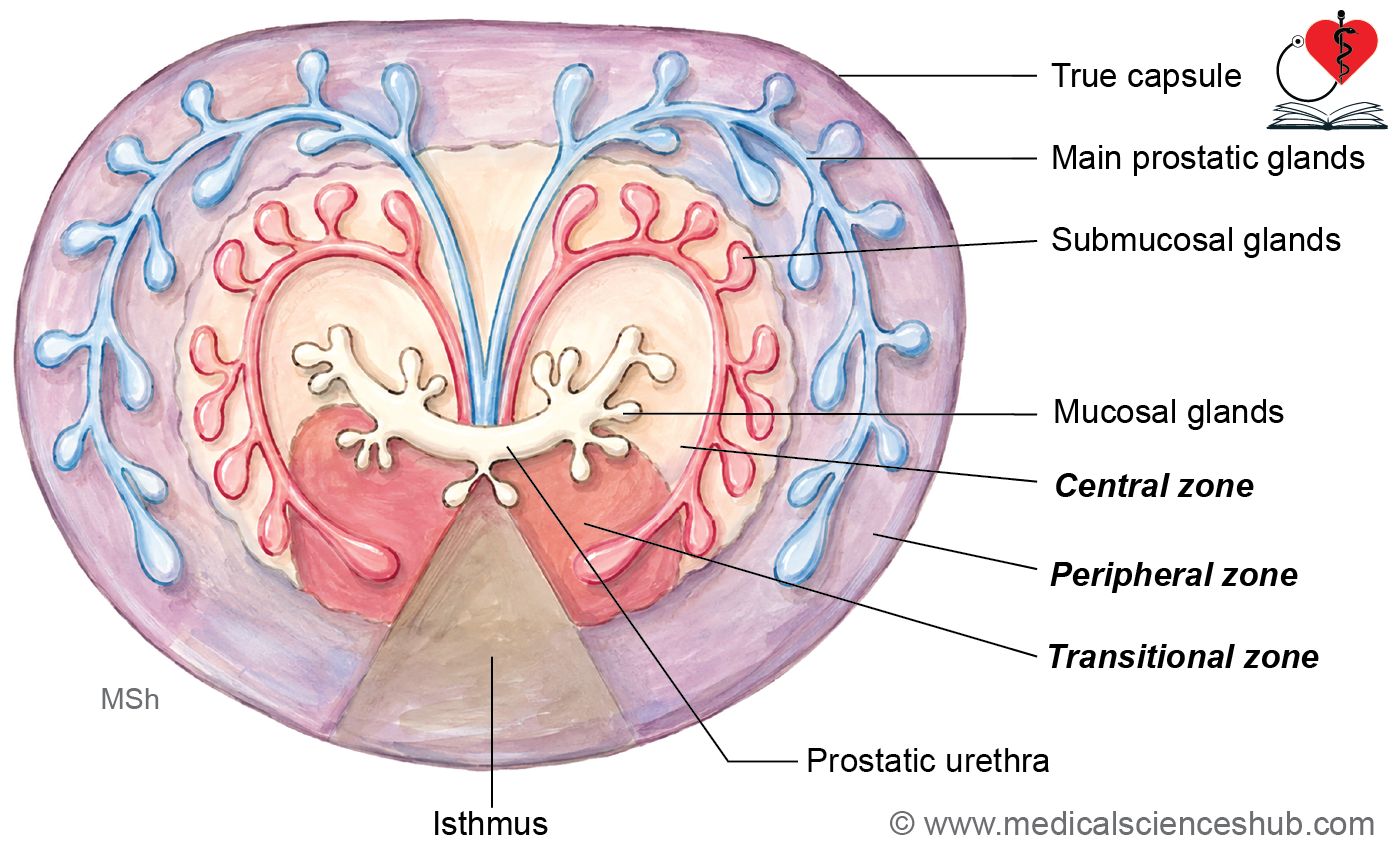
- Its glandular component can be divided into:
- Mucosal glands, which open directly into the prostatic urethra.
- Submucosal glands, located deeper within the gland and draining via ducts.
- Main prostatic glands, situated predominantly in the peripheral region and opening into the urethra.
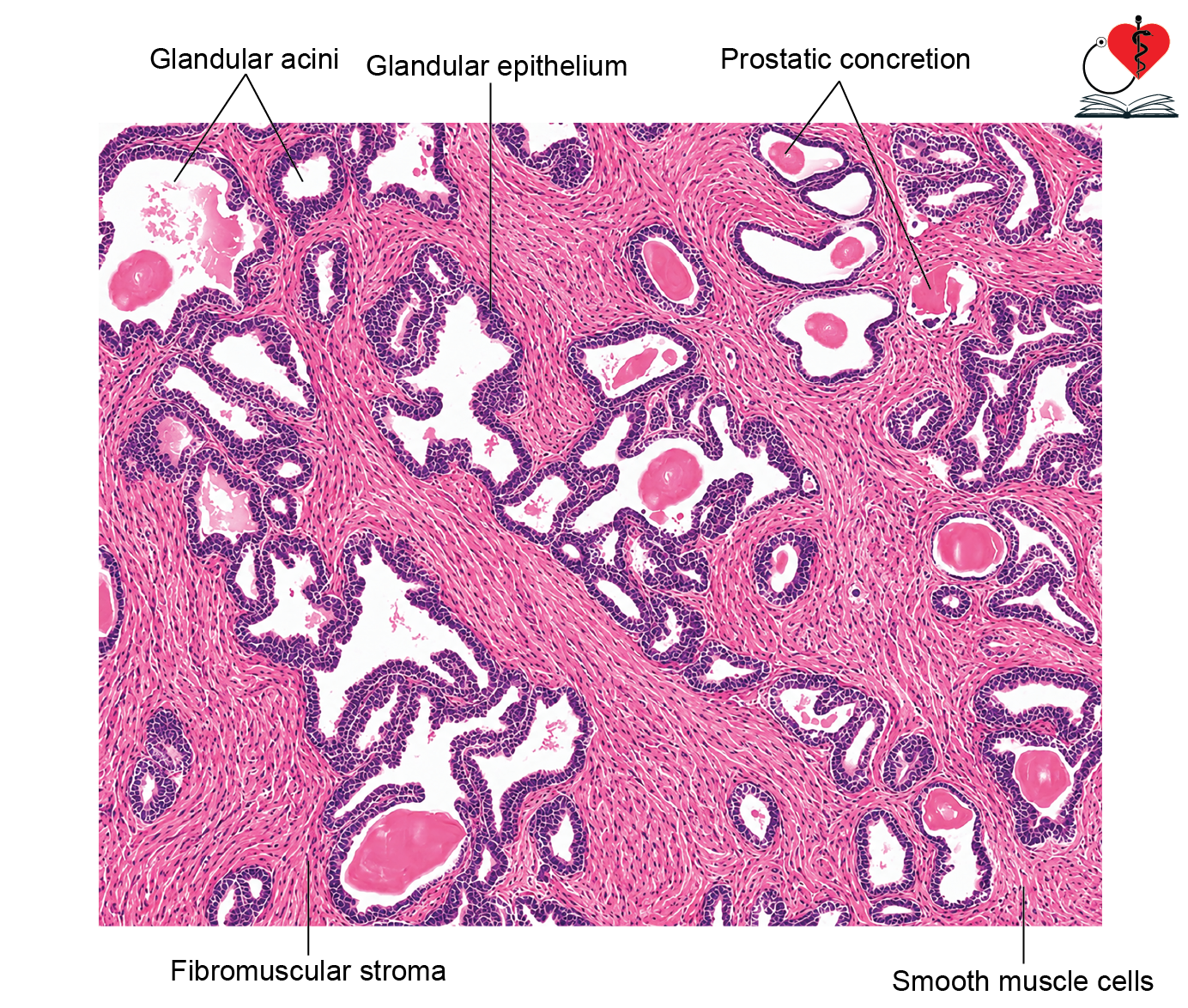
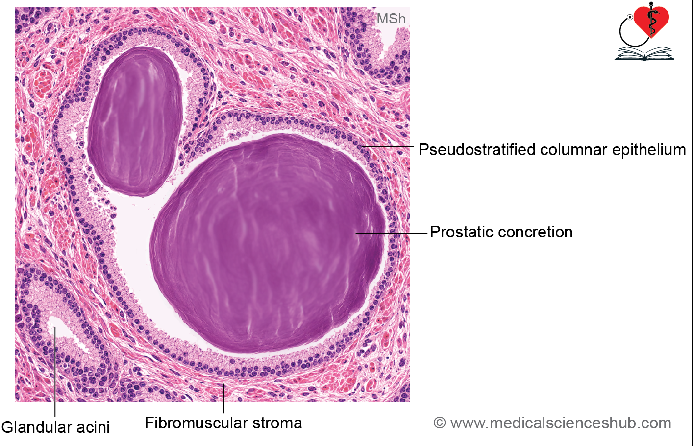
- The secretory alveoli are lined by pseudostratified columnar epithelium.
- Prostatic ducts are lined by stratified columnar epithelium, consisting of superficial columnar cells and deeper cuboidal cells.
Fibromuscular Stroma
- The glandular acini are supported by abundant fibromuscular stroma.
- This stroma contains smooth muscle fibers, collagen, elastic fibers, blood vessels, lymphatics, and nerves.
- Smooth muscle bundles are arranged in multiple directions and aid in expelling prostatic secretions during ejaculation.
Corpora Amylacea (Prostatic Concretions)
- With advancing age, many prostatic acini contain corpora amylacea, which are concentric, eosinophilic accumulations of glycoprotein-rich secretions.
- These structures may undergo calcification and increase in number in older individuals.
Prostatic Urethra
- The upper part of the prostatic urethra is lined by transitional epithelium.
- The lower part is lined by stratified columnar epithelium.
Functions of Prostate
- Prostatic secretions contribute approximately 10–30% of semen volume.
- The fluid contains prostate-specific antigen (PSA), prostatic acid phosphatase (PAP), citric acid, and proteolytic enzymes.
- PSA and fibrinolytic enzymes help liquefy semen after ejaculation, enhancing sperm motility.
- Contraction of the surrounding smooth muscle facilitates the release of prostatic fluid into the urethra.
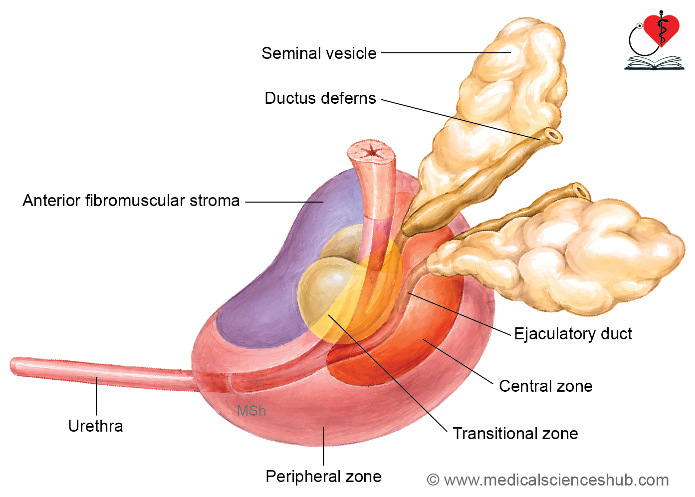
Zones of adult prostatic parenchyma:
- The adult prostate gland is divided into four anatomically and clinically important zones.
- Central Zone
- Surrounds the ejaculatory ducts.
- Constitutes approximately 25% of the glandular tissue.
- Derived embryologically from the mesonephric ducts.
- Relatively resistant to inflammation and malignant transformation.
- Peripheral Zone
- Forms about 70% of the glandular component.
- Located around the central zone and occupies the posterior and lateral portions of the gland.
- Most cases of prostatic adenocarcinoma arise in this region.
- Transitional Zone
- Accounts for roughly 5% of the prostatic parenchyma.
- Surrounds the proximal prostatic urethra.
- Common site of benign prostatic hyperplasia (BPH), which may obstruct urinary flow by compressing the urethra.
- Periurethral Zone
- Composed mainly of mucosal and submucosal glands adjacent to the urethra.
- Frequently becomes involved as benign prostatic hyperplasia progresses.
CLINICAL CORRELATION
- Prostatitis refers to inflammation of the prostate gland and may be acute or chronic in nature.
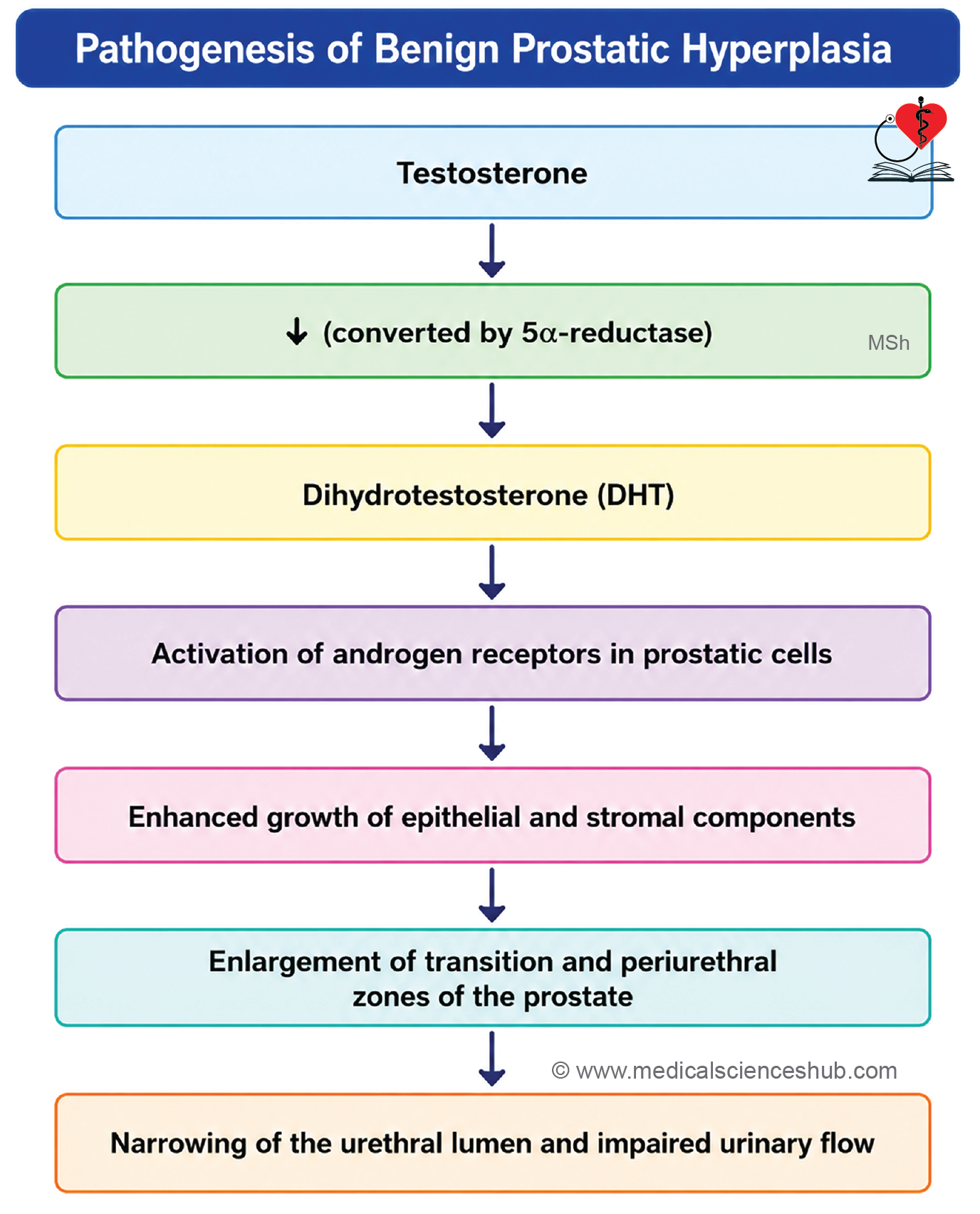
- Benign Prostatic Hyperplasia (BPH) is a non-malignant enlargement of the prostate that commonly affects older men.
- It primarily involves the transitional zone surrounding the prostatic urethra.
- Growth of the gland is influenced by dihydrotestosterone (DHT), a metabolite of testosterone.
- BPH usually develops after 50 years of age and may cause urinary obstruction.
- Management includes:
- α1-adrenergic blockers to improve urine flow.
- 5α-reductase inhibitors to reduce DHT production and prostate size.
- Transurethral resection of the prostate (TURP) in severe or refractory cases.
- Prostatic adenocarcinoma is one of the most common malignancies in men and most frequently arises in the peripheral zone of the prostate.
- The tumor originates from the glandular epithelium of the prostate.
- Clinical Markers
- Prostate-specific antigen (PSA), also known as kallikrein-3, is a glycoprotein enzyme secreted by prostatic epithelial cells.
- Elevated PSA levels may occur in prostate cancer, BPH, prostatitis, and other prostatic disorders; therefore, PSA should be interpreted with clinical findings.
- Prostatic acid phosphatase (PAP) is another prostatic secretion that may show increased levels in certain prostatic diseases, particularly advanced malignancy.
Penis
- The penis is the male copulatory organ and functions in both reproduction and urination.
- It consists of a fixed root attached to the perineum and a free body (shaft).
Structure of Erectile Tissue
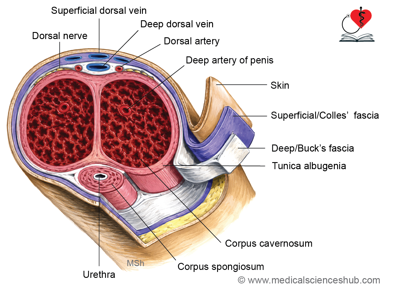
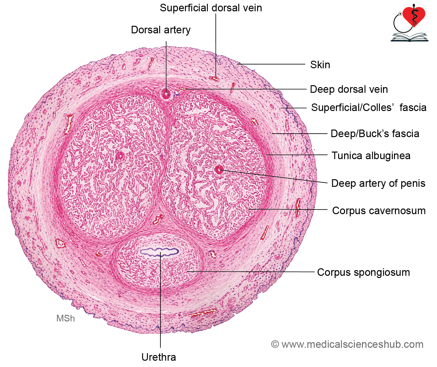
- The penis contains three cylindrical masses of erectile tissue:
- Two dorsal corpora cavernosa.
- One ventral corpus spongiosum, which encloses the penile urethra.
- The corpora cavernosa are composed of endothelial-lined vascular spaces separated by smooth muscle trabeculae and supported by connective tissue rich in elastic and collagen fibers.
- The corpus spongiosum has a similar structure but remains less rigid during erection, helping maintain urethral patency.
Supporting Structures
- A dense connective tissue capsule, the tunica albuginea, surrounds the erectile tissues and partially separates the two corpora cavernosa.
- The tunica albuginea contains inner circular and outer longitudinal fiber layers and is thinner around the corpus spongiosum.
- External to the tunica albuginea lies Buck’s fascia, a layer of connective tissue lacking adipose tissue, followed by thin, hairless skin.
Penile urethra
- The penile (spongy) urethra is mainly lined by pseudostratified columnar epithelium.
- Near the external urethral opening, the lining transitions to stratified squamous epithelium.
- Mucus-secreting glands of Littre are present in the distal part of the penile urethra.
Mechanism of Erection
- Erectile tissue receives blood through helicine arteries, branches of the deep penile arteries.
- Sexual stimulation causes vasodilation, increasing blood flow into the cavernous spaces.
- Expansion of these spaces compresses venous channels against the tunica albuginea, reducing venous outflow.
- The resulting accumulation of blood produces penile erection.
CLINICAL CORRELATION
- Erectile dysfunction is the persistent inability to achieve or maintain an erection and may result from psychological factors, medications, vascular disorders, neurological disease, or systemic illnesses.
Important Questions
- Q. Write a short note on histology of penis..
📝 Test Your Knowledge – Practice MCQs
Attempt the chapter MCQ quiz and assess your understanding of key concepts.
Click to see: Medical Disclaimer and Copyright Notice