
Competencies
- AN52.2 Describe and identify the microanatomical features of female reproductive system: Ovary, uterus, uterine tube, cervix.
- AN52.3 Describe and identify the microanatomical features of corpus luteum.
Introduction
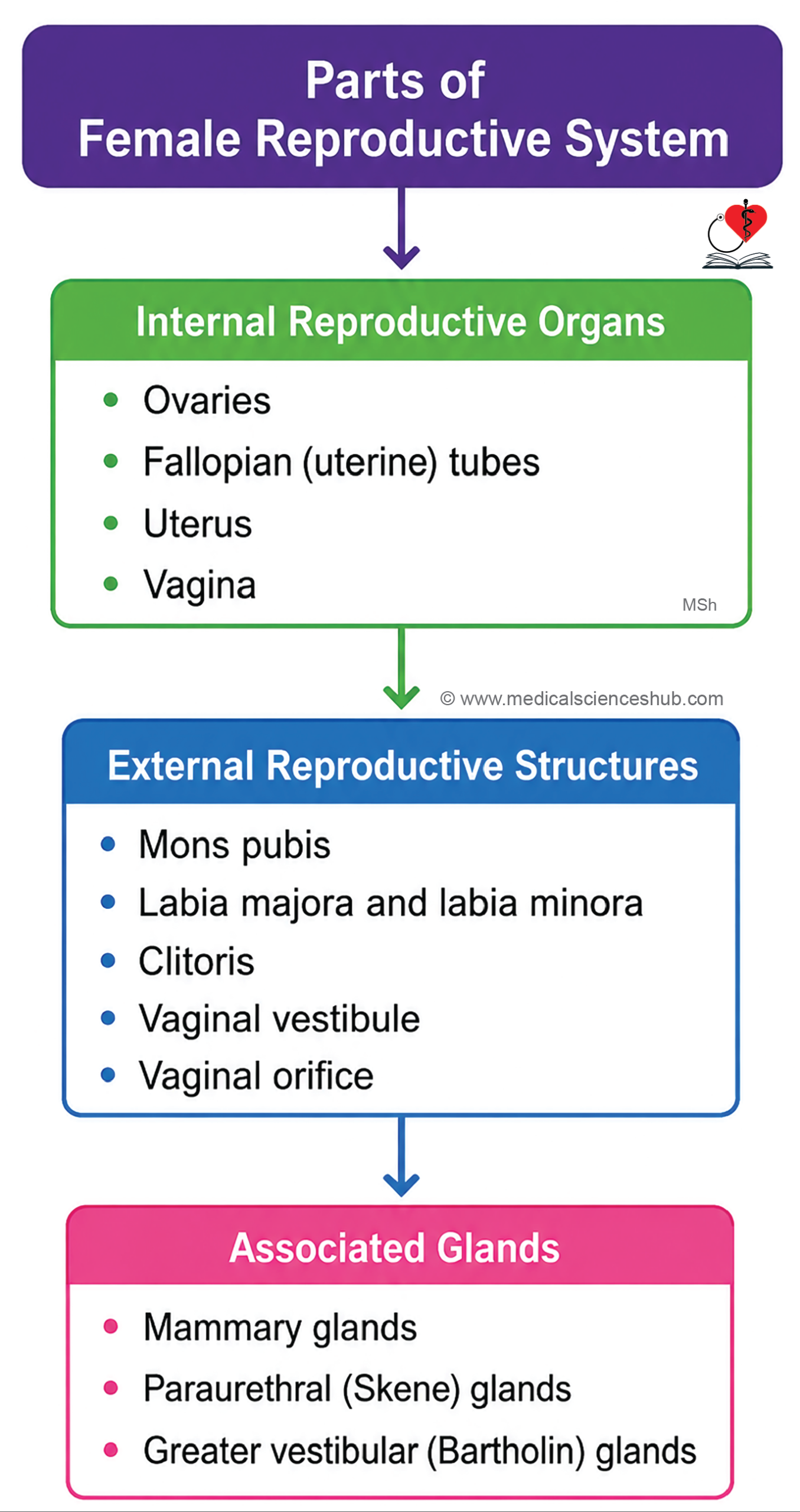
- The female reproductive system includes internal genital organs, external genital organs, and accessory glands.
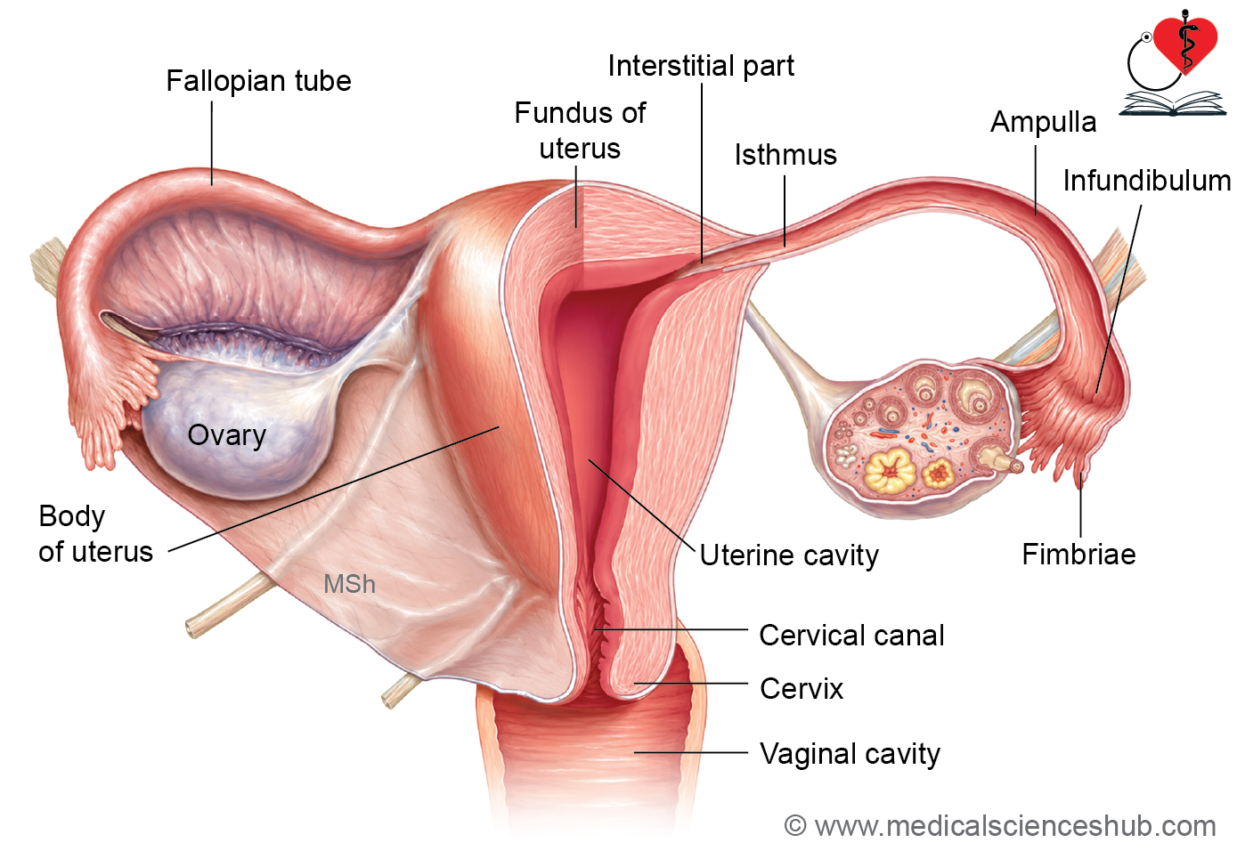
- Internal genital organs, located within the pelvis, include the ovaries, uterine (fallopian) tubes, uterus, and vagina.
- External genital organs, collectively known as the vulva, include the mons pubis, labia majora, labia minora, clitoris, vestibule, and vaginal orifice.
- Accessory reproductive glands include the mammary glands, Skene’s (paraurethral) glands, and Bartholin’s (greater vestibular) glands. Temporary structures associated with pregnancy include the placenta and umbilical cord.
- The menstrual cycle consists of cyclic changes in the ovaries and uterus regulated by reproductive hormones.
- Menarche is the onset of menstruation and typically occurs between 9 and 14 years of age.
- Menopause marks the permanent cessation of menstrual cycles and usually occurs between 45 and 55 years of age.
- The average menstrual cycle lasts approximately 28–30 days, although normal variation exists among individuals.
Ovary
- The ovary is the female gonad and performs two essential functions:
- Gametogenesis: Production and maturation of female gametes (oocytes).
- Hormone secretion: Production of estrogens and progesterone, which regulate reproductive function and the menstrual cycle.
- Ovaries are paired, almond-shaped organs with a pinkish-white appearance.
- Each ovary is attached to the broad ligament and typically measures about 3 cm × 1.5 cm × 1 cm.
General Structure of Ovary
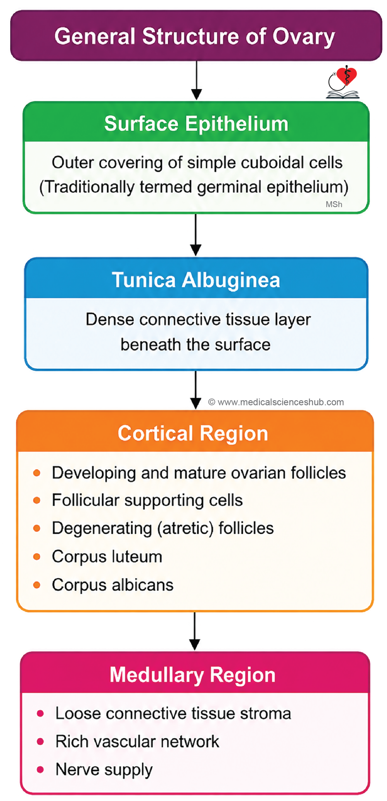
- Each ovary consists of the following components:
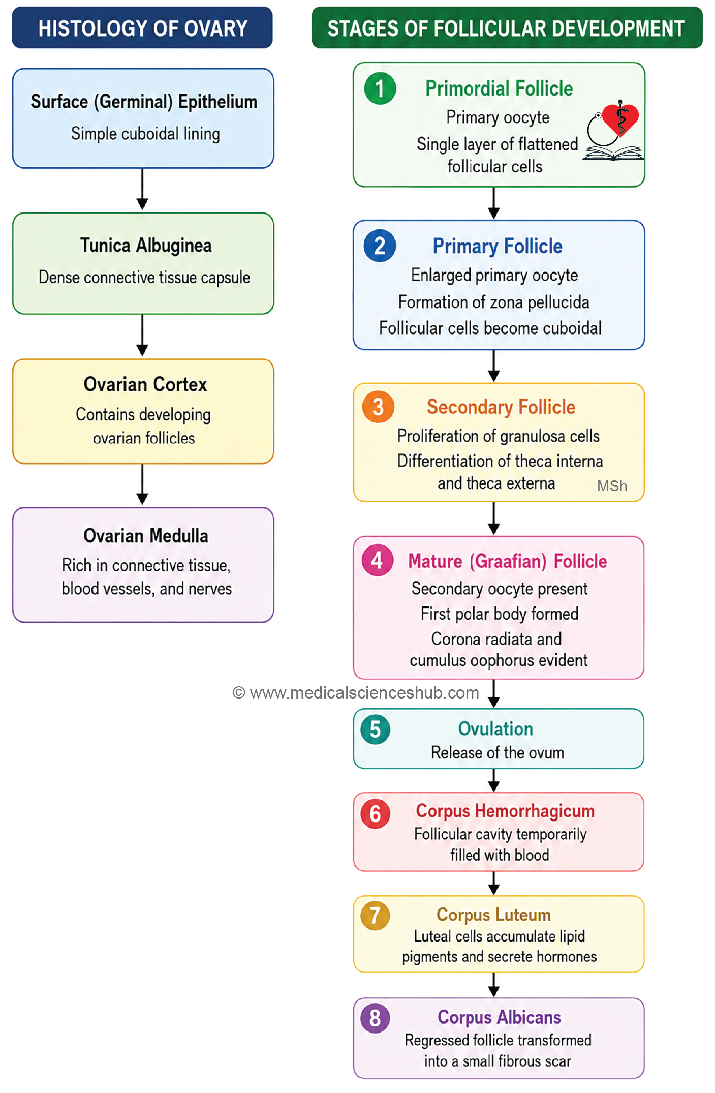
Germinal Epithelium
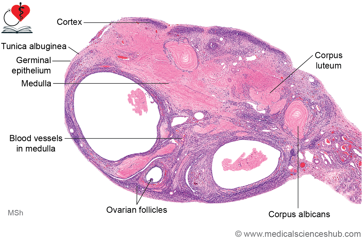
- The ovarian surface is covered by a single layer of simple cuboidal epithelium, traditionally called the germinal epithelium.
- Despite its name, this epithelium does not produce germ cells.
- Female germ cells originate from primordial germ cells that migrate from the yolk sac during embryonic development.
- The surface epithelium is continuous with the mesothelium of the peritoneum.
Tunica Albuginea
- Beneath the surface epithelium lies the tunica albuginea, a thin layer of dense connective tissue.
- This layer is considerably thinner than the tunica albuginea of the testis.
Cortex
- The cortex forms the outer functional region of the ovary, located beneath the tunica albuginea.
- It contains ovarian follicles at various stages of development embedded within a highly cellular stromal tissue.
- Cortical stroma gives rise to supporting follicular structures, including granulosa cells, theca interna, theca externa, corona radiata, and cumulus oophorus.
- The cortex also contains corpus luteum, corpus albicans, and atretic follicles.
Medulla
- The medulla occupies the central part of the ovary.
- It consists mainly of loose connective tissue containing numerous blood vessels, lymphatics, and nerve fibers.
- The hilum of the ovary serves as the entry and exit site for blood vessels and nerves.
- Histologically, the boundary between the cortex and medulla is not sharply defined.
Ovarian Follicles (Oogenesis)
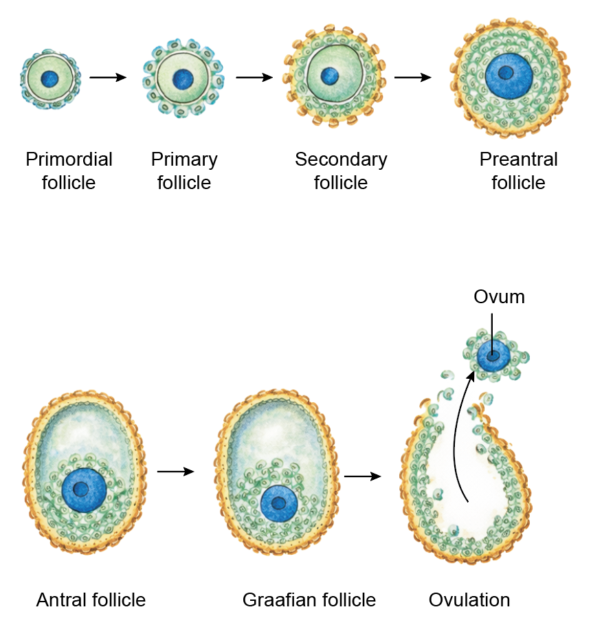
- The ovarian cortex contains follicles at different stages of development, including primordial follicles, primary follicles, secondary follicles, Graafian follicles, corpus luteum, corpus albicans, and atretic follicles.
Oogonia
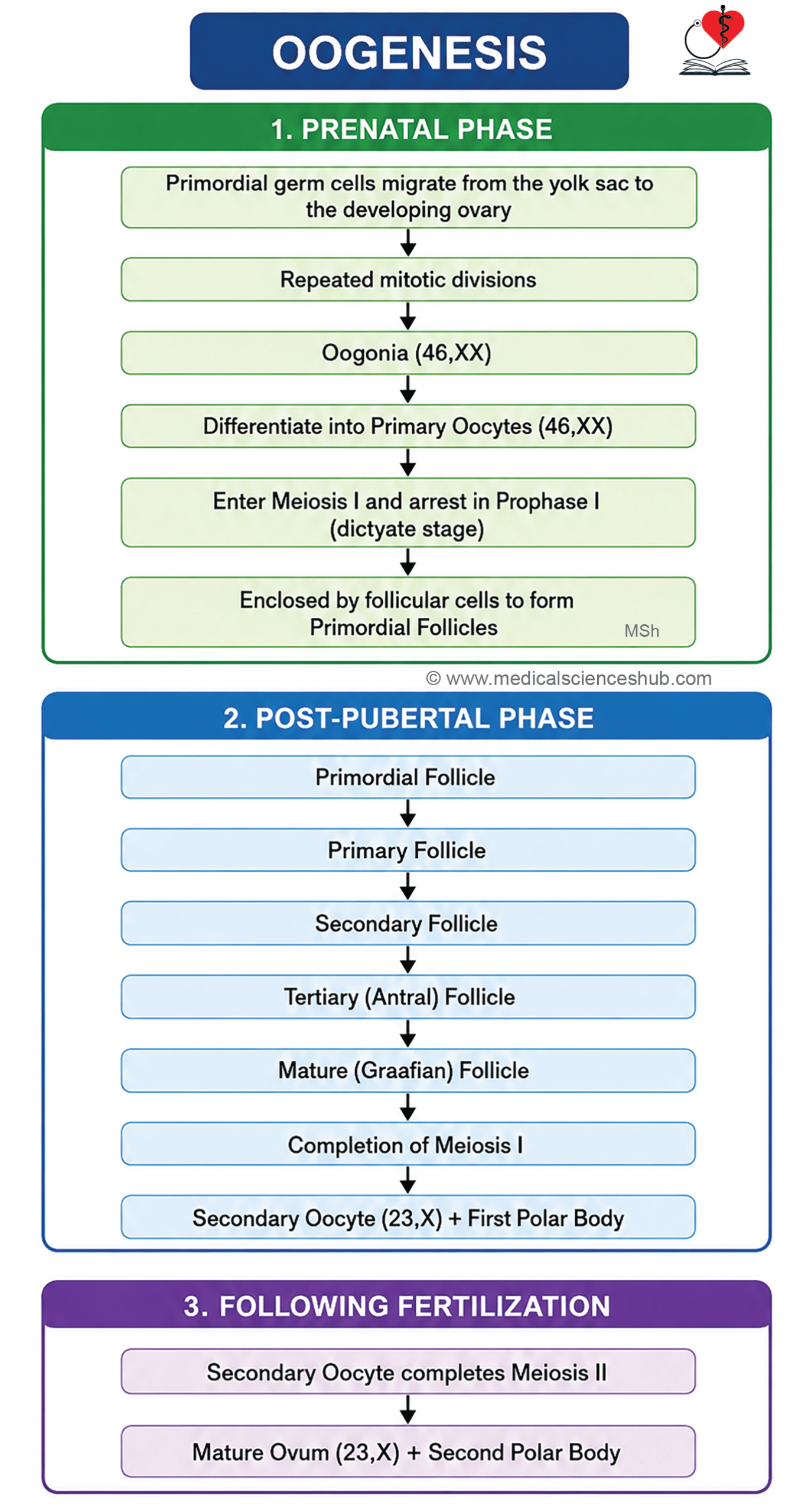
- During fetal life, primordial germ cells differentiate into oogonia.
- Oogonia undergo mitotic divisions and enlarge to form primary oocytes.
- Before birth, primary oocytes enter meiosis I and become arrested in the dictyotene (diplotene) stage of prophase I.
- This arrest is maintained until puberty by factors produced within the follicle.
- At birth, the ovaries contain several hundred thousand primary oocytes, but only about 400–500 are ovulated during reproductive life.
- From birth until puberty, most follicles remain dormant.
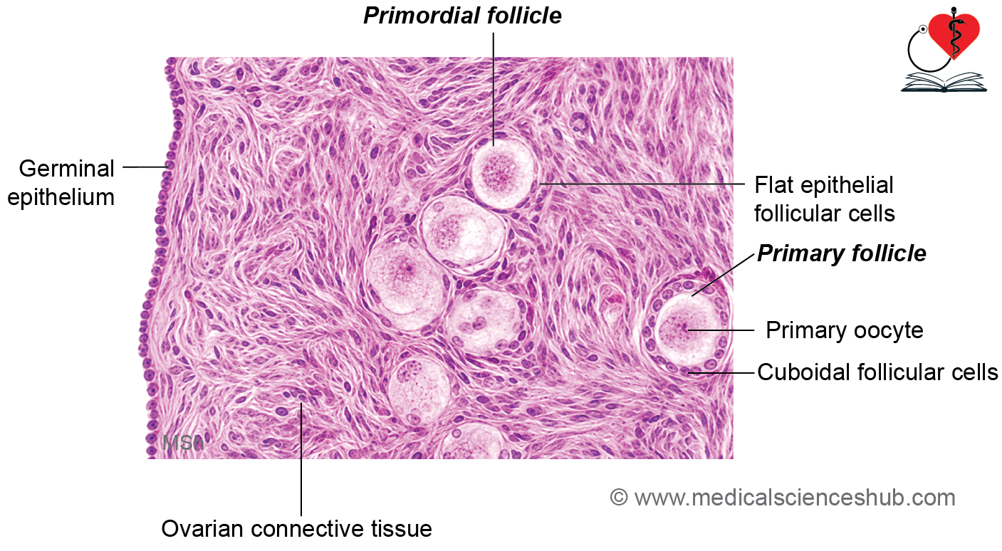
Primordial Follicle
- Primordial follicles are located immediately beneath the tunica albuginea.
- Each follicle contains:
- A primary oocyte with a prominent nucleus and cytoplasmic organelles, including the Balbiani body.
- A single layer of flattened follicular cells surrounding the oocyte.
Primary Follicle
- Growth of the primordial follicle produces a primary follicle.
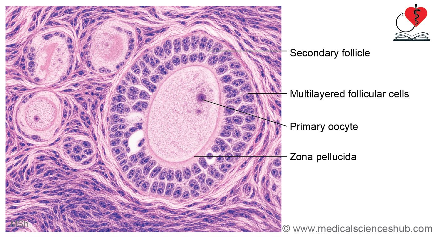
- Follicular cells become cuboidal and proliferate to form the granulosa cell layer.
- The oocyte secretes the zona pellucida, a glycoprotein-rich layer that surrounds it and stains positively with PAS stain.
- Stromal cells differentiate into:
- Theca interna, a vascular endocrine layer.
- Theca externa, an outer connective tissue layer containing collagen fibers and smooth muscle cells.
Secondary Follicle
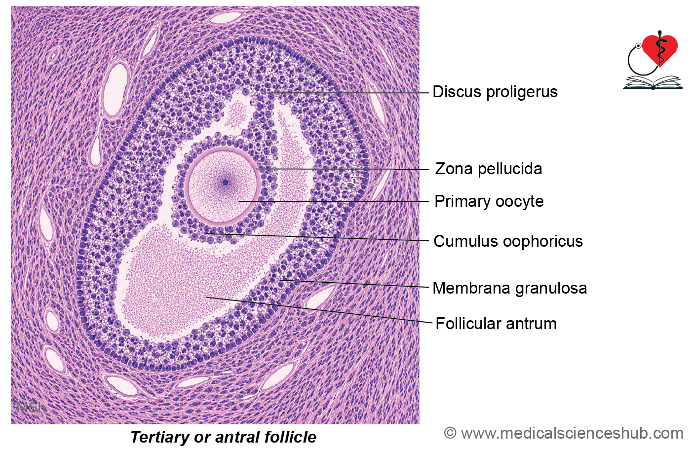
- Continued follicular growth leads to the formation of fluid-filled spaces among granulosa cells.
- These spaces merge to form the antrum, which contains liquor folliculi.
- The oocyte enlarges significantly, reaching approximately 120–125 μm in diameter.
- Granulosa cells surrounding the oocyte form the cumulus oophorus, which later gives rise to the corona radiata.
- Call–Exner bodies, small PAS-positive extracellular deposits rich in glycoproteins and hyaluronic acid, may be present between granulosa cells.
Graafian Follicle
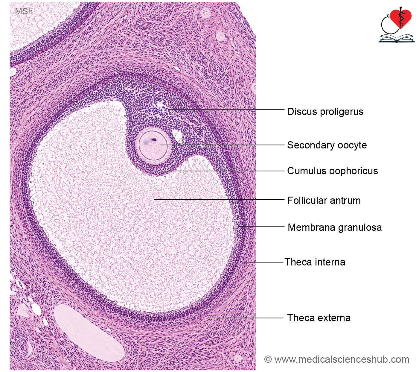
- The Graafian follicle is the fully mature ovarian follicle and may exceed 10 mm in diameter.
- Just before ovulation, the primary oocyte completes meiosis I to form a secondary oocyte and the first polar body.
- The enlarged antrum occupies most of the follicle, while granulosa cells around the oocyte form the corona radiata.
- The mature follicle bulges from the ovarian surface before ovulation.
- Theca interna cells produce androgens, which are converted into estrogens by granulosa cells, supporting follicular growth and maturation.
Ovulation
- Ovulation is the release of a secondary oocyte from a mature Graafian follicle.
- The process is facilitated by:
- Increased pressure from accumulating follicular fluid.
- Proteolytic activity mediated by plasminogen activators, which weaken the follicular wall.
- Prostaglandin-induced contraction of smooth muscle cells in the theca externa.
- A small avascular area called the stigma (macula pellucida) develops on the ovarian surface over the bulging follicle.
- Rupture at this site allows the secondary oocyte, surrounded by the corona radiata, to be released.
- The secondary oocyte remains viable for approximately 24 hours after ovulation.
- It is arrested in metaphase II of meiosis and completes meiosis only if fertilization occurs.
Corpus Luteum/Luteal Gland
- Following ovulation, the ruptured follicle collapses and transforms into the corpus luteum.
- Granulosa and theca interna cells enlarge and accumulate lipid pigments, giving the structure its characteristic yellow color.
- Two main cell types are present:
- Granulosa lutein cells, which predominantly secrete progesterone.
- Theca lutein cells, which mainly produce estrogens.
- The corpus luteum prepares the endometrium for possible implantation and supports early pregnancy if fertilization occurs.
Corpus Albicans
- In the absence of fertilization, the corpus luteum undergoes degeneration.
- Luteal cells regress and are gradually replaced by fibrous connective tissue, forming the corpus albicans.
- This scar-like structure eventually shrinks and becomes inconspicuous within the ovarian cortex.
Follicular Atresia
- Most ovarian follicles do not ovulate and instead undergo follicular atresia.
- Characteristic changes include:
- Apoptosis and degeneration of granulosa cells.
- Enlargement of theca interna cells.
- Removal of degenerating cells by macrophages.
- Replacement of follicular structures by connective tissue.
- A prominent feature of atretic follicles is the formation of a thick, wavy glassy (hyaline) membrane, produced by thickening of the basement membrane between granulosa and theca layers.
Fallopian Tube (Uterine Tubes)
- The fallopian tubes are paired muscular ducts that transport the oocyte from the ovary to the uterus and provide the usual site for fertilization.
- Each tube measures approximately 10–12 cm in length.
Parts of the Fallopian Tube
- Infundibulum: Funnel-shaped lateral end bearing finger-like fimbriae that help capture the ovulated oocyte.
- Ampulla: Longest and widest segment; the most common site of fertilization.
- Isthmus: Narrow, thick-walled medial segment.
- Uterine (intramural/interstitial) part: Traverses the uterine wall and opens into the uterine cavity.
Histology of Fallopian Tube
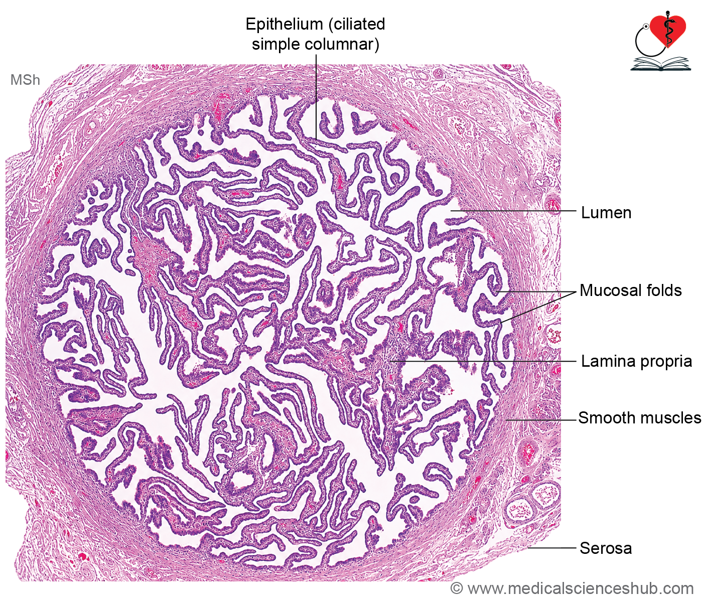
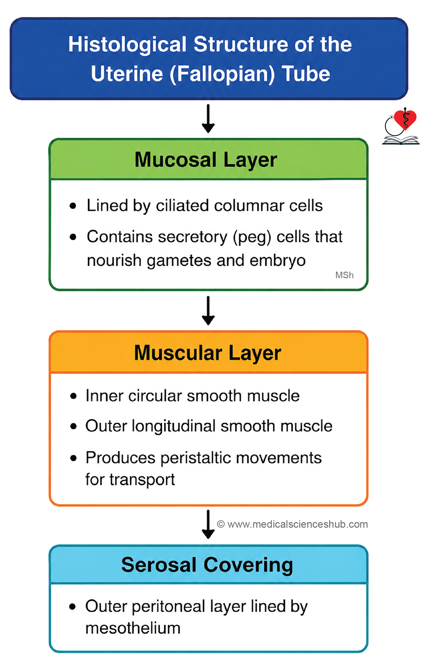
- Wall of fallopian tube shows three layers from inside outwards: Mucosa, muscle layer, and serosa.
Mucosa
- The mucosa forms numerous branching longitudinal folds that project into the lumen, especially in the ampulla.
- It is lined by simple columnar epithelium containing three cell types:
- Ciliated cells: Propel the oocyte and tubal contents toward the uterus through coordinated ciliary movement.
- Secretory (peg) cells: Produce nutritive fluid that supports sperm, oocytes, and early embryos and facilitates sperm capacitation.
- Intercalary cells: Small regenerative cells that function as epithelial stem cells.
- The epithelial lining undergoes cyclical hormonal changes during the menstrual cycle.
- The core of each mucosal fold contains highly vascular connective tissue.
Muscle Layer
- Composed of:
- An inner circular smooth muscle layer.
- An outer longitudinal smooth muscle layer.
- The circular layer is particularly prominent in the isthmus and assists in regulating transport of the oocyte or embryo toward the uterus.
Serosa/Peritoneal Coat
The outer surface is covered by a thin connective tissue layer and mesothelium (visceral peritoneum).
Functions of Fallopian Tube
- Facilitates bidirectional transport, moving sperm toward the ampulla and transporting the oocyte or embryo toward the uterus.
- Serves as the primary site of fertilization.
- Provides nutritive secretions that support gametes and the early developing embryo.
- Unlike most regions of the gastrointestinal tract, the uterine tube lacks both muscularis mucosae and submucosa.
CLINICAL CORRELATION
- The fallopian tube, particularly the ampullary region, is the most common site of ectopic pregnancy.
- Normal implantation occurs within the endometrium of the uterine body, usually in the upper uterine segment.
Uterus
- The uterus is a hollow, muscular reproductive organ located in the pelvic cavity.
- It is typically pear-shaped and is divided into three main regions:
- Fundus: The dome-shaped superior portion located above the openings of the uterine tubes.
- Body: The central and largest part of the uterus.
- Cervix: The lower cylindrical portion that projects into the vagina and is connected to the body by the isthmus.
- The cervical canal communicates with:
- The uterine cavity through the internal os.
- The vagina through the external os.
Histology of Uterus
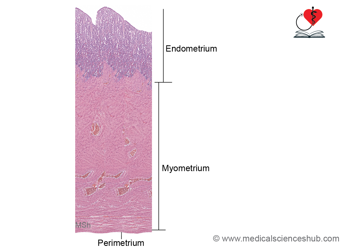
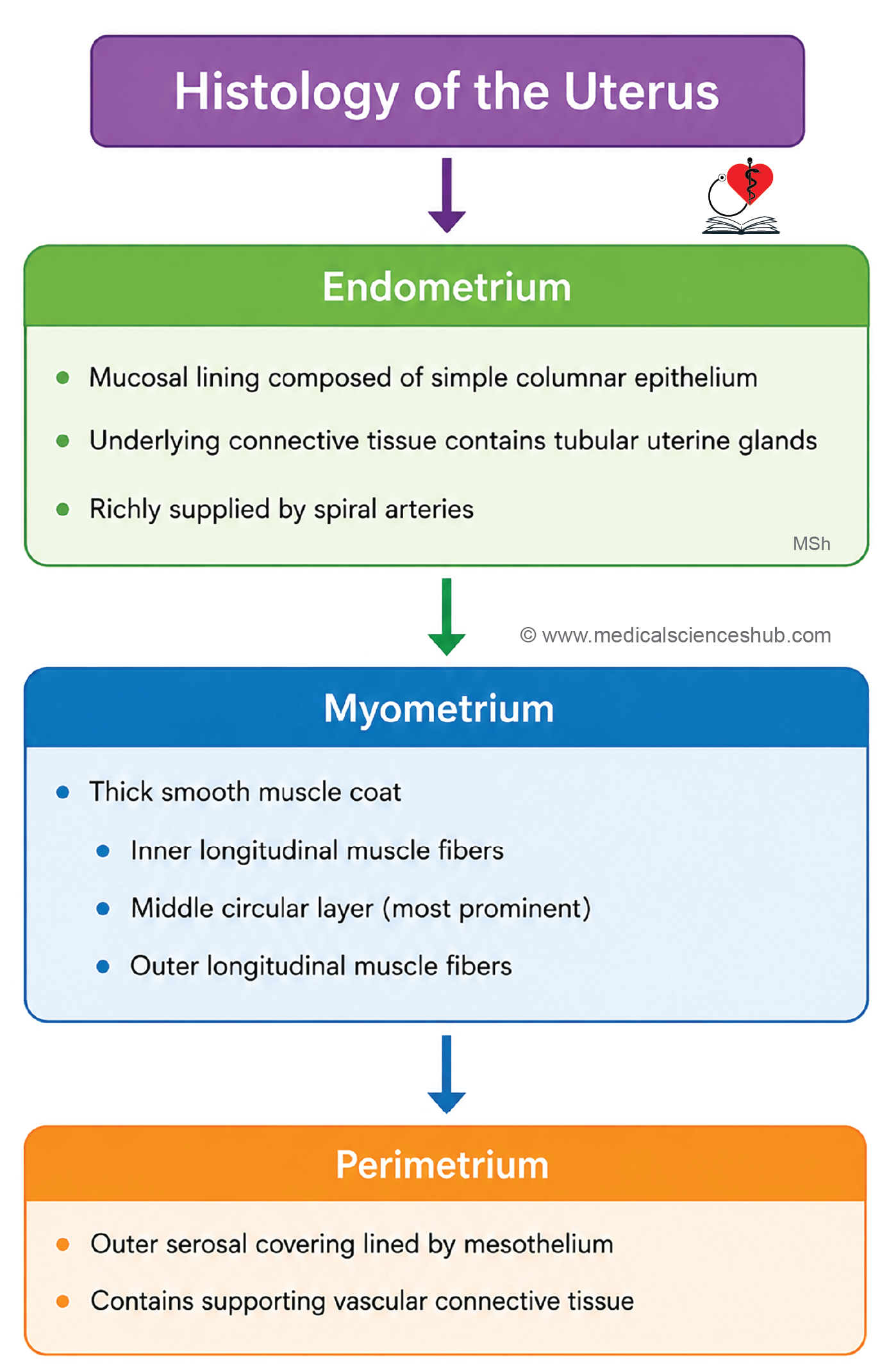
The uterine wall consists of three layers:
1. Endometrium
- The endometrium is the mucosal lining of the uterus.
- It is composed of:
- Simple columnar epithelium.
- Simple tubular uterine glands lined by columnar cells.
- A highly vascular endometrial stroma containing connective tissue, blood vessels, and glands.
- This layer undergoes cyclical changes during the menstrual cycle.
2. Myometrium
- The myometrium is the thickest layer of the uterine wall and is composed of smooth muscle.
- Smooth muscle fibers are arranged in:
- An inner longitudinal layer.
- A middle interlacing layer rich in blood vessels, known as the stratum vasculare.
- An outer longitudinal layer.
- During pregnancy, myometrial cells undergo both hypertrophy and hyperplasia to accommodate fetal growth.
- Contraction of the myometrium, stimulated by oxytocin, plays a key role in labor and childbirth.
3. Perimetrium
- The outer covering of the uterus is the perimetrium, which forms part of the visceral peritoneum.
- It consists of:
- An outer mesothelial layer (simple squamous epithelium).
- An inner layer of vascular connective tissue.
Strata of Endometrium
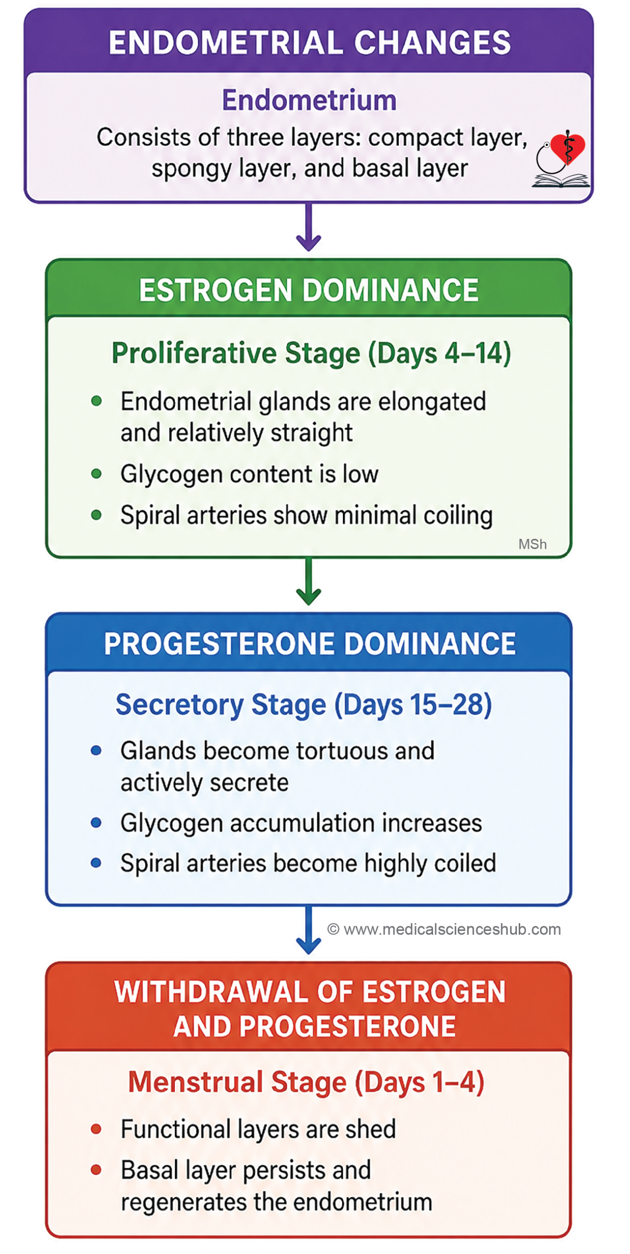
- The endometrium is organized into three layers:
- Stratum compactum: The superficial, densely cellular layer containing the upper portions of uterine glands.
- Stratum spongiosum: The intermediate layer composed of loose connective tissue and dilated glandular segments.
- Stratum basale: The deepest layer adjacent to the myometrium, containing regenerative cells responsible for renewal of the endometrium after menstruation.
Histological Changes in Endometrium
- The endometrium undergoes cyclical structural changes during the menstrual cycle under the influence of ovarian hormones.
Proliferative Phase
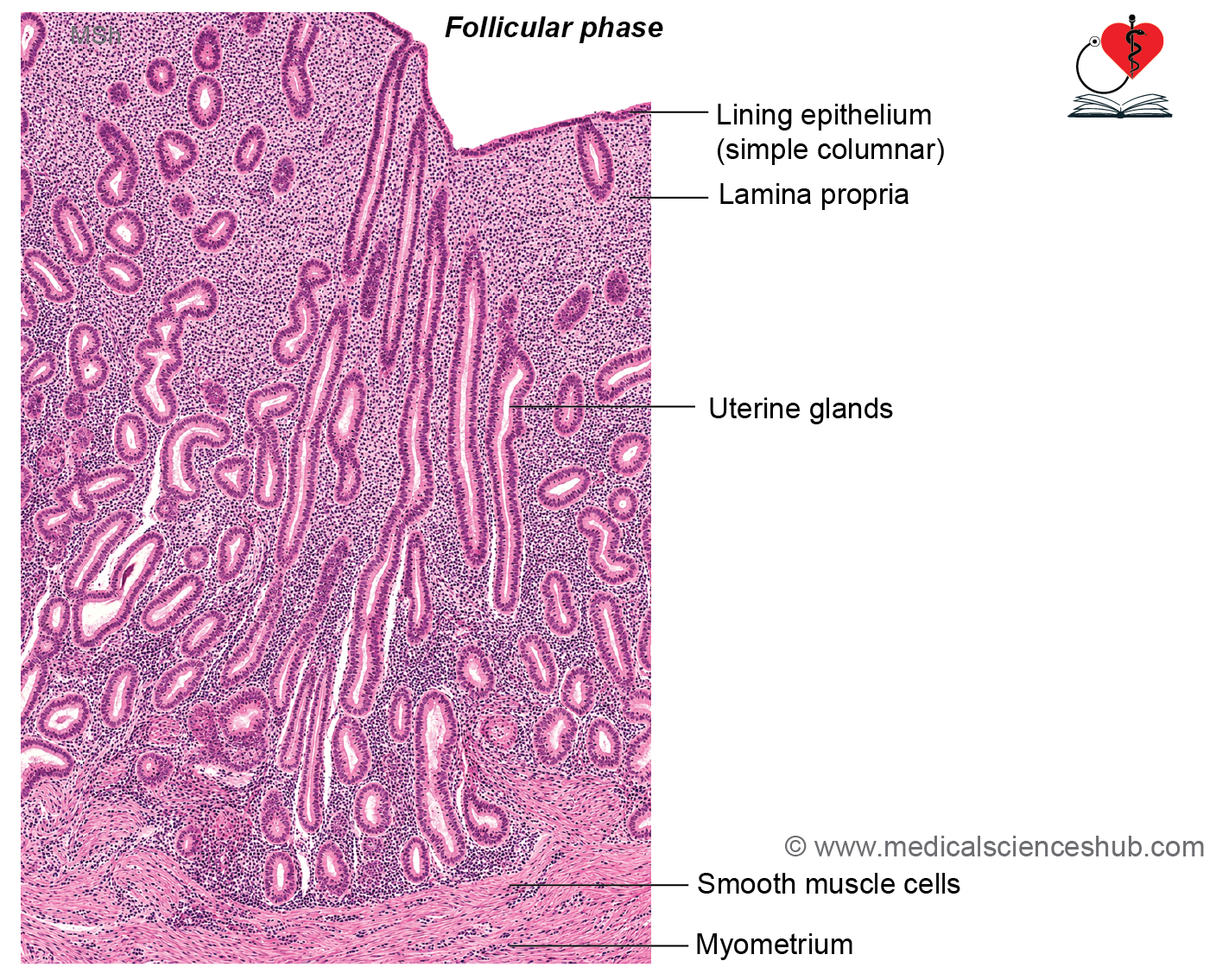
- This phase is primarily regulated by estrogen and involves regeneration of the endometrium from the stratum basale following menstruation.
- Surface epithelium and uterine glands proliferate rapidly.
- Uterine glands appear straight, narrow, and tubular.
- Endometrial stroma increases in volume, causing the endometrium to thicken to approximately 3–4 mm.
- Spiral arteries elongate and extend into the developing functional layer.
Menstrual Phase
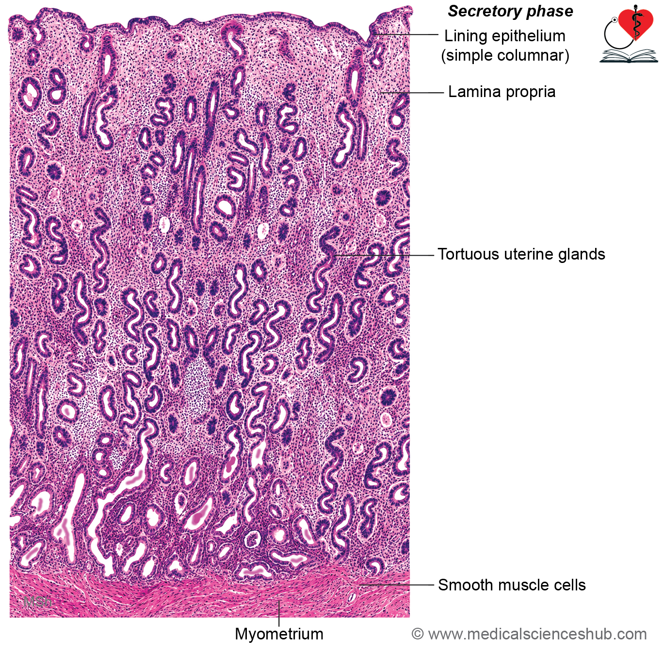
- This phase is predominantly controlled by progesterone released from the corpus luteum.
- Uterine glands become highly coiled and tortuous, producing a characteristic saw-toothed appearance.
- Glandular cells accumulate glycogen and secrete nutrient-rich fluid to support potential implantation.
- Spiral arteries become more convoluted and vascularity increases.
- Stromal edema develops due to fluid accumulation.
- Stromal cells enlarge and accumulate glycogen and lipids, producing the decidual reaction.
Menstrual Phase
- Withdrawal of estrogen and progesterone due to corpus luteum regression causes constriction of spiral arteries.
- Reduced blood flow leads to ischemia, tissue breakdown, and shedding of the functional layer of the endometrium.
- Prostaglandins and proteolytic enzymes contribute to menstrual bleeding.
- The stratum basale remains intact because it receives blood from straight arteries and serves as the source for endometrial regeneration in the next cycle.
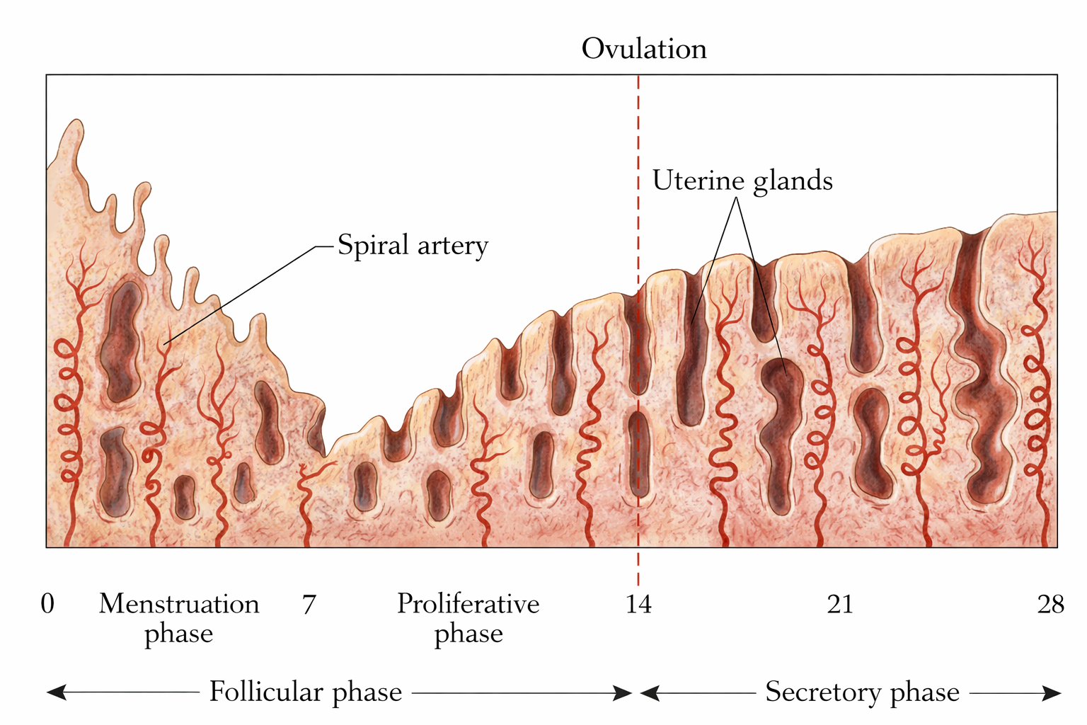
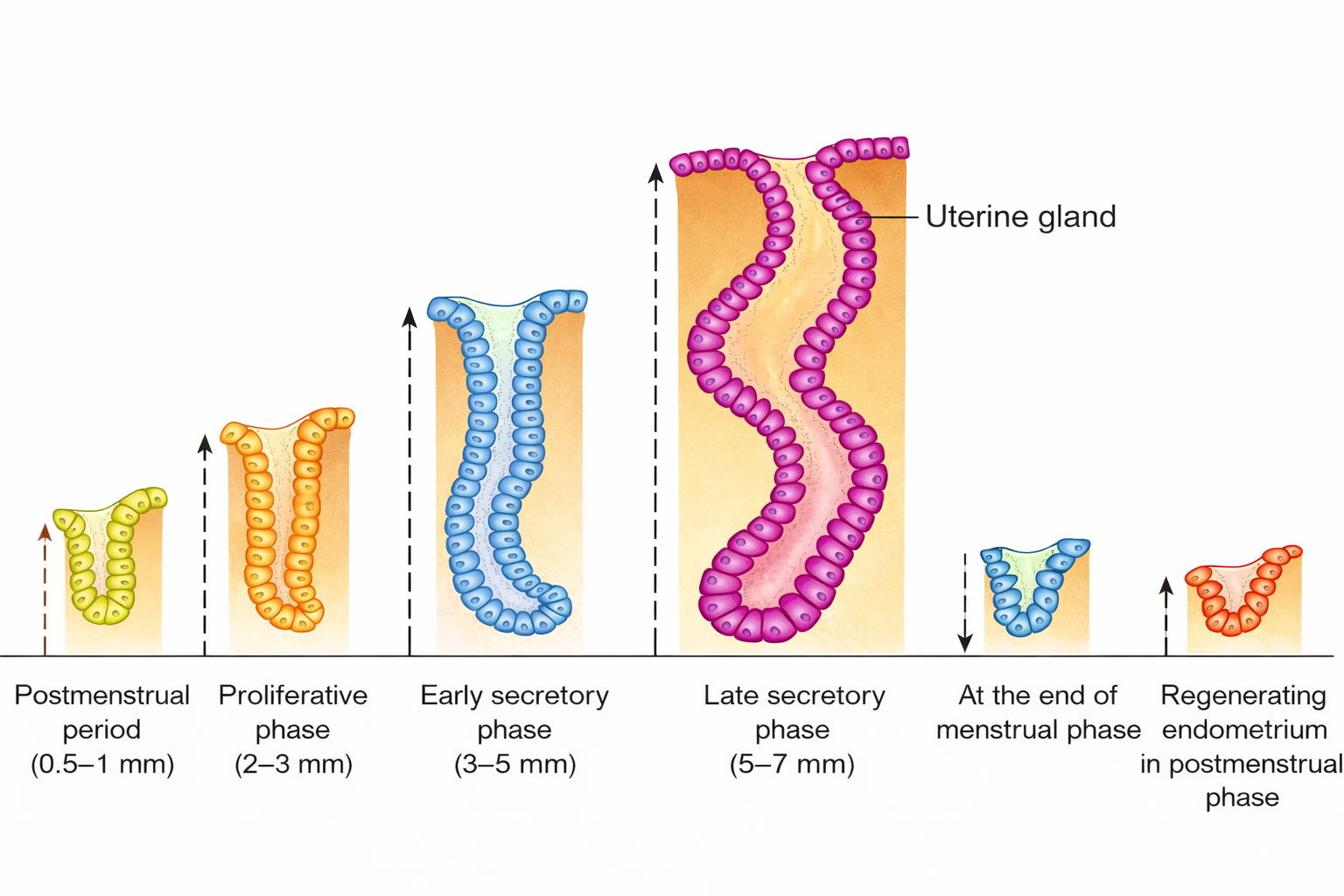
Table 20.1: Differences between endometrium of proliferative and secretory phases
| Feature | Proliferative (Follicular/Preovulatory) Phase | Secretory (Luteal/Postovulatory) Phase |
|---|---|---|
| Dominant hormone | Estrogen | Progesterone |
| Endometrial thickness | Approximately 3–4 mm | Approximately 6–7 mm |
| Endometrial layers | Initially, the endometrium is mainly represented by the stratum basalis, with regeneration of the functional layer occurring during this phase | All layers of the endometrium, including the functional layer, are fully developed |
| Uterine glands | Long, relatively straight glands with narrow lumina and wide spacing between adjacent glands | Highly coiled, corkscrew-shaped glands that are closely packed and possess wider lumina |
| Glandular secretions | Minimal secretory activity | Abundant secretory activity; glandular lumina may contain accumulated secretions |
| Decidual change | Absent | Present, particularly in the stromal cells |
| Intracellular glycogen | Absent or minimal | Markedly increased due to progesterone stimulation |
| Spiral arteries | Slightly tortuous | Highly coiled and elongated |
| Stromal appearance | Dense cellular stroma with active cell proliferation | Edematous stroma with prominent predecidual changes |
| Primary purpose | Regeneration and growth of the endometrium following menstruation | Preparation of the endometrium for implantation of a fertilized ovum |
Cervix Of Uterus
- The cervix is the lower cylindrical portion of the uterus and contains a narrow passage known as the cervical canal.
- The cervical canal communicates with the uterine cavity through the internal os and opens into the vagina through the external os.
- The portion of the cervix projecting into the vagina is called the portio vaginalis (ectocervix).
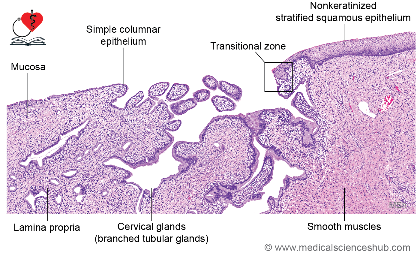
Histology of Cervix
- Cervix shows endocervix (mucous membrane) and muscle layer.
Endocervix
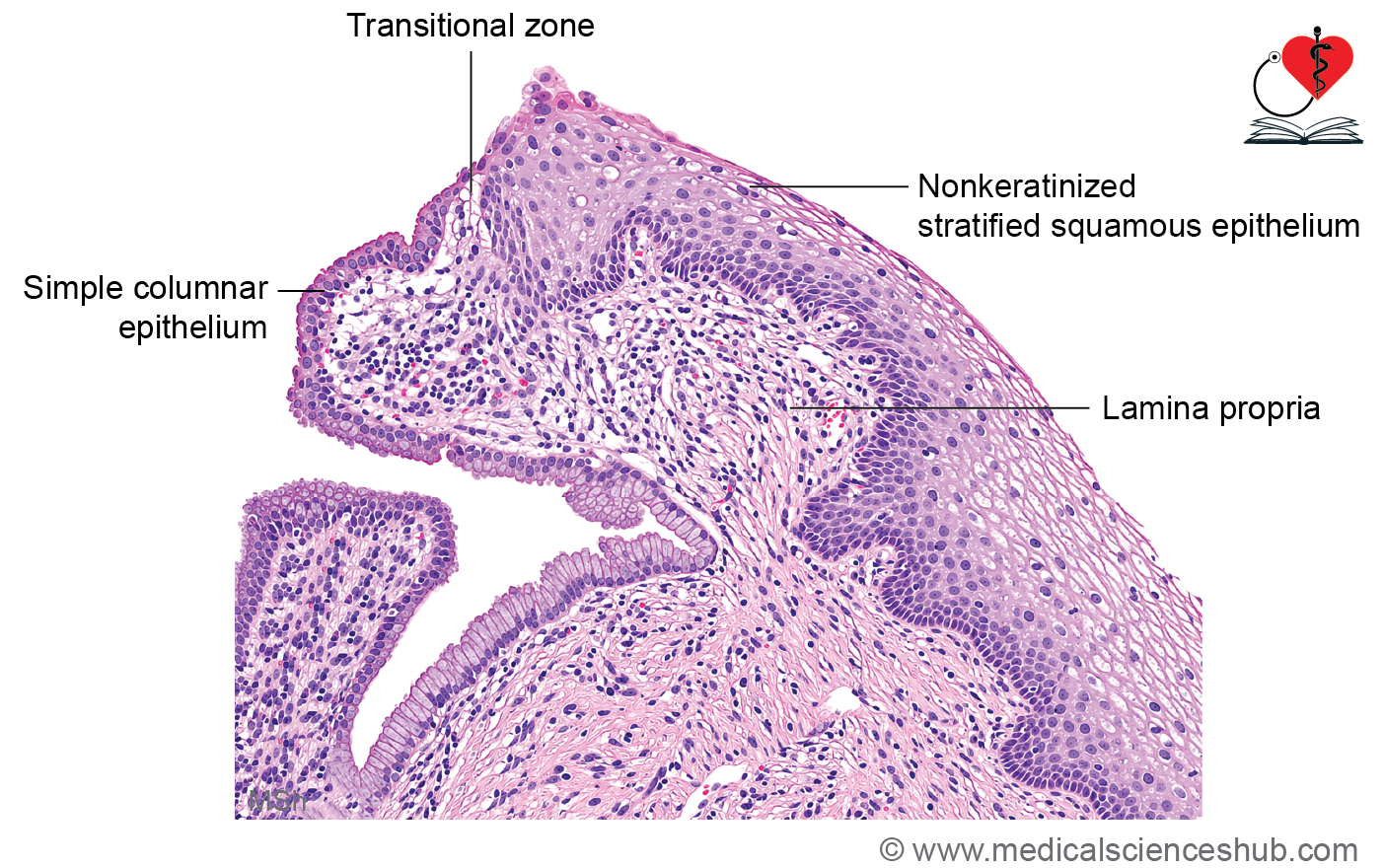
- The cervical canal is lined by simple columnar mucus-secreting epithelium.
- This epithelium rests on a loose connective tissue lamina propria.
- The lamina propria contains branched tubular cervical glands that secrete mucus rich in antimicrobial substances, including lysozyme.
Cervical Stroma
- Beneath the mucosa lies a thick layer of fibroelastic connective tissue containing a relatively small amount of smooth muscle.
- The abundant elastic fibers allow cervical expansion and dilation during childbirth.
Ectocervix (Portio Vaginalis)
- The ectocervix is lined by nonkeratinized stratified squamous epithelium, continuous with the vaginal epithelium.
- The junction between the columnar epithelium of the endocervix and the squamous epithelium of the ectocervix forms the transformation zone, an important site for cervical pathology.
CLINICAL CORRELATION
- The properties of cervical mucus vary during the menstrual cycle:
- Around ovulation, mucus becomes thin and less viscous, facilitating sperm passage.
- During other phases, it becomes thicker and less permeable to sperm.
- Nabothian cysts develop when cervical gland ducts become obstructed, leading to retention of glandular secretions.
- A Pap smear (Papanicolaou smear) involves microscopic examination of exfoliated cervical epithelial cells and is widely used for screening and early detection of precancerous and cancerous cervical lesions.
Vagina
- The vagina is a fibromuscular, elastic canal that extends from the cervix to the external genital region.
- It is approximately 8–10 cm long and serves as the female copulatory organ and birth canal.
Histology of Vagina
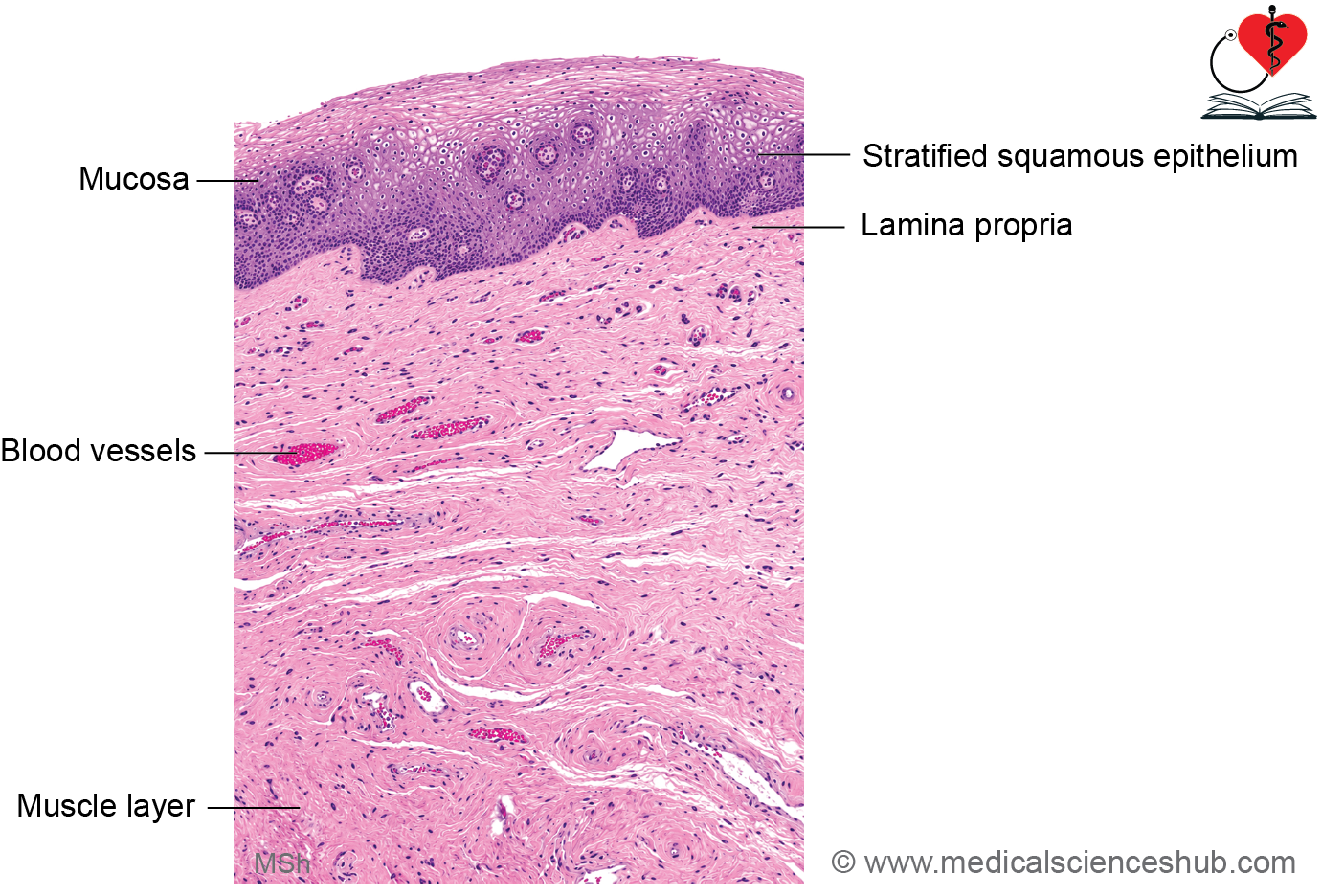
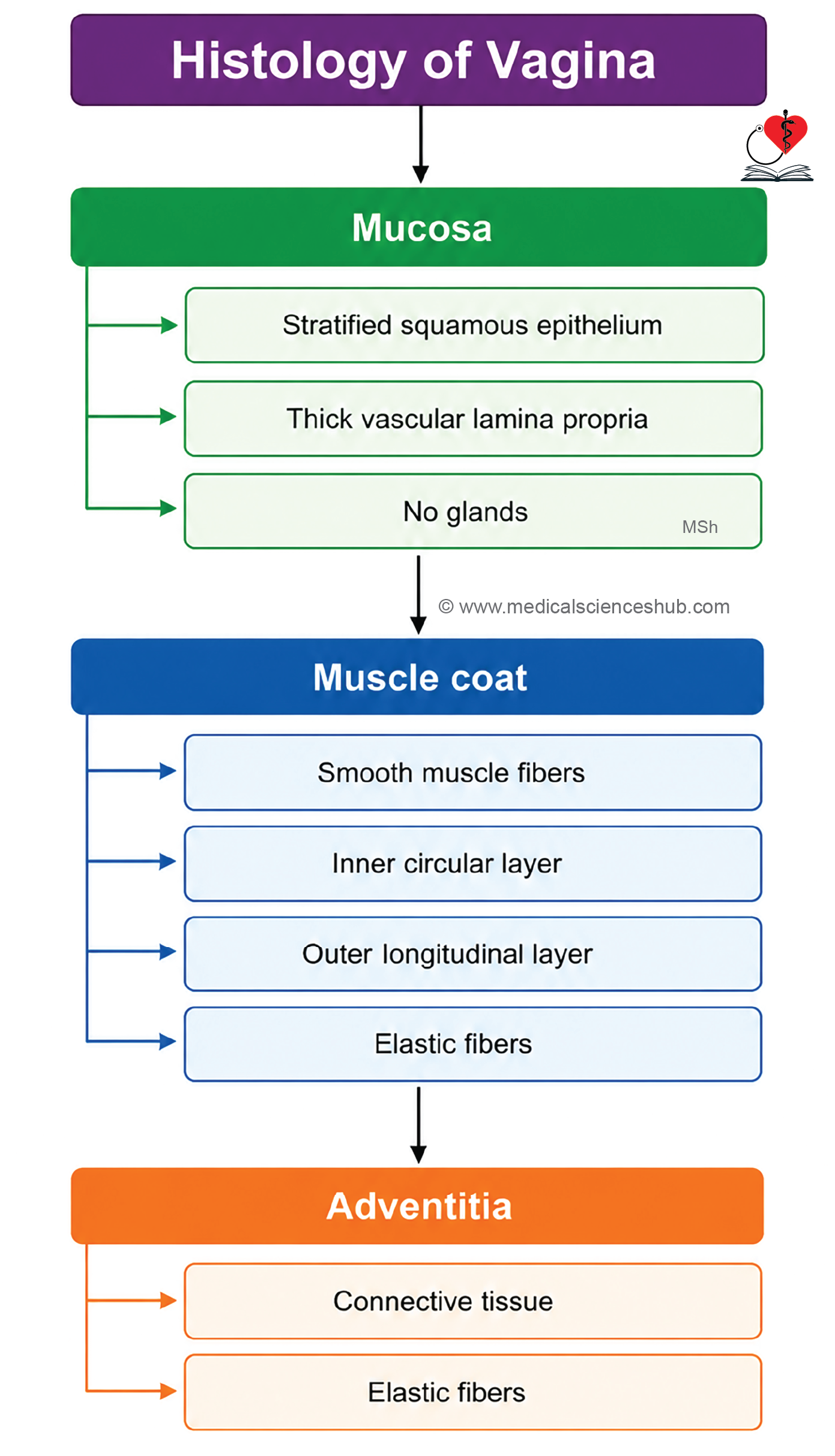
- Vagina has three layers: Mucosa, muscle layer, and adventitia.
Mucosa
- The mucosa forms numerous transverse folds (rugae) that allow distension.
- It is lined by nonkeratinized stratified squamous epithelium, which may show slight keratinization in certain conditions.
- During the follicular phase, epithelial cells accumulate glycogen under estrogenic influence.
- Glycogen content decreases during the secretory phase.
- The lamina propria is broad, highly vascular, and rich in elastic connective tissue.
- The vaginal mucosa lacks glands; lubrication is mainly derived from cervical mucus and transudation from blood vessels.
Muscular Layer
- The muscular coat consists of smooth muscle arranged in:
- An inner circular layer.
- An outer longitudinal layer.
- Numerous elastic fibers contribute to vaginal elasticity.
- The lower end of the vagina is supported by fibers of the bulbospongiosus muscle and other pelvic floor muscles.
Adventitia
- The outermost layer is adventitia, composed of connective tissue containing blood vessels, lymphatics, nerves, and elastic fibers, which anchor the vagina to surrounding structures.
Important Questions
- Write a short note on histology of ovary.
- Write a short note on histology of fallopian tube.
- List the differences between endometrium of proliferative and secretory phases.
- Write a short note on histology of vagina.
📝 Test Your Knowledge – Practice MCQs
Attempt the chapter MCQ quiz and assess your understanding of key concepts.
Click to see: Medical Disclaimer and Copyright Notice