
Competency
- AN52.2 Describe and identify the microanatomical features of placenta and umbilical cord.
Introduction
- The mammary gland, placenta, and umbilical cord are essential structures associated with reproduction and development. The mammary gland supports neonatal nutrition, while the placenta facilitates maternal–fetal exchange and endocrine functions. The umbilical cord connects the fetus to the placenta, enabling transport of oxygen, nutrients, and waste.
Mammary Gland
- The mammary gland is a modified sweat gland and a characteristic feature of mammals.
- It develops from embryonic mammary ridges (milk lines).
- In males, the gland remains rudimentary, whereas in females it enlarges after puberty under the influence of ovarian hormones.
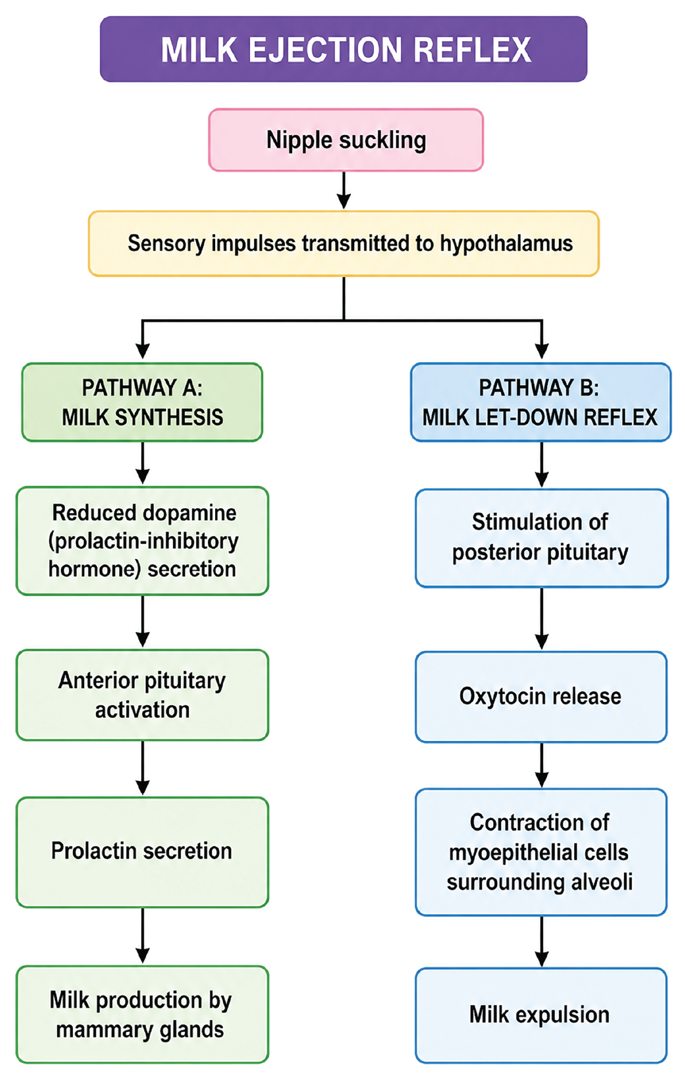
- Prolactin stimulates milk production, while oxytocin triggers milk ejection during lactation.
- Mammary glands are located within the superficial fascia of the anterior chest wall.
External Features
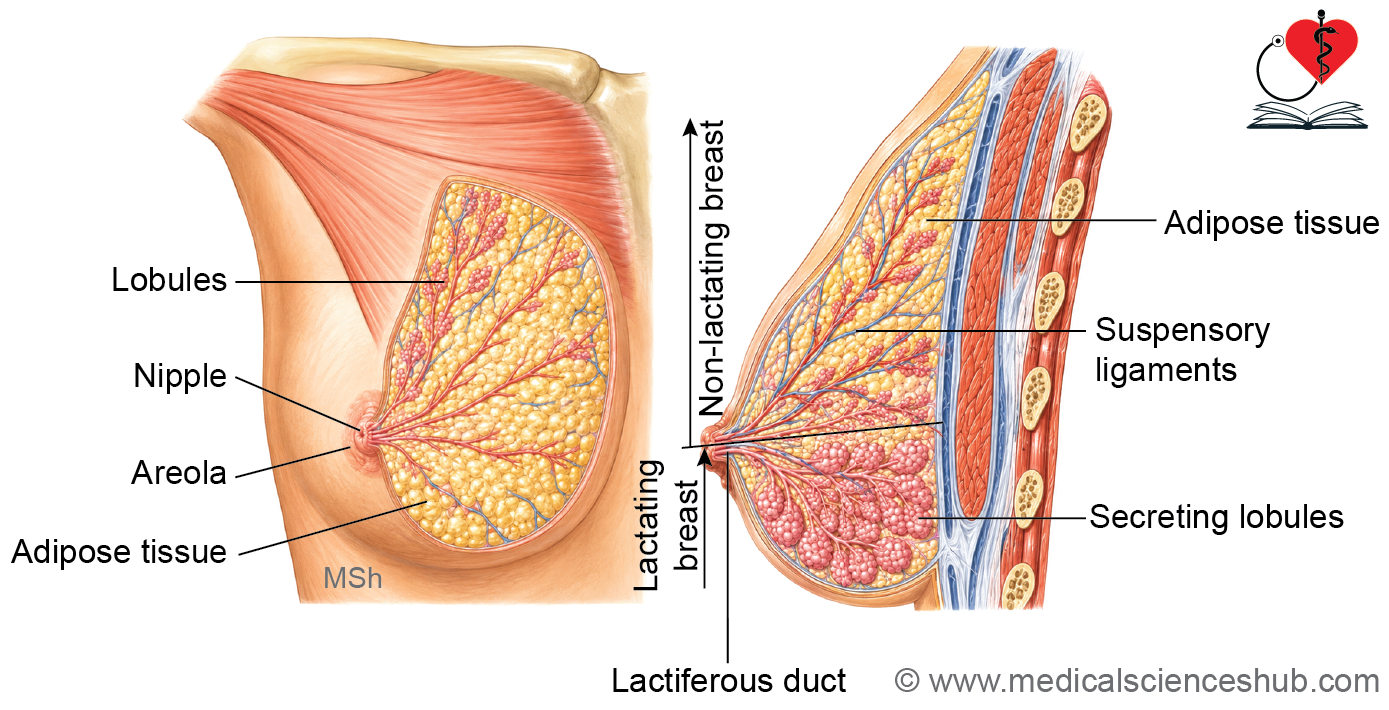
- The areola is the pigmented skin surrounding the nipple.
- The nipple contains openings of the lactiferous ducts.
- Montgomery tubercles are small elevations on the areola formed by enlarged sebaceous glands.
- The gland is supported by suspensory ligaments (Cooper’s ligaments), which connect the skin to the underlying fascia.
Internal Structure
- The gland consists of approximately 15–20 lobes separated by dense connective tissue.
- Each lobe is subdivided into multiple lobules containing secretory alveoli.
- Every lobe drains through a lactiferous duct, which opens onto the nipple.
- Near the nipple, each duct expands to form a lactiferous sinus, a temporary reservoir for milk.
- The duct system branches into interlobular ducts, intralobular ducts, and finally alveolar ductules, which drain clusters of milk-secreting alveoli.
- The histological appearance of the mammary gland varies according to its functional state, particularly during pregnancy and lactation.
Histology of Non-lactating (Inactive) Gland
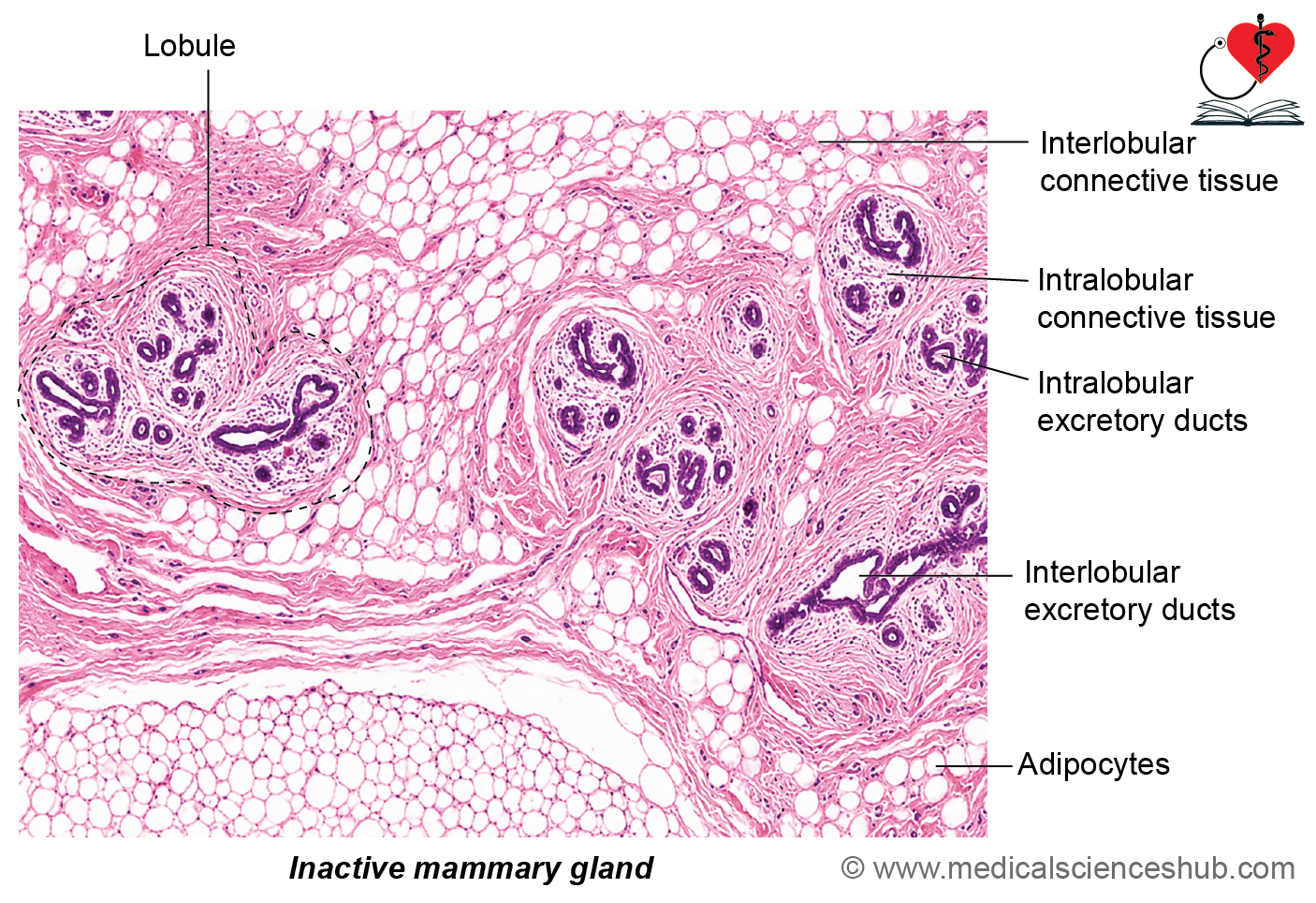
- The inactive mammary gland is composed predominantly of ducts and ductal branches embedded within connective tissue.
- Each lobule contains:
- Intralobular ducts lined by simple cuboidal epithelium and supported by a few myoepithelial cells.
- Rudimentary or absent alveoli, appearing as small clusters or cords of epithelial cells.
- The intralobular connective tissue is loose, cellular, and typically lacks adipose tissue.
- Adjacent lobules are separated by dense interlobular connective tissue, which may contain fat.
Histology of Lactating (Active) Gland
- During pregnancy, estrogen and progesterone stimulate extensive growth and differentiation of the mammary gland.
- These hormonal changes result in:
- Proliferation of ducts and secretory acini.
- Enlargement and increased number of alveoli.
- Expansion of glandular tissue within lobules.
- Increased adipose tissue in interlobular regions.
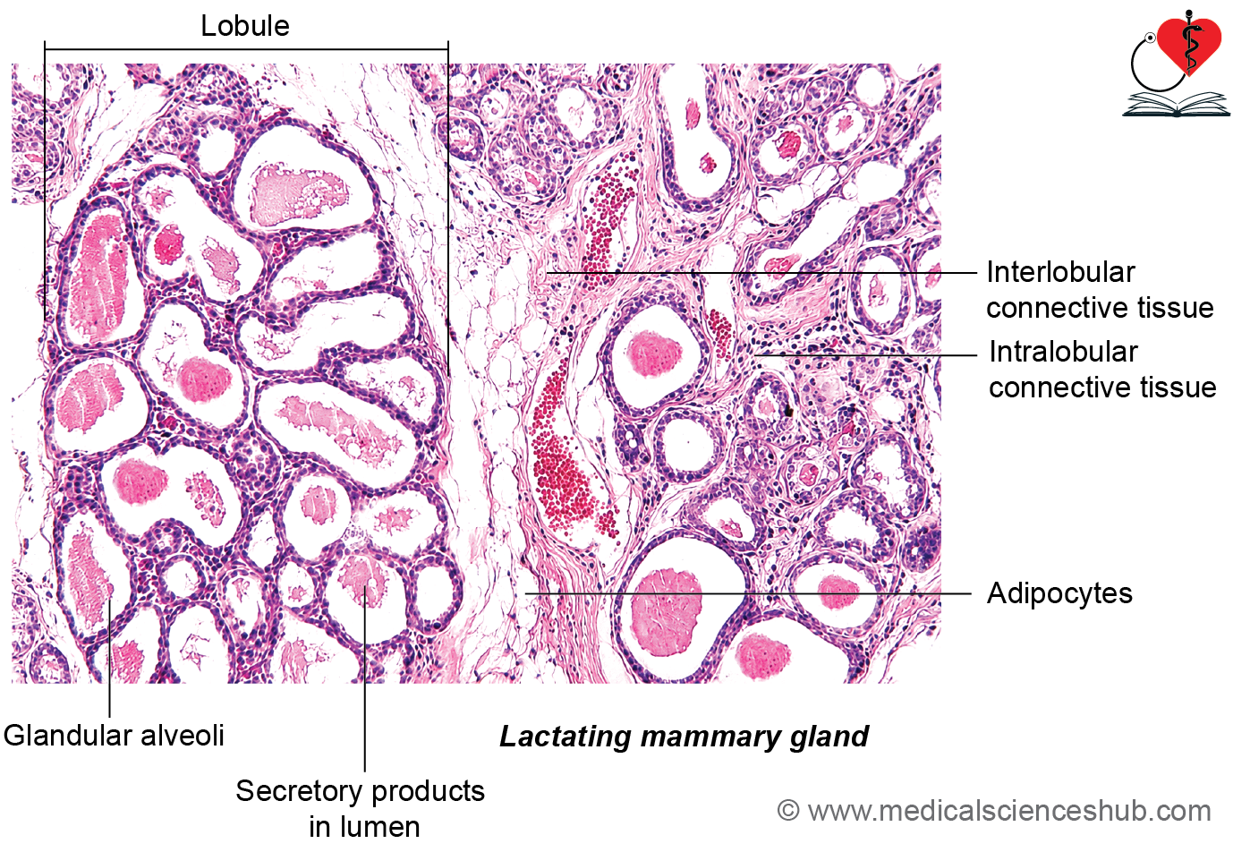
- The gland becomes dominated by numerous secretory alveoli, reducing the relative amount of connective tissue.
Microscopic Features of Lactating Alveoli
- Alveoli are lined by low cuboidal to low columnar secretory epithelial cells resting on a basal lamina.
- Surrounding the basal lamina are contractile myoepithelial cells, which assist in milk ejection.
- Alveolar lumina often contain accumulated secretory material, including milk components.
- Secretory cells typically show:
- Basally located rough endoplasmic reticulum, producing basophilic staining.
- Apical secretory vesicles, lipid droplets, and a prominent Golgi apparatus, contributing to eosinophilic staining.
- Myoepithelial cells are difficult to identify in routine H&E-stained sections but play a crucial role in lactation.
Duct System of Mammary Gland
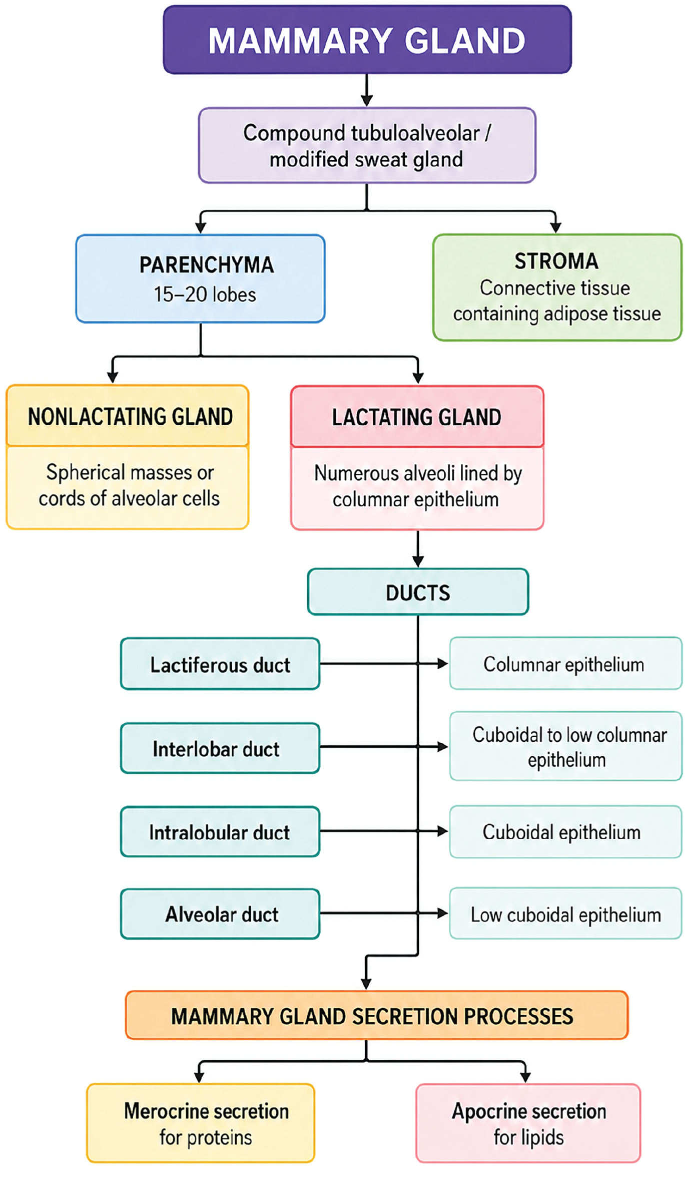
- Alveolar ductules are lined by low cuboidal epithelium.
- Intralobular ducts are lined by cuboidal to low columnar epithelial cells.
- Lactiferous ducts are lined predominantly by columnar epithelium.
- Near the nipple, the ductal epithelium gradually transitions from simple columnar to pseudostratified columnar and finally to stratified squamous epithelium.
- Mammary ducts are surrounded by myoepithelial cells and supported by a basal lamina, which aid in milk expulsion.
Functions Mode of Milk Secretion
The mammary gland utilizes two mechanisms for milk production:
- Merocrine secretion: Milk proteins are released by exocytosis of secretory vesicles without loss of cytoplasm.
- Apocrine secretion: Lipid droplets are discharged along with a small portion of apical cytoplasm and plasma membrane.
CLINICAL CORRELATION
- Colostrum is the yellowish secretion produced during the first few days after childbirth.
- It is rich in antibodies, proteins, and vitamins.
- It provides important passive immunity to the newborn.
- Breast carcinoma arises from the epithelial lining of ducts or secretory alveoli and is one of the most common cancers in women. Early detection improves prognosis.
- Fibroadenoma is a common benign tumor of the breast.
- Estrogen primarily stimulates growth and branching of mammary ducts, whereas progesterone promotes development of secretory alveoli and lobules.
Placenta
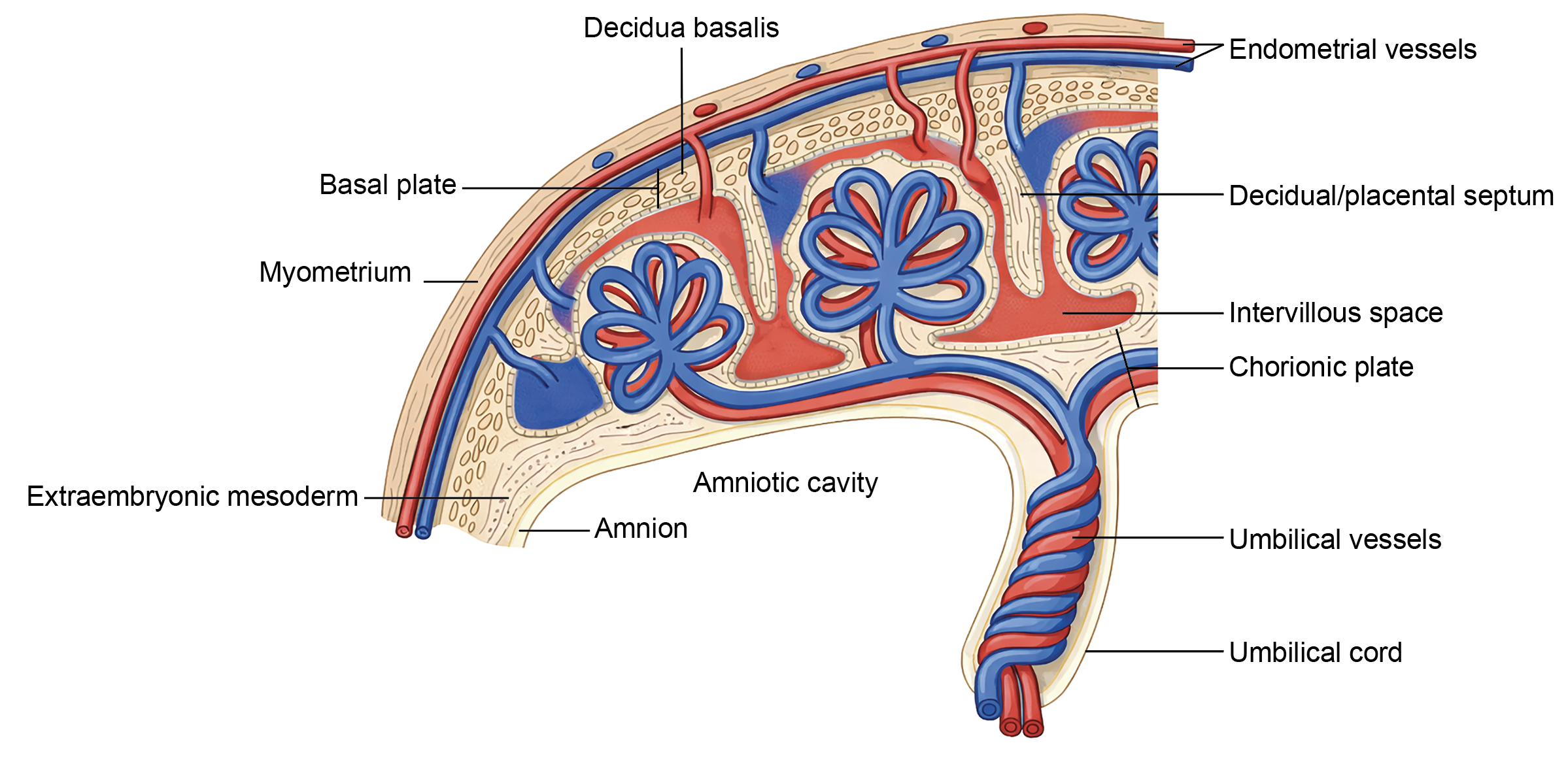
- The placenta is a temporary fetomaternal organ that supports fetal growth by facilitating the exchange of nutrients, gases, and waste products between the mother and fetus.
- It develops within the uterus during pregnancy.
- The mature placenta is typically disc-shaped and weighs approximately 500–600 g at term.
Surfaces of the Placenta
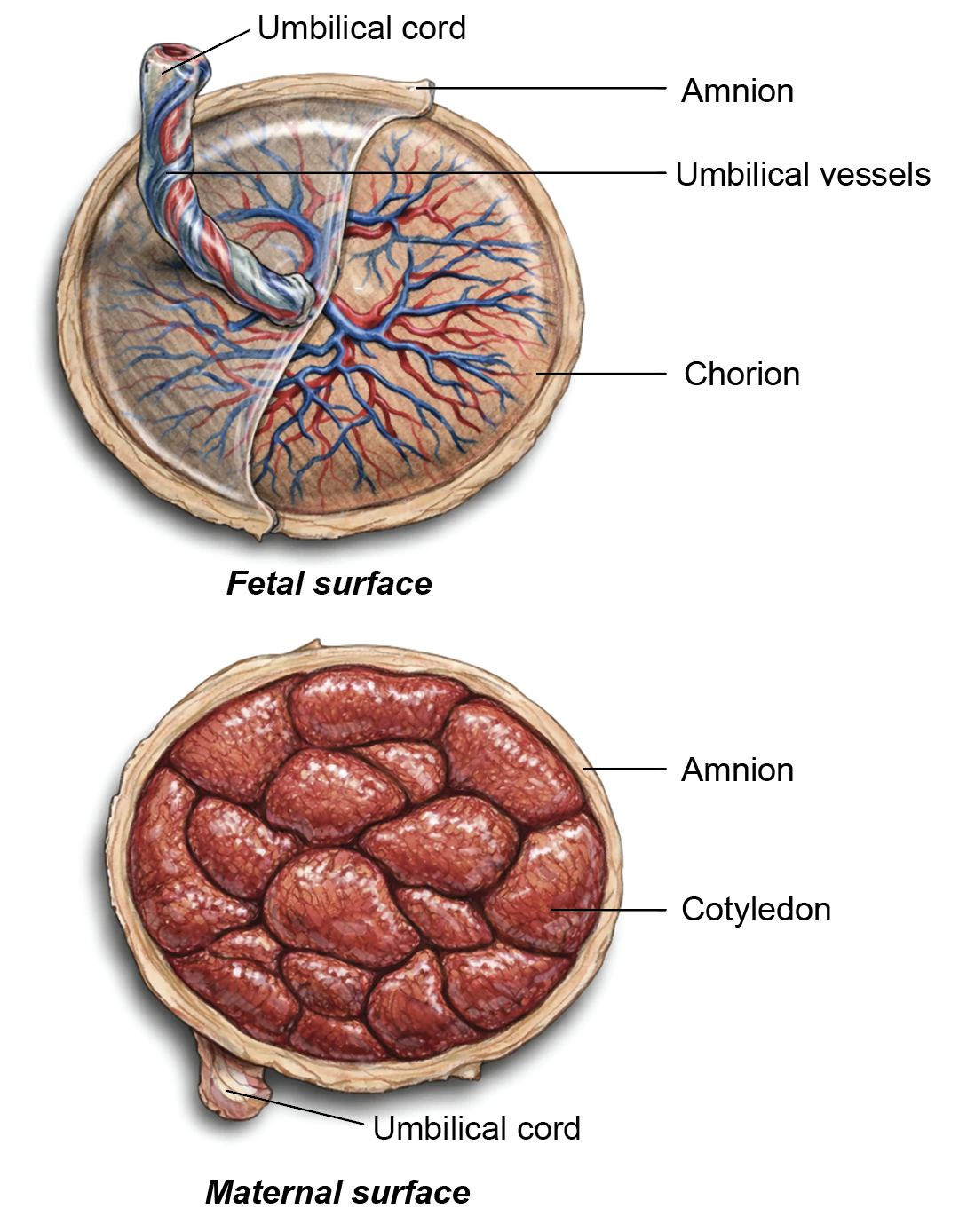
- Fetal surface:
- Smooth and shiny in appearance.
- Covered by the amnion.
- Umbilical vessels branch beneath this surface.
- Maternal surface:
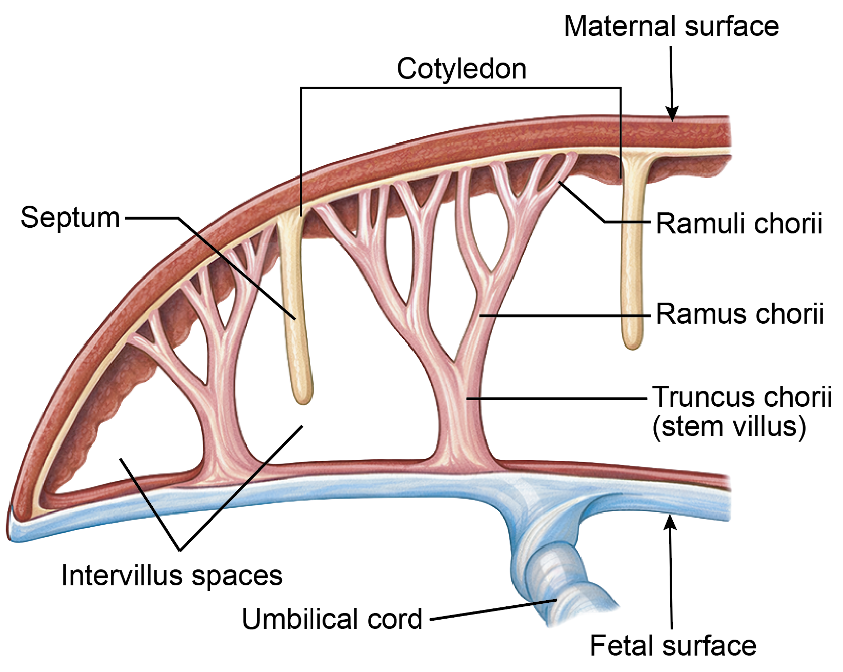
- Rough and divided into 15–20 lobules called cotyledons.
Components of Placenta
The placenta consists of:
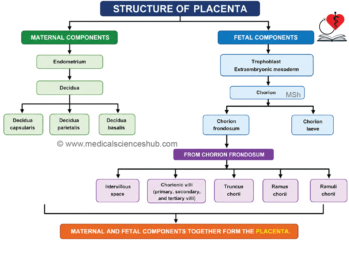
- A fetal component: the chorion.
- A maternal component: the decidua basalis.
Decidua
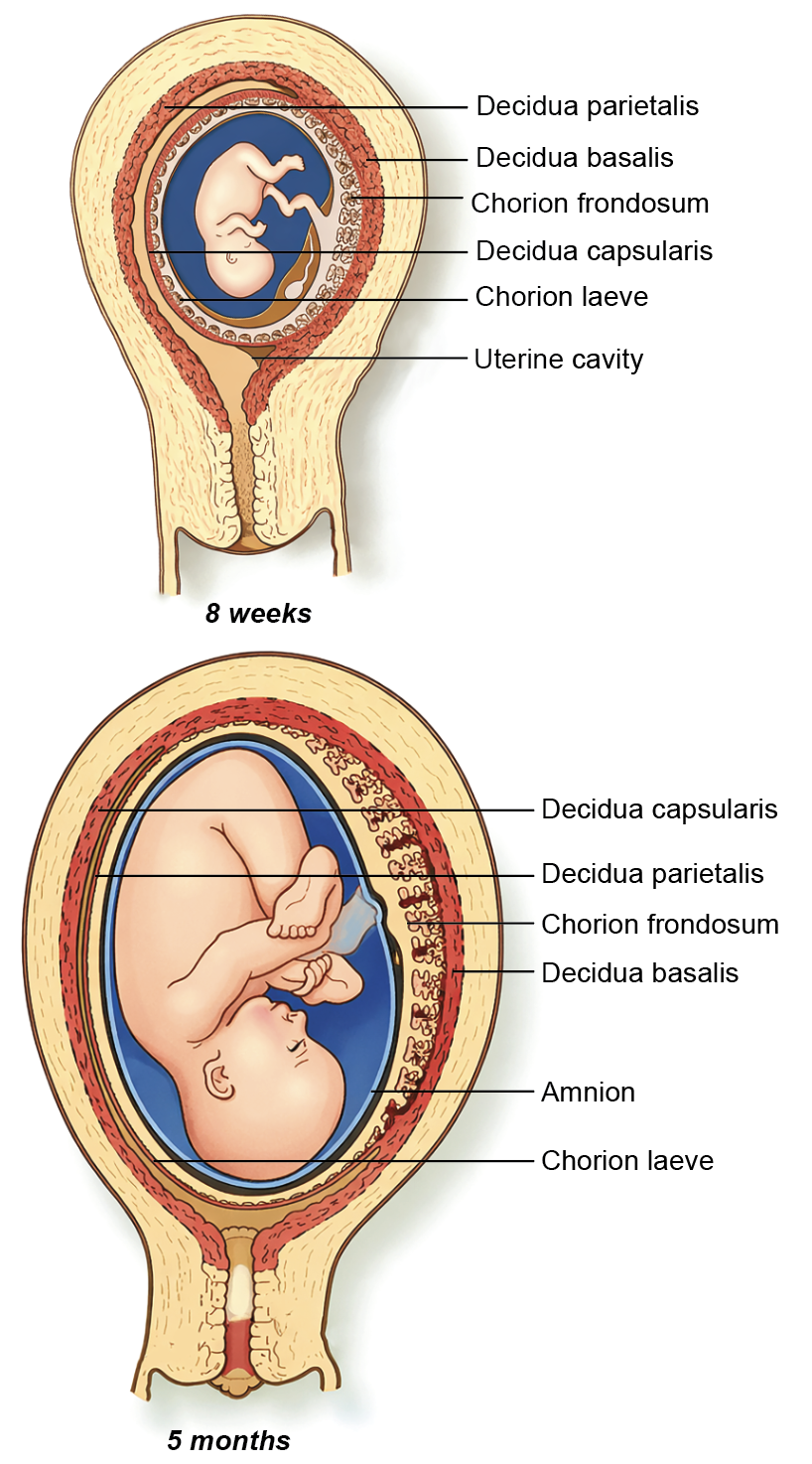
- Following implantation, the uterine endometrium undergoes transformation and is termed the decidua.
- It is divided into:
- Decidua basalis: Lies beneath the implanted embryo and contributes directly to placental formation.
- Decidua capsularis: Surrounds the implanted conceptus.
- Decidua parietalis: Lines the remainder of the uterine cavity.
- During decidualization, endometrial stromal cells enlarge and become rounded, forming decidual cells.
- Wedge-shaped extensions of decidual tissue project into the placenta as placental septa, partially dividing it into cotyledons.
Chorion and Trophoblast
- The blastocyst consists of an outer trophoblast and an inner embryoblast.
- The embryoblast gives rise to the embryo and associated embryonic structures.
- The trophoblast differentiates into:
- Cytotrophoblast (inner cellular layer).
- Syncytiotrophoblast (outer multinucleated layer).
- The chorion is formed by trophoblastic layers together with extraembryonic mesoderm.
- The chorion has two regions:
- Chorion frondosum: The villous, placental part adjacent to the decidua basalis.
- Chorion laeve: The smooth, non-villous part facing the decidua capsularis.
Placental Villi
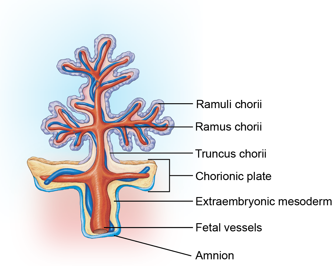
- The chorion frondosum gives rise to major stem villi, which repeatedly branch to form an extensive villous tree.
- The spaces surrounding the villi are called intervillous spaces and are filled with maternal blood, allowing maternal-fetal exchange.
Stages of Villous Development
- Primary villi consist of an inner cytotrophoblast covered by an outer syncytiotrophoblast.
- Secondary villi develop when extraembryonic mesoderm grows into the core of primary villi.
- Tertiary villi form when fetal blood vessels develop within the mesodermal core, establishing the fetal placental circulation.
Placental Barrier
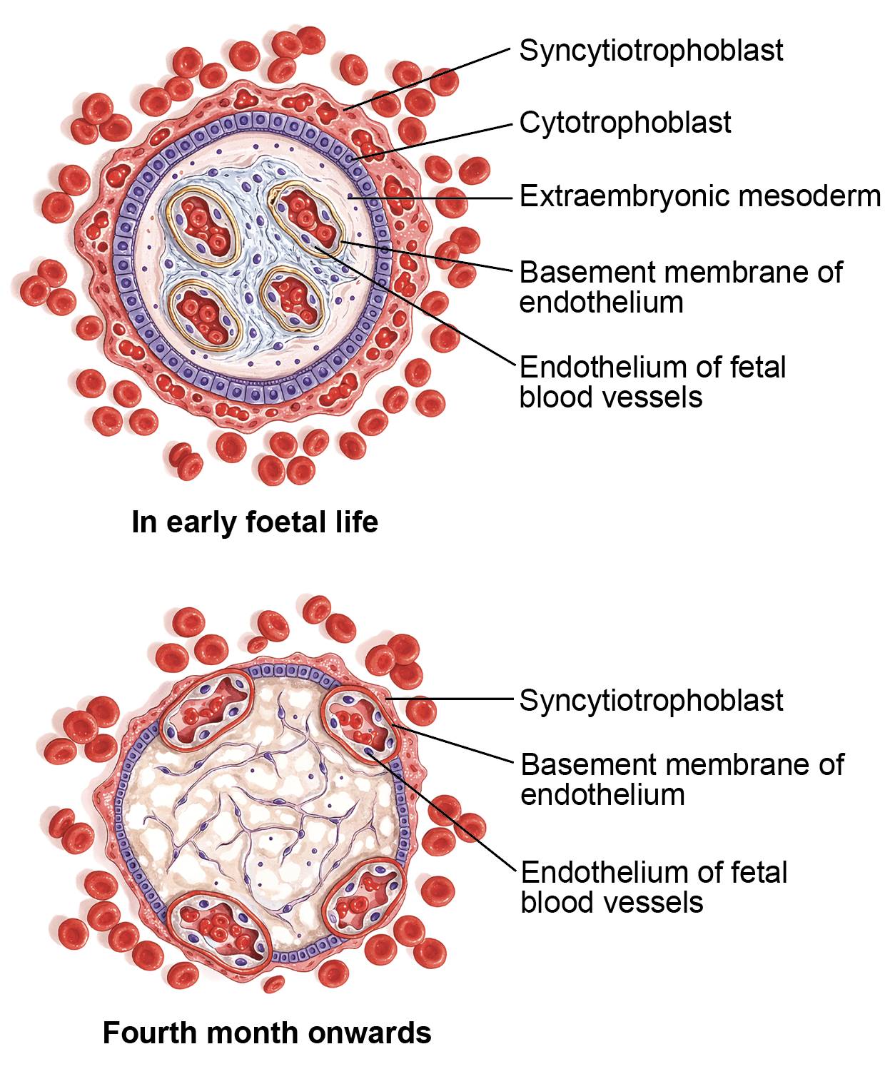
- The placental barrier separates maternal and fetal blood while permitting the exchange of oxygen, nutrients, waste products, antibodies, and selected molecules.
- Components of the placental barrier include:
- Syncytiotrophoblast
- Cytotrophoblast and its basal lamina
- Extraembryonic mesoderm (connective tissue core)
- Basal lamina of fetal capillaries
- Endothelium of fetal capillaries
- This barrier helps maintain separation of the two circulations while supporting efficient maternal-fetal exchange throughout pregnancy.
Histology of Placenta
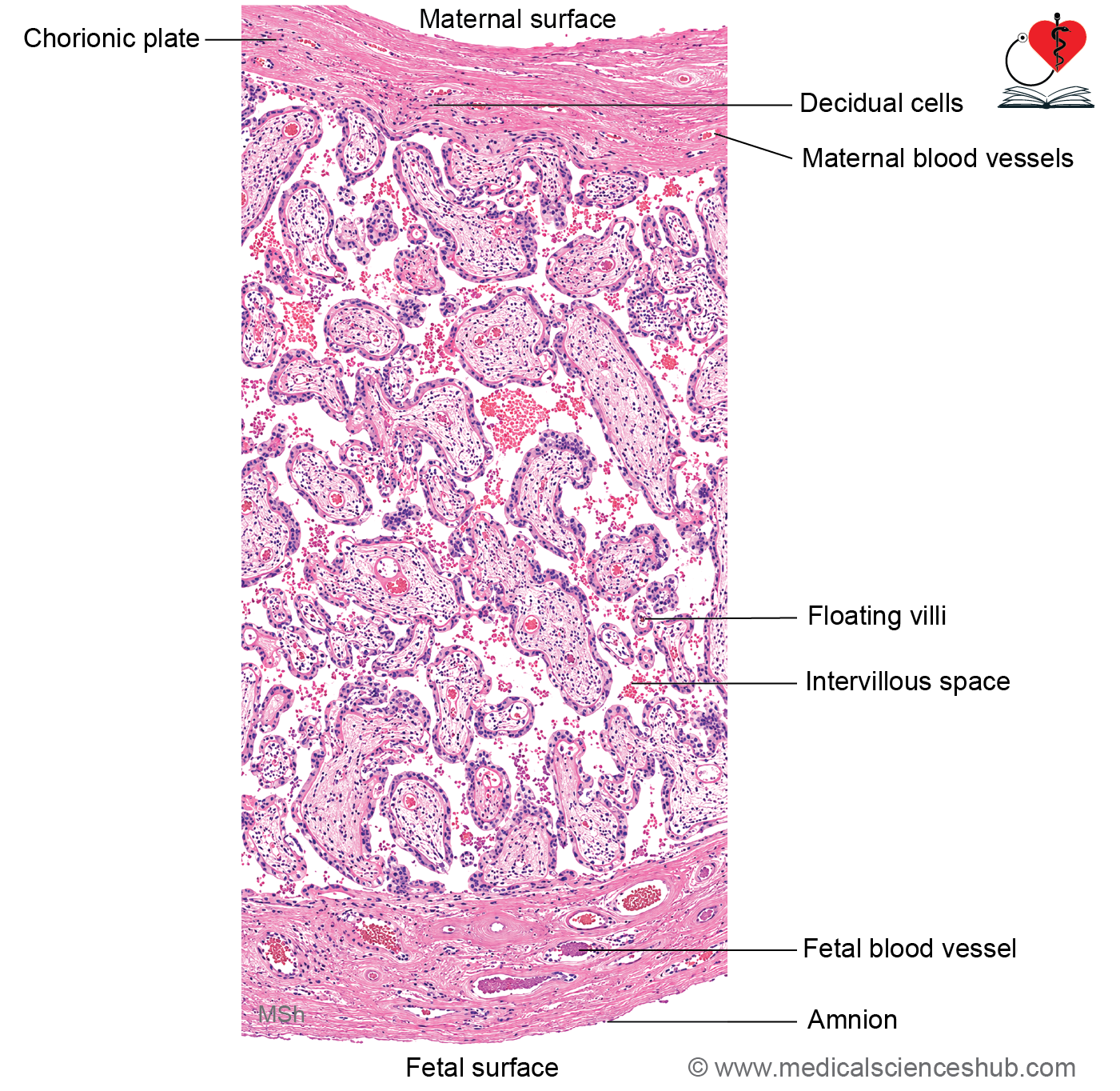
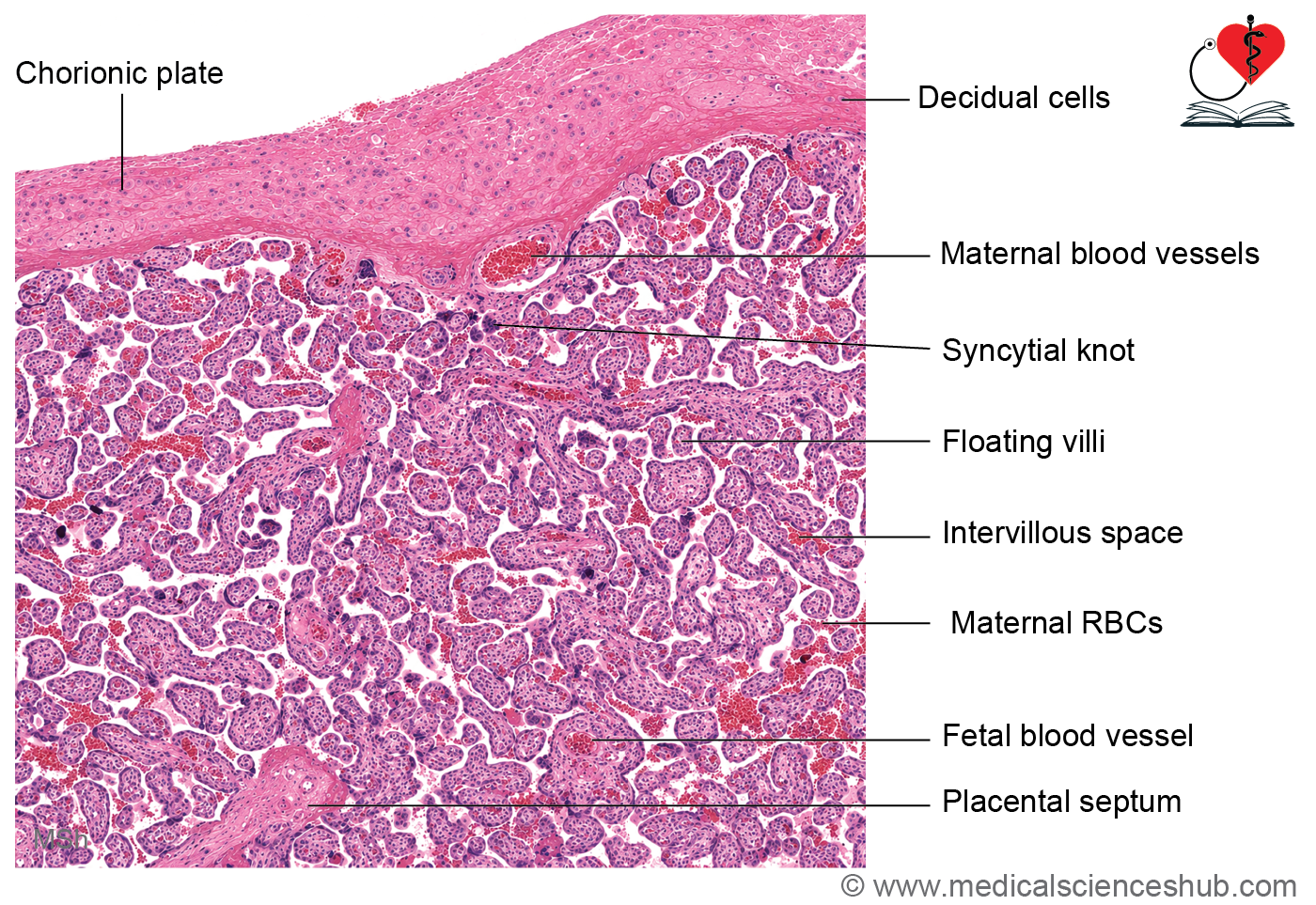
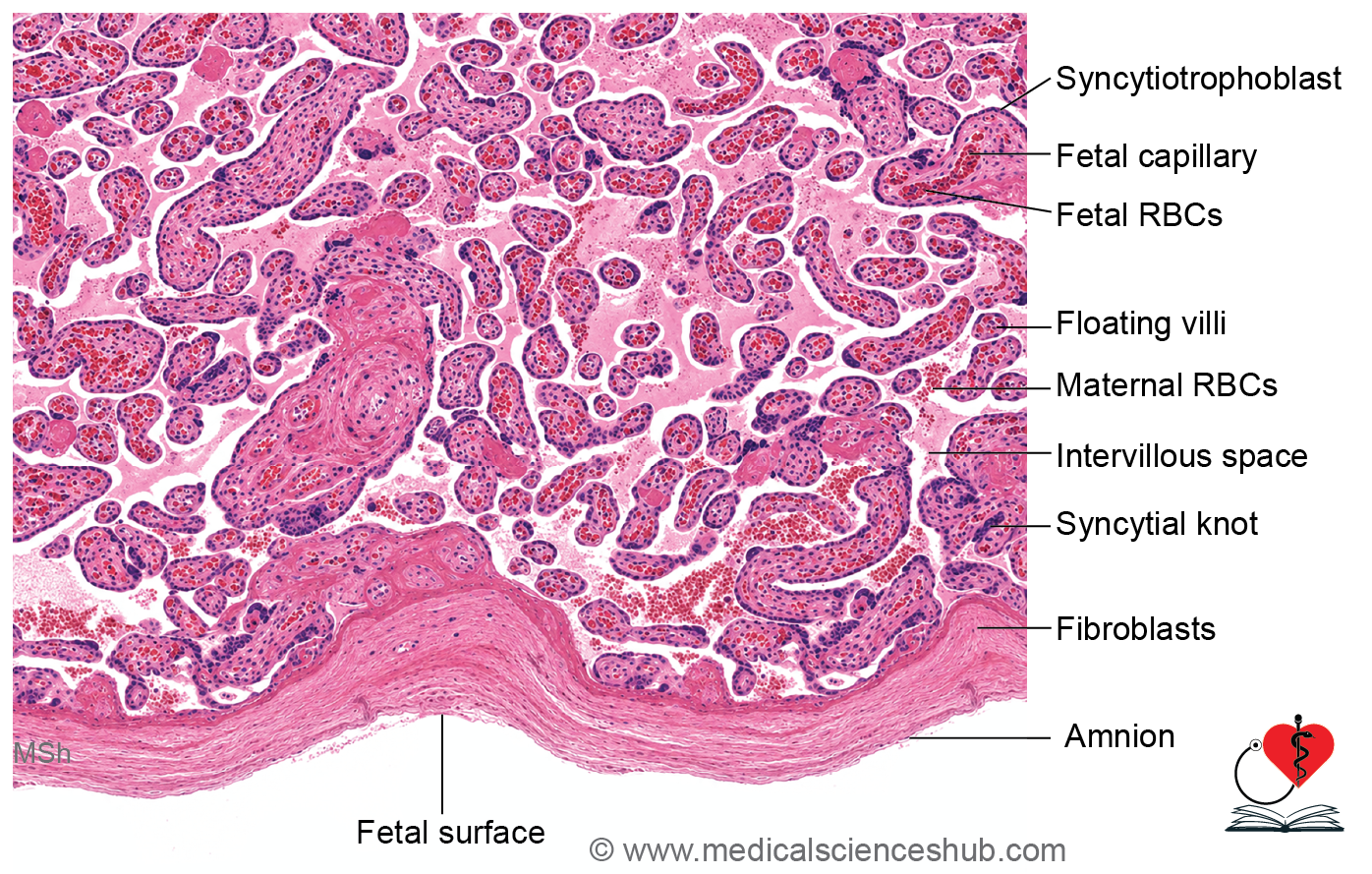
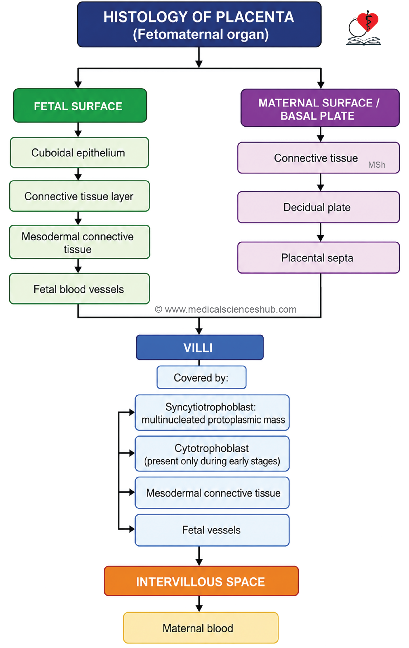
- The placenta extends from the smooth fetal surface, covered by the amnion, to the rough maternal surface formed by the decidua basalis.
- The amnion consists of simple cuboidal epithelium supported by underlying connective tissue.
- Beneath the amnion lies extraembryonic mesodermal connective tissue. Together, these layers form the chorionic plate.
- The chorionic plate contains fetal blood vessels, although arteries and veins are not always easily distinguished histologically.
- Most of the placental tissue is composed of chorionic villi of varying sizes suspended within maternal blood-filled intervillous spaces.
- On the maternal side, the basal plate consists of connective tissue and clusters of decidual cells, which often appear epithelial-like under the microscope.
- Placental septa project from the basal plate into the placenta, partially dividing it into cotyledons. These septa do not contain fetal blood vessels.
Microscopy of Villi
- Chorionic villi are surrounded by intervillous spaces containing maternal blood.
- The outer surface of each villus is covered by syncytiotrophoblast, a multinucleated cellular layer lacking distinct cell boundaries.
- Aggregates of syncytiotrophoblastic nuclei form syncytial knots, which are common in mature placentas.
- The syncytiotrophoblast bears numerous microvilli that increase the surface area available for maternal-fetal exchange.
- Cytotrophoblast cells are prominent in early placental development and lie beneath the syncytiotrophoblast. Their number decreases as the placenta matures.
- The core of each villus contains:
- Loose connective tissue and extracellular matrix
- Hofbauer cells (fetal macrophages)
- Fetal blood vessels
- Smaller terminal villi often consist of a very thin trophoblastic covering closely associated with fetal capillaries, facilitating efficient exchange of gases, nutrients, and waste products.
- Both fetal vessels within villi and maternal blood in intervillous spaces contain blood cells, but the two circulations normally remain separated by the placental barrier.
Functions of Placenta
- The placenta performs several essential functions that support fetal growth and development.
- Gaseous exchange occurs across the placental barrier by simple diffusion. Efficient oxygen transfer is facilitated by the high oxygen affinity of fetal hemoglobin and its relatively high concentration in fetal blood.
- The placenta transports nutrients, including glucose, amino acids, fatty acids, vitamins, and electrolytes, from maternal to fetal circulation.
- Metabolic waste products such as urea, uric acid, and creatinine are transferred from fetal blood into maternal blood for elimination.
- Passive immunity is provided through the transfer of maternal IgG antibodies across the placenta. These antibodies protect the newborn against several infectious diseases during early life.
- The placental barrier limits the passage of many microorganisms and harmful substances; however, some viruses, drugs, and fetal cells can cross this barrier.
- The placenta serves as a temporary storage site for glycogen, iron, calcium, and other nutrients required for fetal development.
- As an important endocrine organ, the placenta synthesizes and secretes several hormones, including:
- Human chorionic gonadotropin (hCG)
- Progesterone
- Estrogens
- Human placental lactogen (hPL)
- These hormones help maintain pregnancy, support fetal growth, and prepare the maternal body for lactation and childbirth.
Umbilical Cord
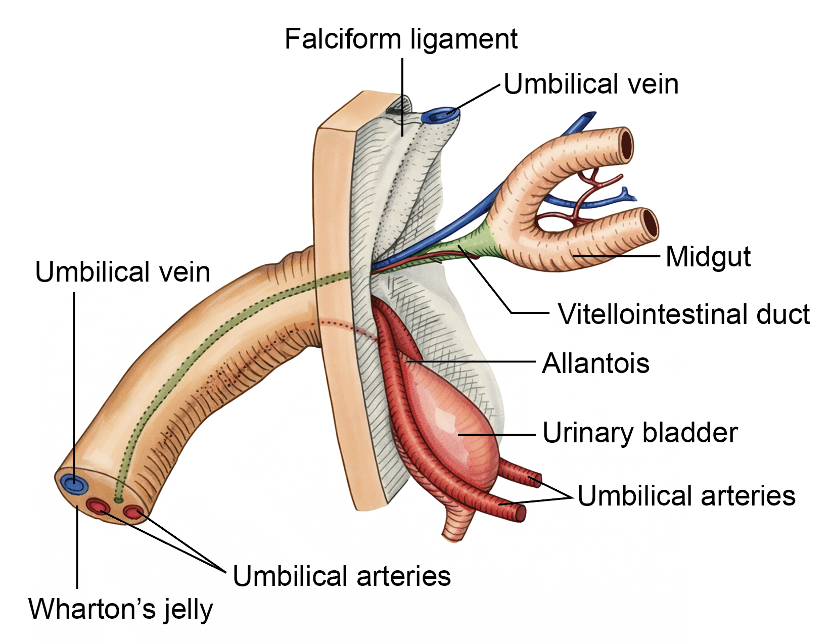
- The umbilical cord forms the vital connection between the fetus and the placenta, enabling exchange of nutrients, gases, and waste products.
- It typically measures 50–60 cm in length and about 1–1.5 cm in diameter at term.
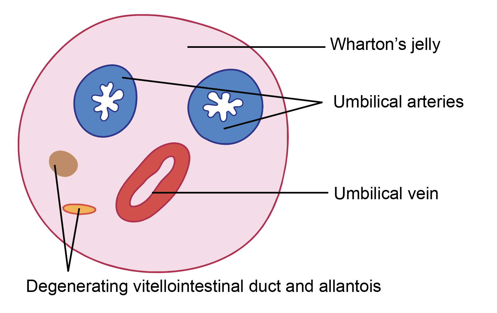
- The cord contains one umbilical vein, which carries oxygenated and nutrient-rich blood from the placenta to the fetus, and two umbilical arteries, which return deoxygenated blood from the fetus to the placenta.
- All vessels are embedded within Wharton’s jelly, a specialized mucoid connective tissue that protects them from compression and kinking.
Histology of Umbilical Cord
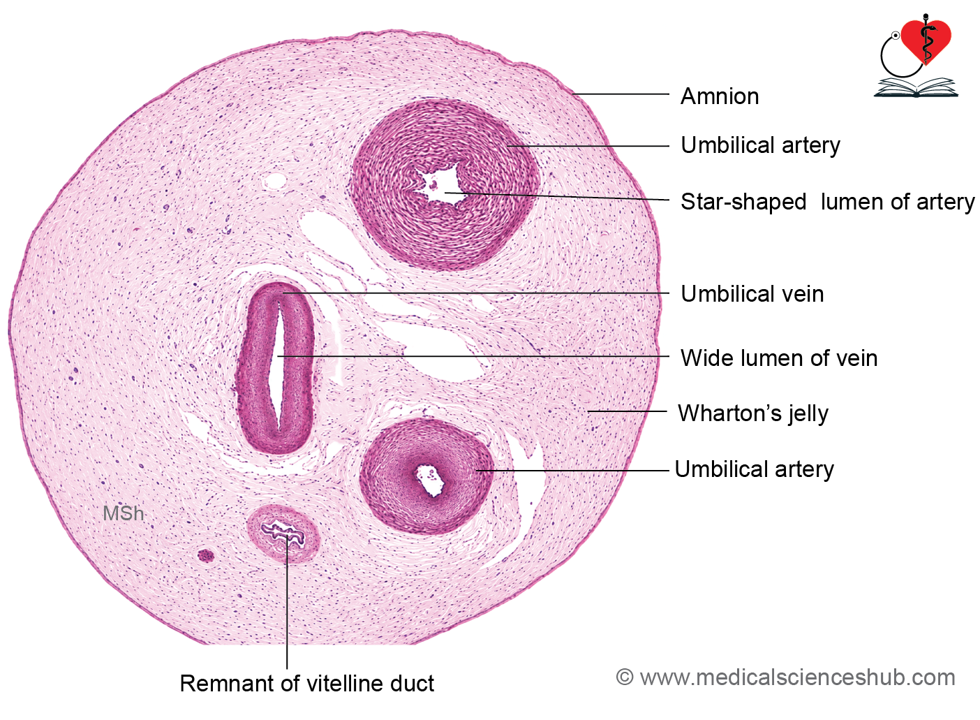
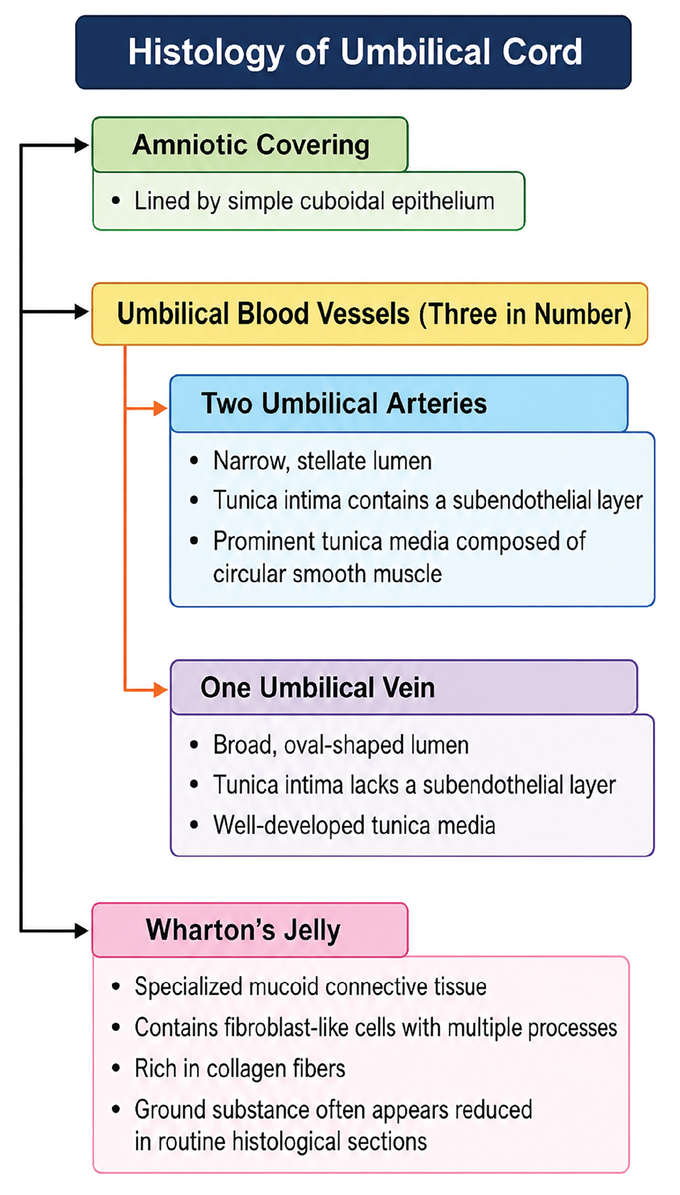
The outer surface of the umbilical cord is covered by amnionic epithelium, consisting of simple squamous to low cuboidal cells.
Umbilical Artery
- The wall contains tunica intima and tunica media.
- The tunica intima is lined by endothelium and contains subendothelial connective tissue folds that contribute to the characteristic narrow, often irregular lumen.
- The tunica media is thick and rich in smooth muscle cells with elastic fibers, helping regulate blood flow.
Umbilical Veins
- The umbilical vein also contains a tunica intima and tunica media.
- It is lined by simple squamous endothelium and has a wider, more regular lumen than the arteries.
- The tunica media contains circularly arranged smooth muscle cells and elastic tissue.
Wharton’s Jelly/Mucoid Connective Tissue
- Wharton’s jelly is a gelatinous connective tissue composed of fibroblast-like cells, collagen fibers, and abundant ground substance rich in glycosaminoglycans.
- It provides mechanical support and protects umbilical vessels from compression during fetal movements and childbirth.
Important Questions
- Q. Write a short note on histology of mammary gland.
- Write a short note on histology of umbilical cord.
📝 Test Your Knowledge – Practice MCQs
Attempt the chapter MCQ quiz and assess your understanding of key concepts.
Click to see: Medical Disclaimer and Copyright Notice