
Competencies
- AN43.2 Identify, describe and draw the microanatomy of: Pituitary gland, thyroid, parathyroid gland.
- AN52.1 Describe and identify the microanatomical features of gastrointestinal system: Suprarenal gland.
Introduction
- The endocrine system consists of glands and tissues that secrete hormones directly into the bloodstream to regulate the functions of target cells, tissues, and organs.
- Hormonal signaling generally produces slower but longer-lasting effects than the nervous system.
- Endocrine glands are typically highly vascular, ensuring efficient release and transport of hormones throughout the body.
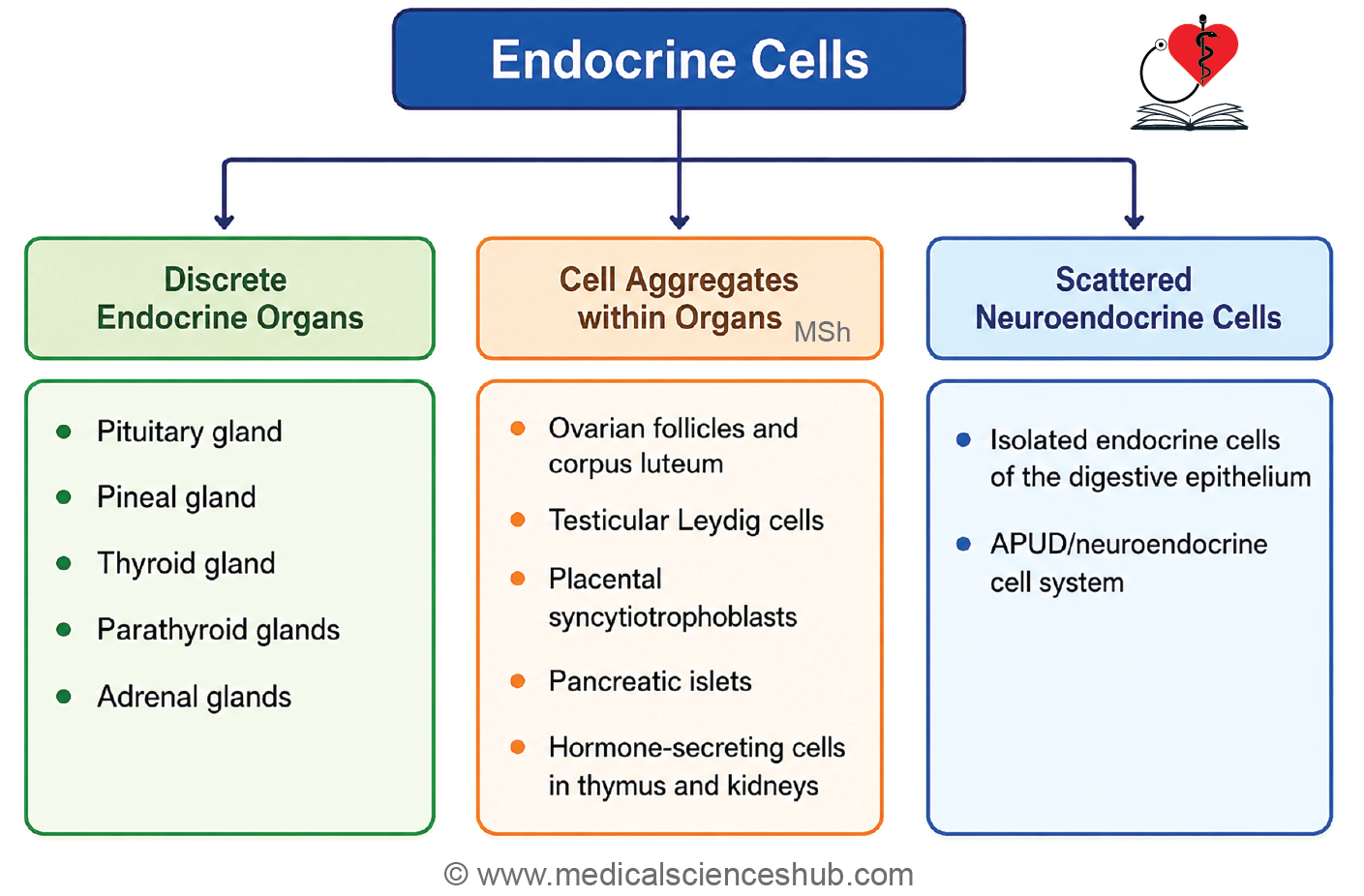
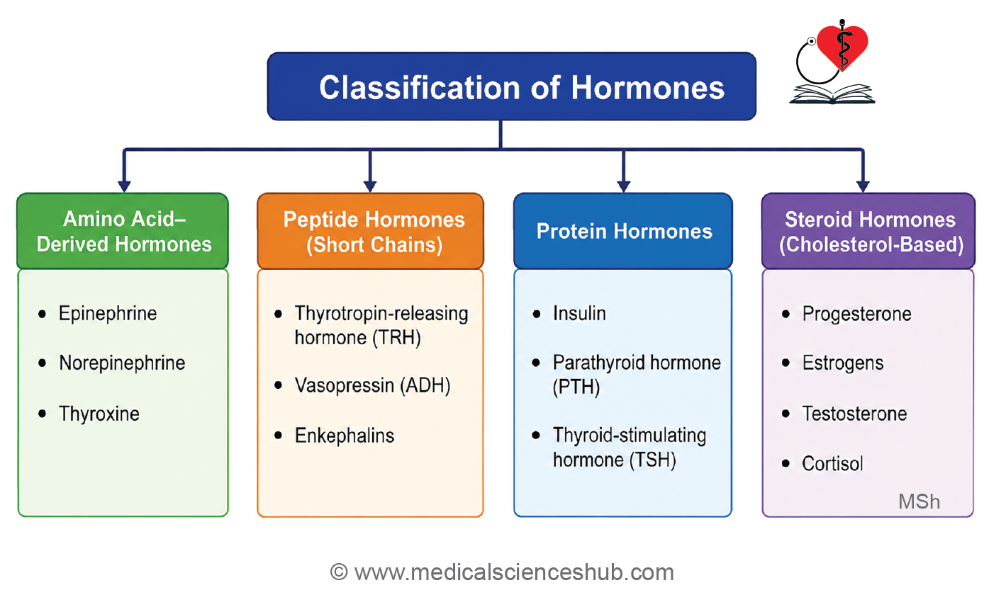
Pituitary Gland (Hypophysis Cerebri)
- The pituitary gland (hypophysis cerebri) is a small endocrine organ located within the sella turcica (hypophyseal fossa) of the sphenoid bone.
- It is approximately the size of a pea and is connected to the hypothalamus by the infundibulum (pituitary stalk).
- The pituitary gland regulates the activity of many endocrine organs and is therefore often referred to as the master endocrine gland.
Parts/Subdivisions of Pituitary Gland
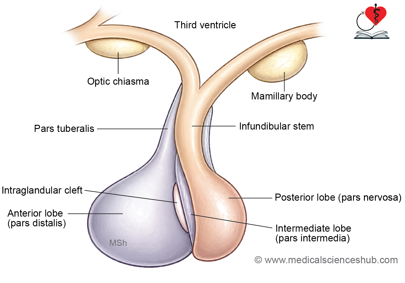
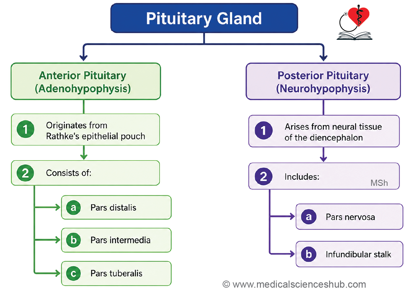
- The gland is composed of two functionally distinct parts:
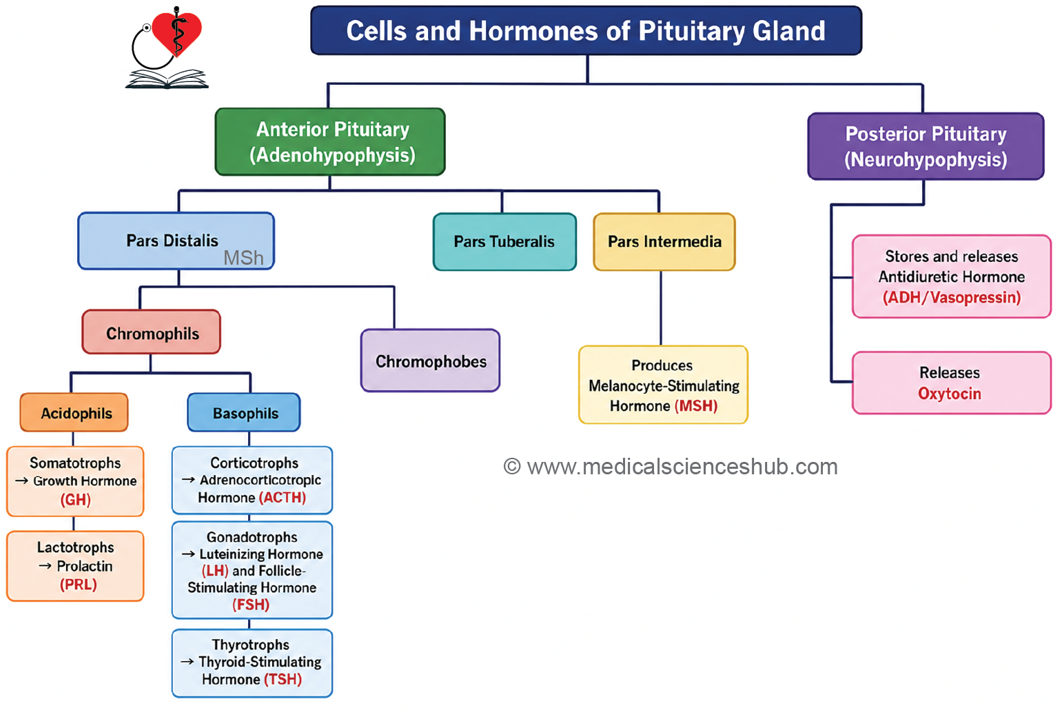
Adenohypophysis (Anterior Pituitary)
- Derived from glandular epithelial tissue.
- Consists of:
- Pars distalis (largest and most active region)
- Pars intermedia
- Pars tuberalis, which surrounds the infundibulum
Neurohypophysis (Posterior Pituitary)
- Derived from neural tissue and functions in hormone storage and release.
- Consists of:
- Pars nervosa
- Infundibulum containing hypothalamo-hypophyseal nerve fibers
- Together, these components coordinate endocrine regulation through close interaction between the hypothalamus and pituitary gland.
Hypothalamo-hypophyseal portal system
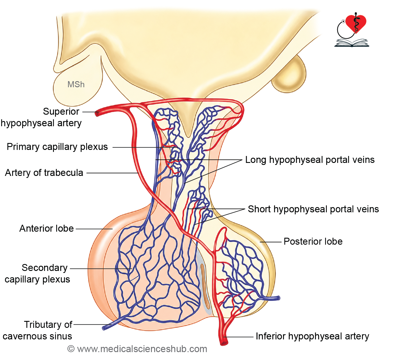
- The pituitary gland receives its blood supply from branches of the internal carotid artery:
- Superior hypophyseal arteries supply the median eminence, infundibulum, and pars tuberalis.
- Inferior hypophyseal arteries supply the neurohypophysis.
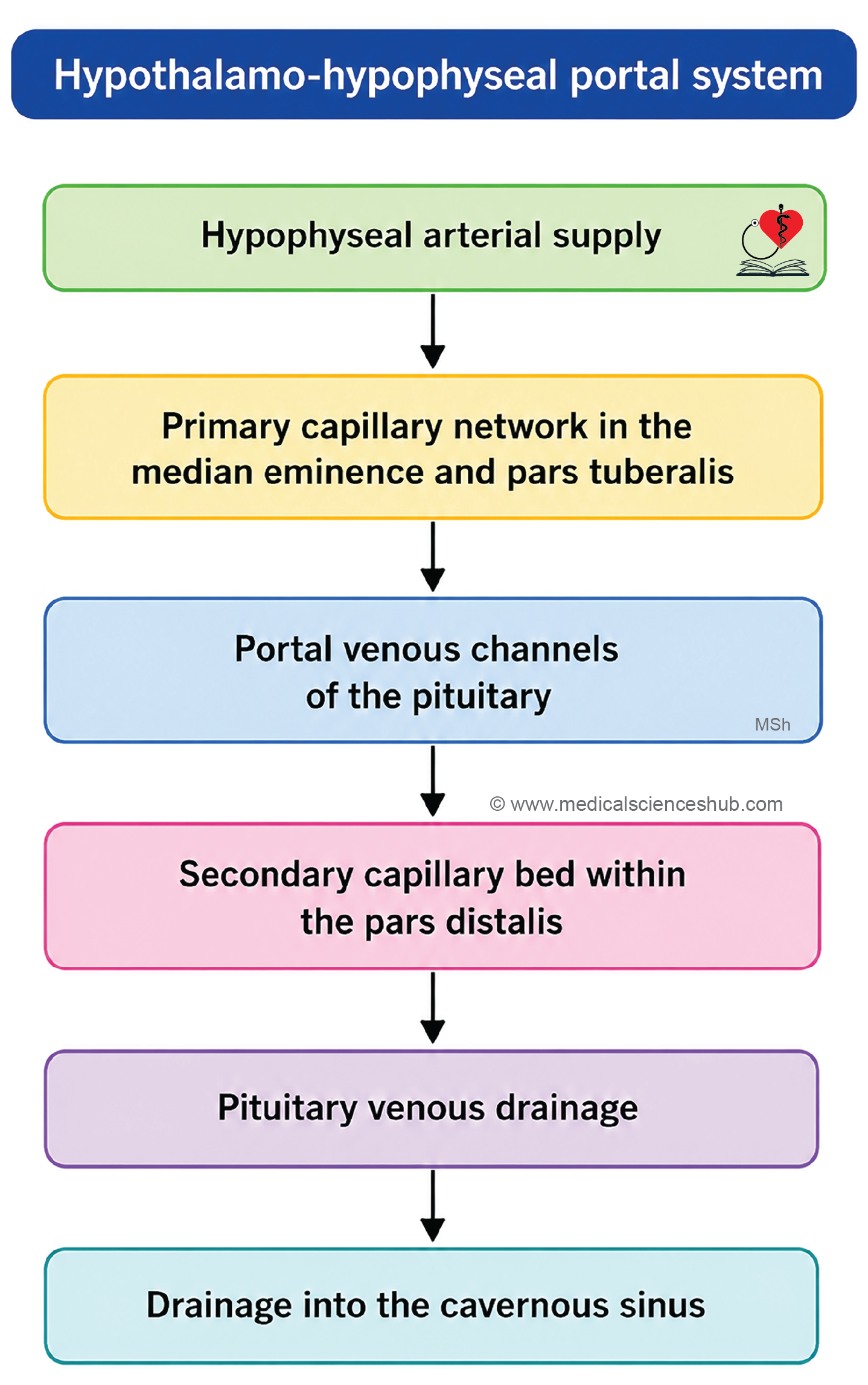
- The superior hypophyseal arteries form a primary fenestrated capillary plexus in the median eminence and infundibular region.
- Blood from this plexus drains into hypophyseal portal veins, which descend to the anterior pituitary.
- These portal veins form a secondary capillary plexus within the pars distalis.
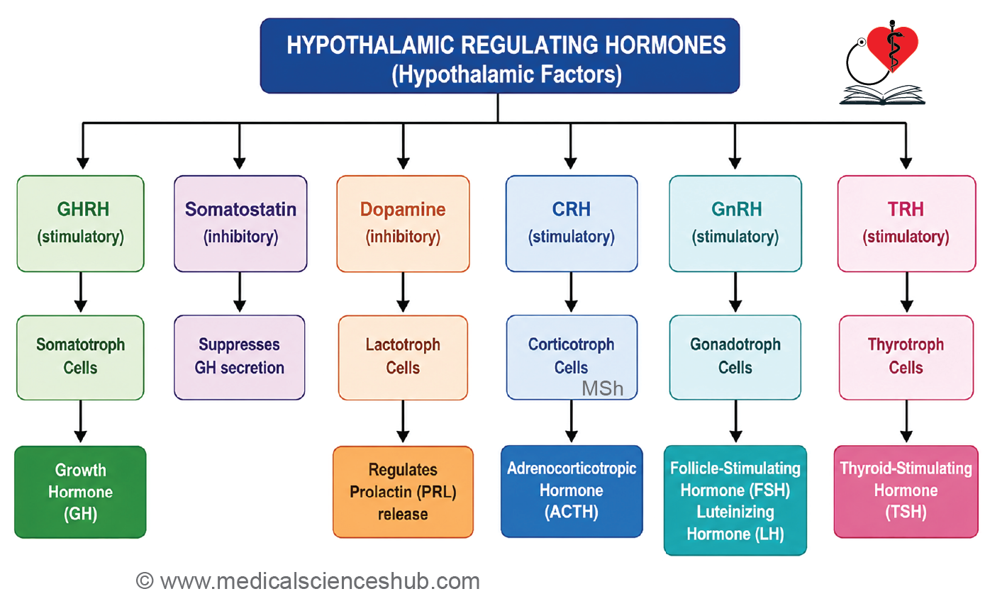
- This specialized vascular system transports hypothalamic releasing and inhibiting hormones directly to the anterior pituitary, enabling precise endocrine regulation.
- Venous blood from the pituitary ultimately drains into the cavernous sinus through hypophyseal veins.
Histology of Pituitary Gland
Adenohypophysis
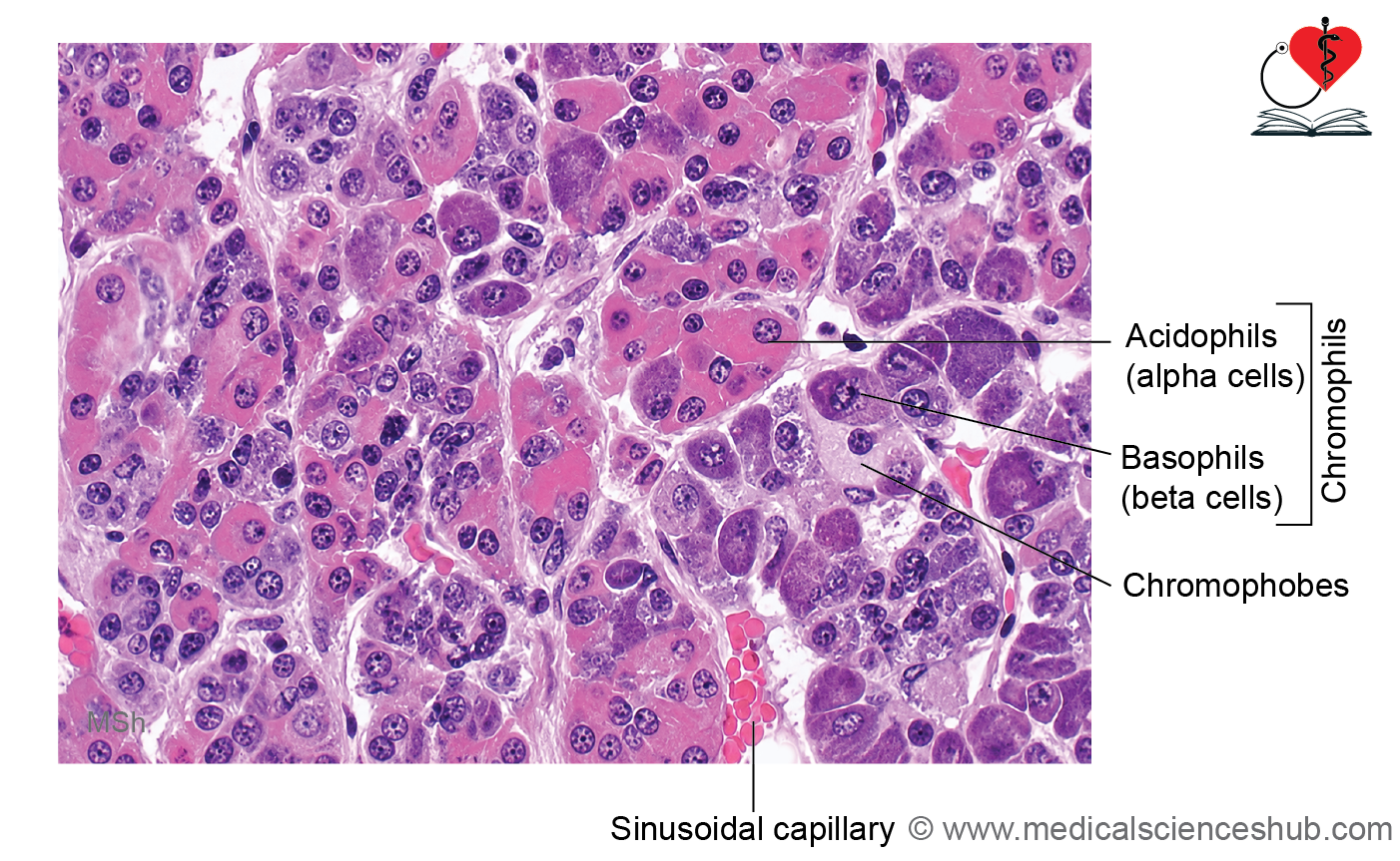
- The adenohypophysis is composed of cords and clusters of endocrine cells separated by numerous fenestrated sinusoidal capillaries.
- On routine H&E staining, cells are classified into:
- Chromophils (granule-containing, strongly staining cells)
- Chromophobes (poorly stained cells with few visible granules)
Chromophils
- Acidophils (~40%):
- Stain pink with eosin.
- Include:
- Somatotrophs: Secrete growth hormone (GH), which regulates body growth. Excess GH causes gigantism in children and acromegaly in adults.
- Lactotrophs (mammotrophs): Produce prolactin (PRL), which stimulates milk production. These cells enlarge during pregnancy and lactation.
- Basophils (~10%):
- Stain with basic dyes and are often PAS-positive.
- Include:
- Corticotrophs: Secrete adrenocorticotropic hormone (ACTH) derived from pro-opiomelanocortin (POMC). POMC also gives rise to MSH, β-endorphin, and related peptides.
- Gonadotrophs: Produce follicle-stimulating hormone (FSH) and luteinizing hormone (LH).
- Thyrotrophs: Secrete thyroid-stimulating hormone (TSH).
- Chromophobes (~50%):
- Exhibit weak staining due to a lack of prominent secretory granules.
- May represent degranulated or inactive endocrine cells.
Pars intermedia
- Located between the pars distalis and pars nervosa.
- Poorly developed in humans.
- Contains colloid-filled cystic spaces that represent remnants of Rathke’s pouch.
- Cells are mainly basophils and chromophobes.
- Produces small amounts of melanocyte-stimulating hormone (MSH), which influences melanin synthesis.
Pars tuberalis
- Forms a sleeve around the infundibular stalk.
- Highly vascular and associated with the primary capillary plexus of the hypothalamo-hypophyseal portal system.
- Contains small clusters of endocrine cells, including both acidophils and basophils.
- Contributes to communication between the hypothalamus and anterior pituitary through its rich vascular network.
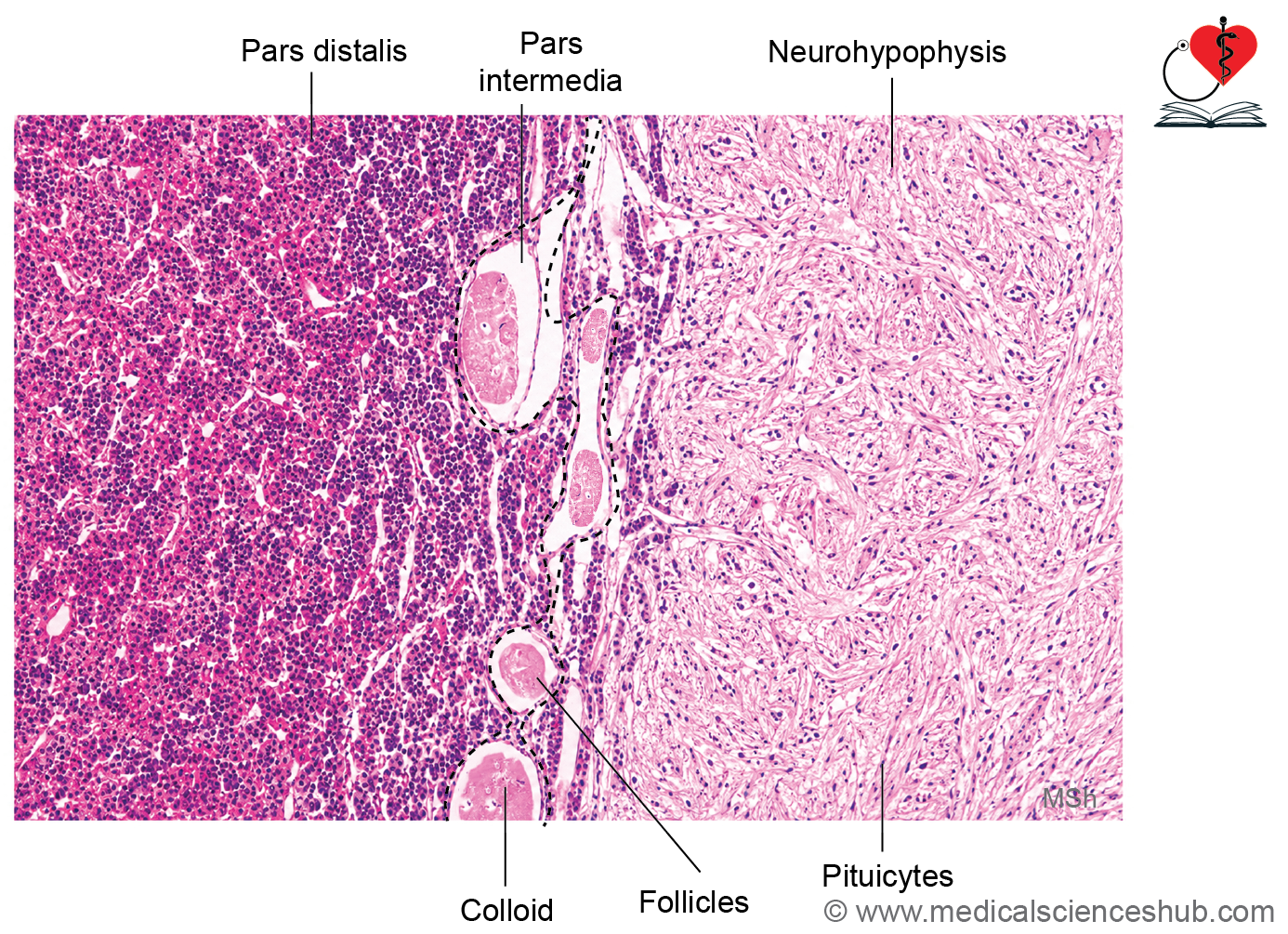
Neurohypophysis/Posterior Lobe of Pituitary Gland
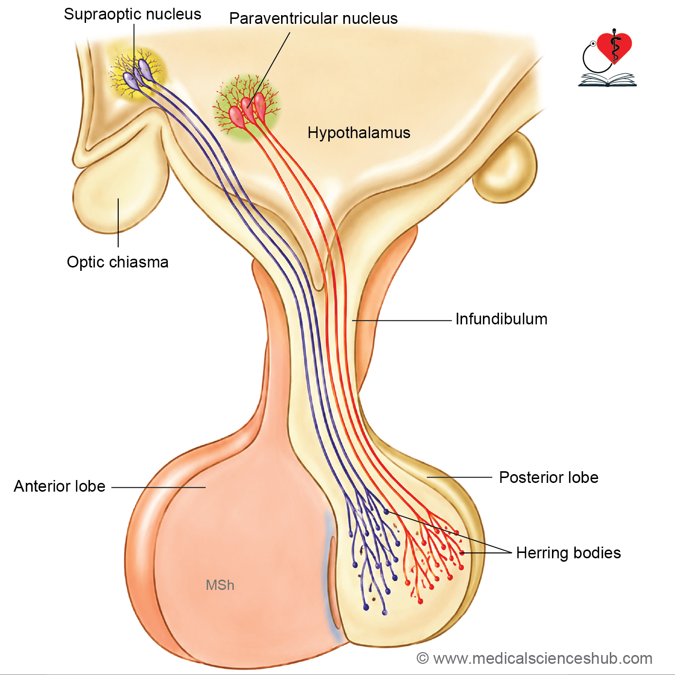
- The neurohypophysis consists of the pars nervosa and infundibulum.
- It contains approximately 100,000 unmyelinated axons arising from neurosecretory neurons located in the supraoptic and paraventricular nuclei of the hypothalamus.
- These axons extend directly into the posterior pituitary without forming synapses and terminate near fenestrated capillaries.
- The posterior pituitary functions primarily as a storage and release site for hypothalamic hormones rather than synthesizing hormones itself.
Pituicytes
- Pituicytes are specialized glial cells that support axons and capillaries within the neurohypophysis.
- They resemble astrocytes and possess elongated cytoplasmic processes.
- Histologically, their cytoplasm is difficult to distinguish from surrounding nerve fibers on routine H&E staining.
Herring Bodies
- Herring bodies are dilated accumulations of neurosecretory granules within axon terminals.
- They store hormones before release into the bloodstream.
- On H&E staining, they appear as eosinophilic (pink) structures.
Hormones Released by the Neurohypophysis
- Oxytocin:
- Stimulates contraction of uterine smooth muscle during labor.
- Promotes contraction of mammary myoepithelial cells, facilitating milk ejection during lactation.
- Antidiuretic hormone (ADH) / Vasopressin:
- Increases water reabsorption in the kidney collecting ducts.
- Reduces urine output and helps maintain blood volume and arterial blood pressure.
- These hormones are synthesized in the hypothalamus and transported along axons to the posterior pituitary for storage and release.
CLINICAL CORRELATION
- Gigantism results from excessive secretion of growth hormone (GH) before closure of the epiphyseal growth plates. It is characterized by excessive but proportionate skeletal growth, marked increase in height, enlargement of bones, and expansion of the thoracic cage.
- Acromegaly occurs when GH excess develops after epiphyseal closure in adulthood. Typical features include enlargement of the hands and feet, coarse facial appearance, prominent supraorbital ridges, mandibular enlargement, and prognathism (forward projection of the lower jaw).
- Diabetes insipidus is caused by deficient secretion or action of antidiuretic hormone (ADH). It is characterized by the production of large volumes of dilute urine (polyuria) and excessive thirst (polydipsia) due to impaired renal water conservation.
Thyroid Gland
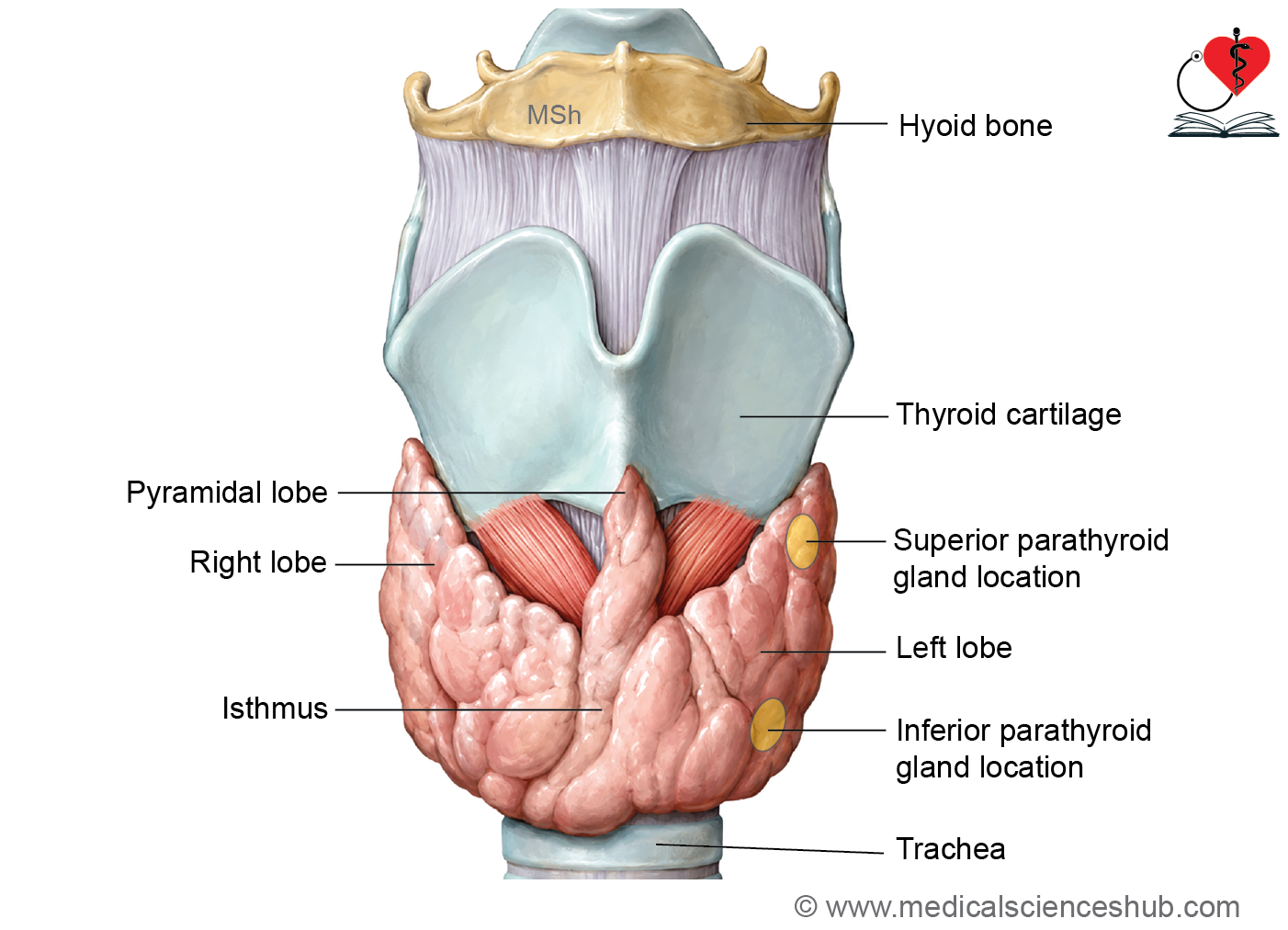
- The thyroid gland is located in the lower anterior region of the neck and consists of right and left lobes connected by an isthmus.
- It typically weighs about 20–30 g and is generally larger in females. Its size may increase slightly during menstruation and pregnancy because of hormonal influences.
- Thyroid follicular cells and follicles develop from the endoderm of the thyroglossal duct, which originates from the primitive pharynx.
- Parafollicular (C) cells, which secrete calcitonin, are derived from the ultimobranchial bodies and later become incorporated into the thyroid gland.
Histology of Thyroid Gland
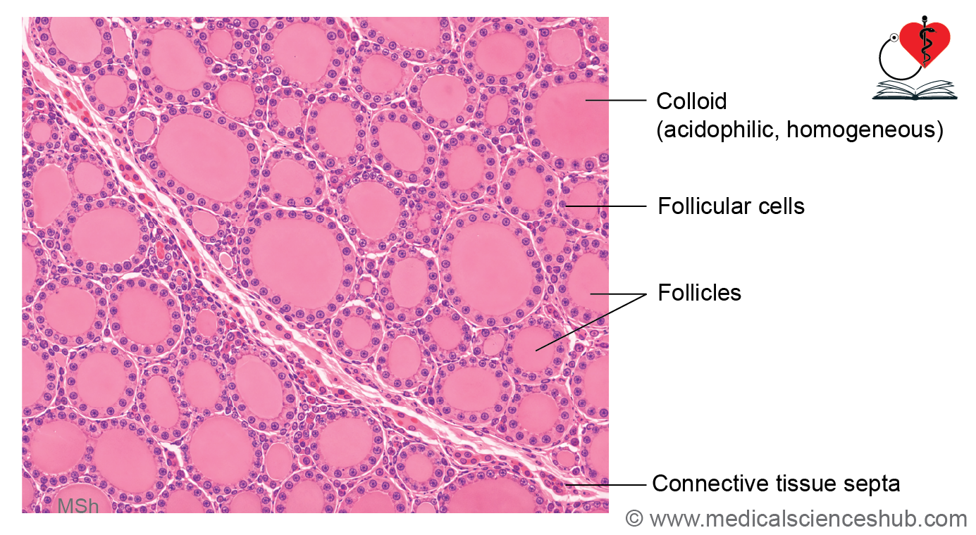
- The thyroid gland is enclosed by a thin fibrous connective tissue capsule.
- Connective tissue septa extend inward from the capsule and subdivide the gland into multiple lobules.
- Each lobule contains numerous thyroid follicles, which are spherical structures measuring approximately 0.2–1.0 mm in diameter.
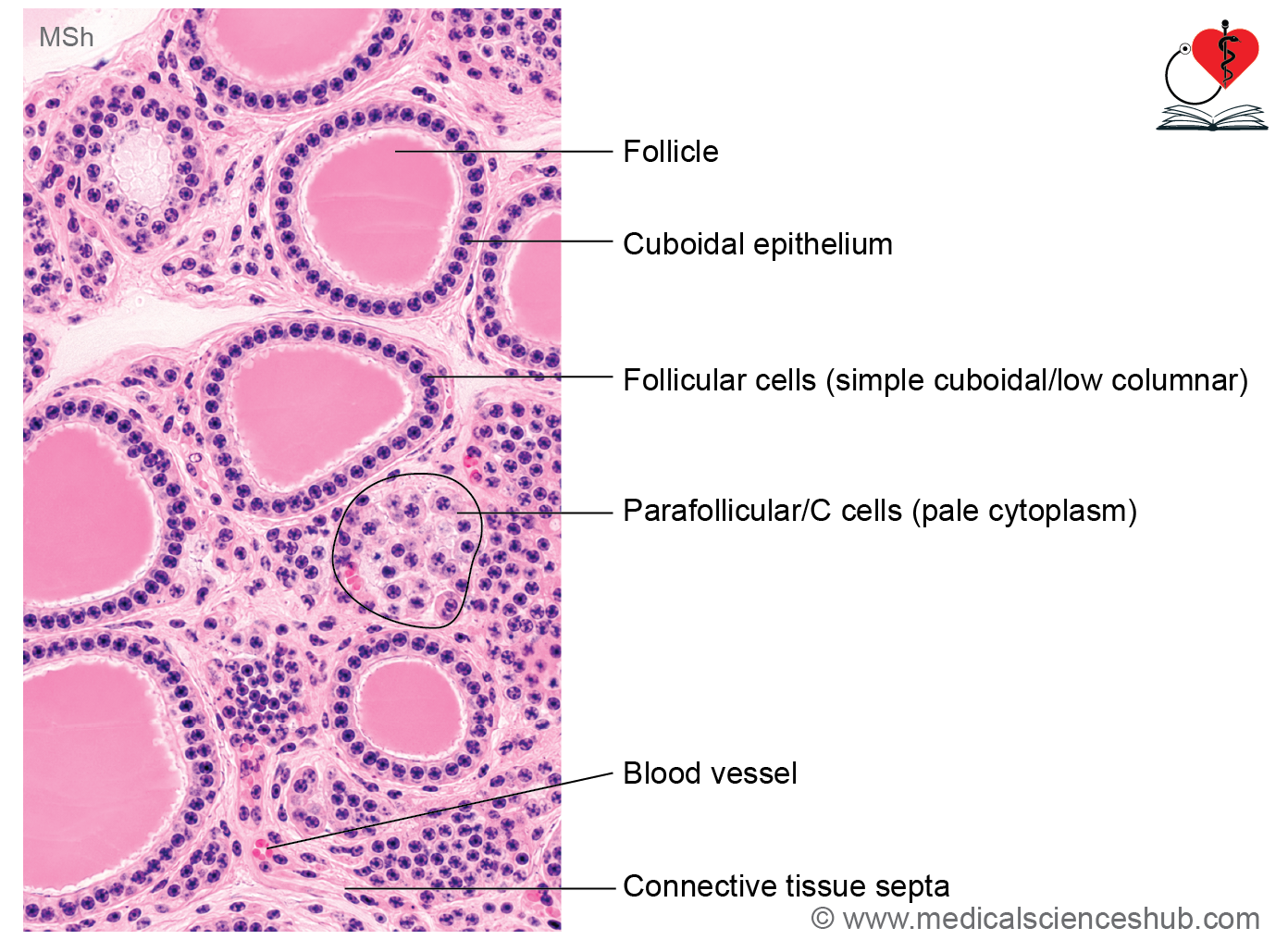
- Follicles are lined by simple cuboidal to low columnar follicular cells resting on a basement membrane.
- The central cavity of each follicle contains colloid, a homogeneous eosinophilic material that serves as a storage form of thyroid hormones.
- Follicular cells possess lightly basophilic cytoplasm and a centrally placed spherical nucleus, often with one or two nucleoli.
- The interfollicular connective tissue contains abundant blood capillaries, lymphatic vessels, and supporting connective tissue fibers.
- Parafollicular (C) cells are located either within the follicular basal lamina or in the connective tissue between follicles.
- C cells are polyhedral cells with pale cytoplasm and eccentric oval nuclei. They do not contact the follicular lumen and are often difficult to identify on routine light microscopy.
- Ultrastructurally, C cells contain numerous secretory granules.
Functional Significance
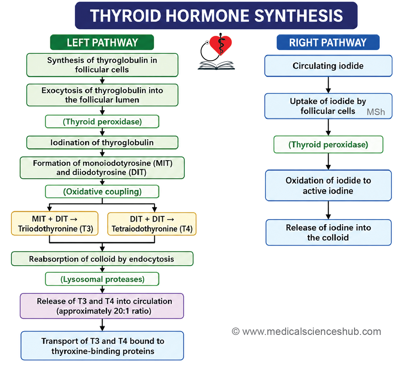
- Follicular cells synthesize and secrete the thyroid hormones triiodothyronine (T3) and thyroxine (T4).
- Parafollicular (C) cells secrete calcitonin, a hormone involved in calcium homeostasis.
Follicular Cells and Activity of Thyroid Gland
- Follicular cells are responsible for the synthesis and secretion of the thyroid hormones triiodothyronine (T3) and thyroxine (T4).
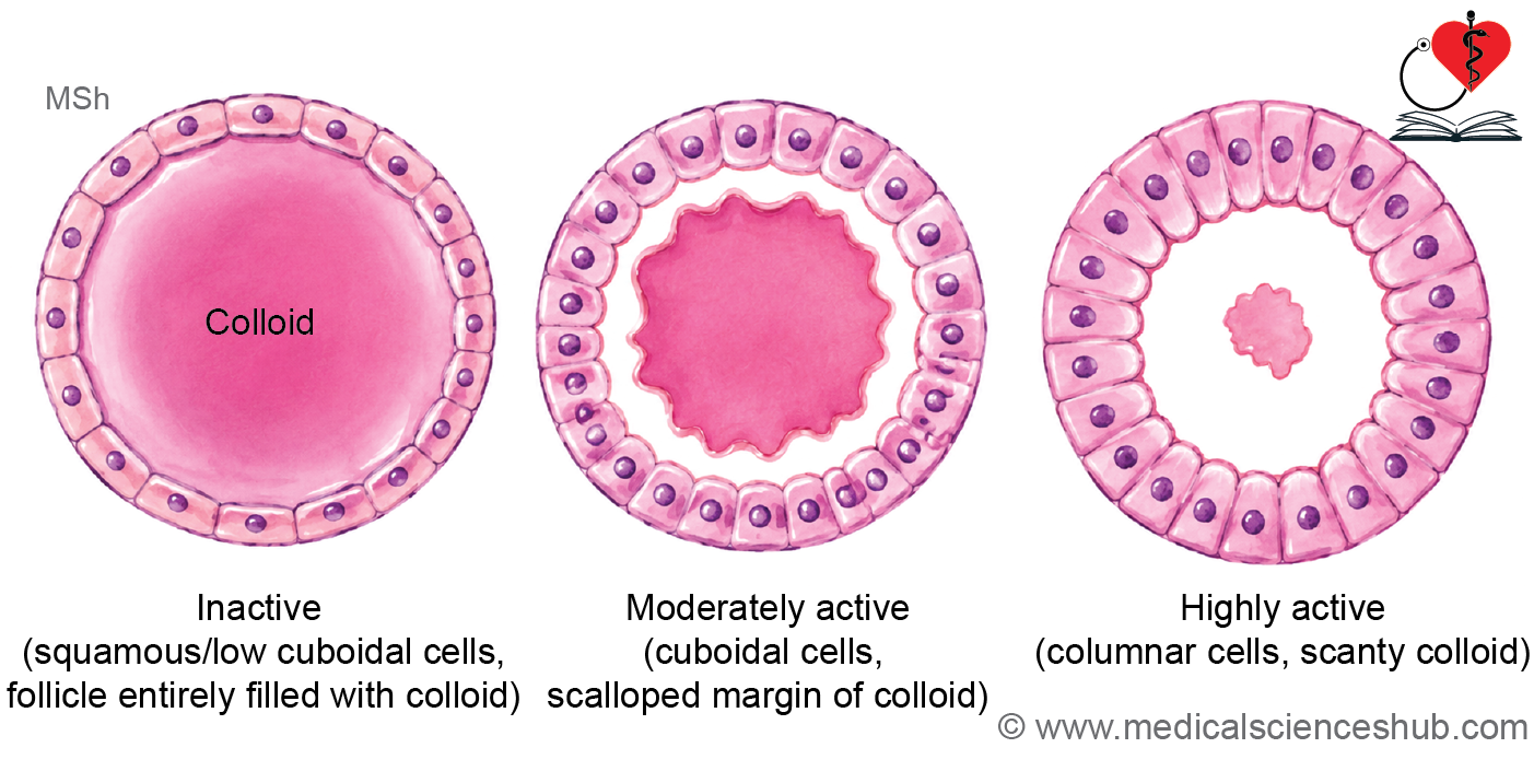
- The morphology of follicular cells varies according to the functional activity of the thyroid gland.
- Inactive follicles are lined by flattened squamous epithelial cells and contain abundant colloid, indicating low secretory activity.
- Under stimulation by thyroid-stimulating hormone (TSH), follicles become active. The lining cells change to cuboidal or low columnar epithelium, sometimes becoming tall columnar, and exhibit large vesicular nuclei.
- Active follicles contain less colloid because stored hormone precursors are actively being utilized and released.
- Scalloping of colloid, characterized by irregular or “moth-eaten” margins, is a histological sign of increased thyroid activity and is commonly observed in Graves disease.
Functions of Thyroid Gland
- The thyroid gland secretes T3, T4, and calcitonin.
- T3 and T4 regulate basal metabolic rate, energy utilization, growth, and tissue differentiation.
- During fetal life and early childhood, thyroid hormones are essential for normal central nervous system (CNS) development and overall body growth.
- Calcitonin, produced by parafollicular (C) cells, helps lower blood calcium levels by promoting calcium deposition in bone and reducing osteoclast-mediated bone resorption.
CLINICAL CORRELATION
- Goiter refers to enlargement of the thyroid gland, regardless of the underlying cause.
- Hyperthyroidism (thyrotoxicosis) results from excessive production of thyroid hormones, leading to an increased metabolic rate.
- Common causes of hyperthyroidism include Graves disease (the most frequent cause), toxic multinodular goiter, toxic thyroid adenoma, and thyroid-stimulating autoantibodies.
- Hypothyroidism is characterized by deficient thyroid hormone production or reduced responsiveness of target tissues to thyroid hormones.
- In infancy and childhood, severe hypothyroidism can cause cretinism, resulting in impaired physical and neurological development.
- In adults, hypothyroidism may lead to myxedema, which is characterized by lethargy, mental slowing, and non-pitting connective tissue edema.
- The most common causes of hypothyroidism are iodine deficiency and Hashimoto thyroiditis, an autoimmune disorder of the thyroid gland.
Parathyroid Glands
- The parathyroid glands are four small endocrine glands, typically arranged as two superior and two inferior glands, located on the posterior surface of the thyroid gland within its capsule.
- The superior parathyroid glands develop from the fourth pharyngeal pouch, whereas the inferior parathyroid glands arise from the third pharyngeal pouch.
- Each gland is oval in shape, approximately the size of a split pea, and weighs about 50 mg.
Histology of Parathyroid Gland
- Each gland is enclosed by a thin connective tissue capsule.
- Delicate connective tissue septa extend inward, dividing the gland into poorly defined lobules.
- The parenchyma is highly vascular and contains numerous sinusoidal capillaries.
- Two principal cell types are present:
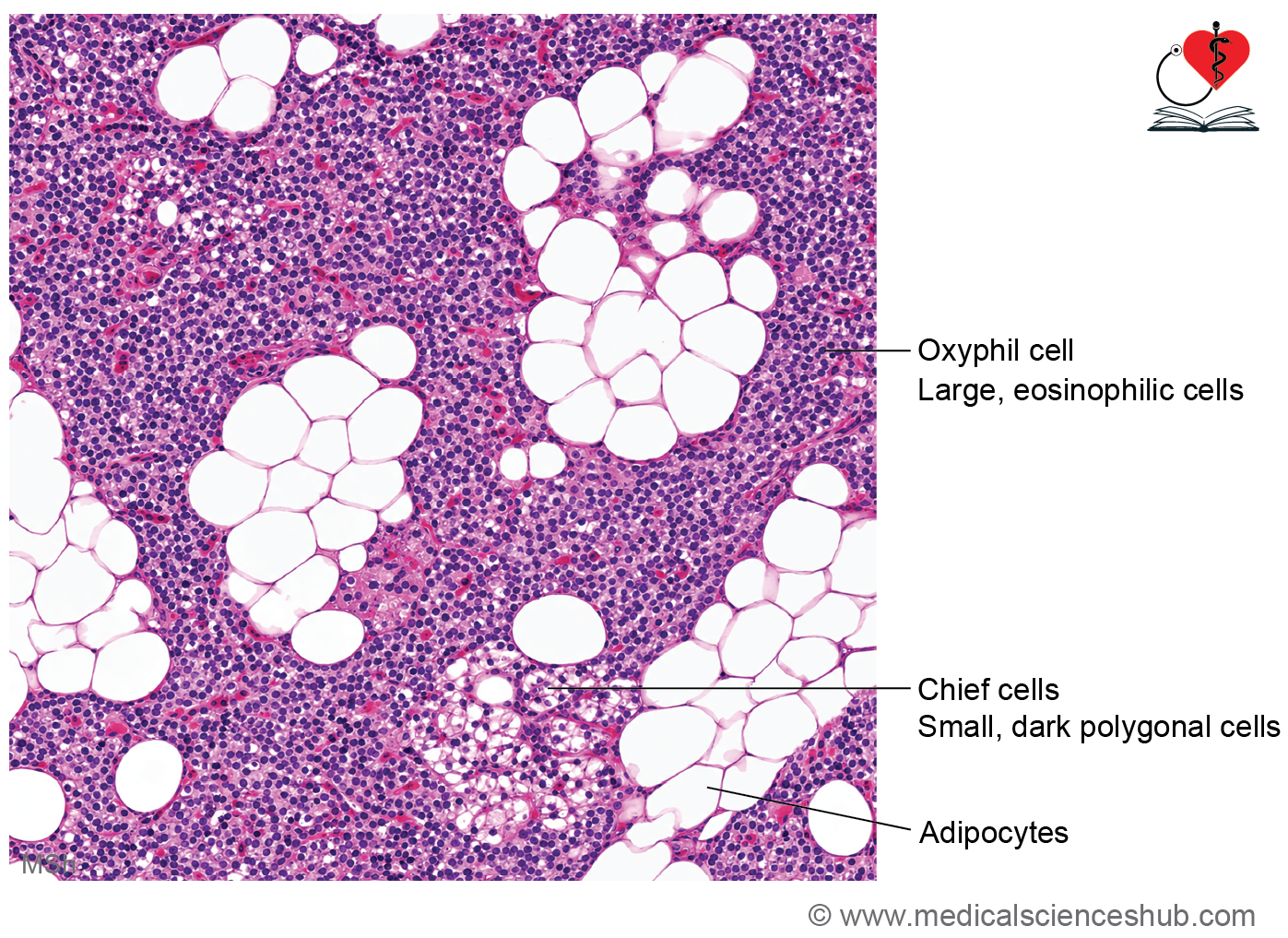
- Chief (principal) cells
- Oxyphil cells
Chief Cells
- Chief cells are the predominant cell type and are responsible for the secretion of parathyroid hormone (PTH).
- They are small polygonal cells with centrally placed nuclei and moderately basophilic cytoplasm.
- Based on glycogen and lipofuscin content, chief cells may appear as dark, light, or clear cells.
- Ultrastructurally, they contain abundant rough endoplasmic reticulum, Golgi complexes, and secretory granules.
Oxyphil Cells
- Oxyphil cells are larger and less numerous than chief cells.
- They occur singly or in small clusters.
- Their intensely eosinophilic cytoplasm results from a high mitochondrial content.
- These cells contain few organelles involved in secretion, and their precise function remains uncertain.
Functions of Parathyroid Gland
- PTH increases blood calcium concentration by:
- Stimulating bone resorption through activation of osteoclast-mediated processes.
- Enhancing calcium reabsorption in the kidneys.
- Promoting renal conversion of vitamin D to its active form, 1,25-dihydroxyvitamin D₃.
- Increasing intestinal absorption of calcium through vitamin D–mediated mechanisms.
- Increasing phosphate excretion by the kidneys.
- PTH and calcitonin act in opposite ways to maintain calcium homeostasis.
Table 22.1: Hormones of thyroid and parathyroid gland
| Gland | Hormone | Secreting Cells | Major Functions |
|---|---|---|---|
| Thyroid Gland | Thyroxine (T₄) and Triiodothyronine (T₃) | Follicular cells | Regulate basal metabolic rate, promote normal growth and development, and are essential for maturation of the fetal and postnatal nervous system. |
| Thyroid Gland | Calcitonin | Parafollicular (C) cells | Lowers blood calcium concentration by inhibiting osteoclastic bone resorption and promoting calcium deposition in bone. |
| Parathyroid Gland | Parathyroid Hormone (PTH) | Chief (Principal) cells | Raises blood calcium concentration by stimulating bone resorption, increasing renal calcium reabsorption, and enhancing intestinal calcium absorption through activation of vitamin D. |
CLINICAL CORRELATION
- Hyperparathyroidism is characterized by excessive secretion of parathyroid hormone (PTH), resulting in elevated blood calcium levels (hypercalcemia).
- Hypoparathyroidism results from reduced PTH secretion and leads to decreased blood calcium levels (hypocalcemia).
- Clinical manifestations of hypocalcemia include tetany, muscle cramps, and paresthesia.
- Chvostek’s sign is facial muscle twitching elicited by tapping the facial nerve.
- Trousseau’s sign is carpal spasm induced by inflating a blood pressure cuff, indicating latent tetany associated with hypocalcemia.
Adrenal/Suprarenal Gland
- The paired adrenal glands are located on the superior poles of the kidneys within the perirenal fat.
- Each gland weighs approximately 5–7 g.
Histology of Adrenal Glands
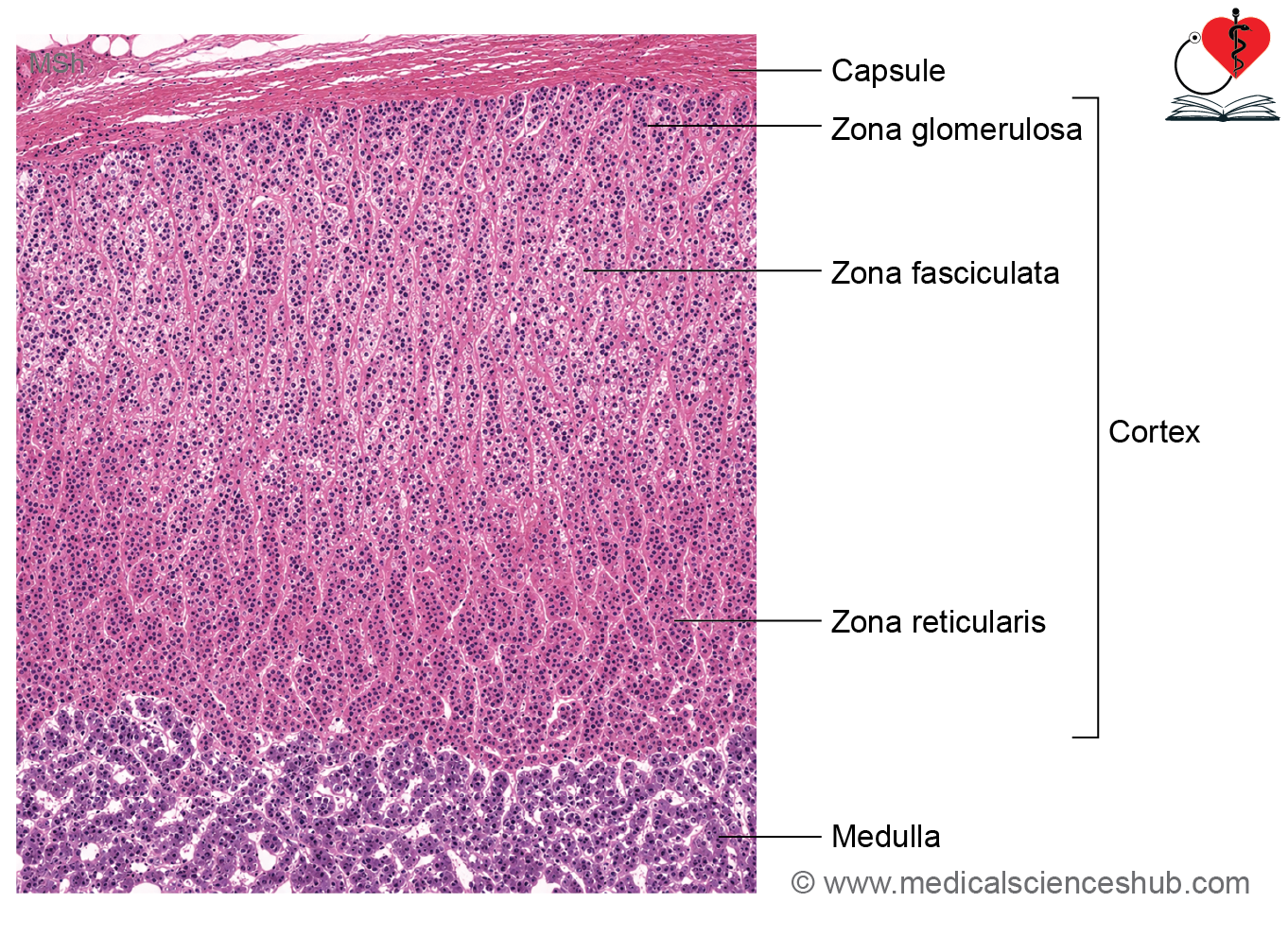
- The gland is enclosed by a thin connective tissue capsule, from which delicate septa extend into the gland.
- Histologically, the adrenal gland consists of:
- Adrenal cortex (about 90% of the gland)
- Adrenal medulla (about 10% of the gland)
- The cortical region is approximately ten times larger than the medulla.
Adrenal Cortex
The cortex is divided into three concentric zones:
- Zona glomerulosa (outermost; ~15%)
- Zona fasciculata (middle; ~80%)
- Zona reticularis (innermost; ~5%).
Zona Glomerulosa
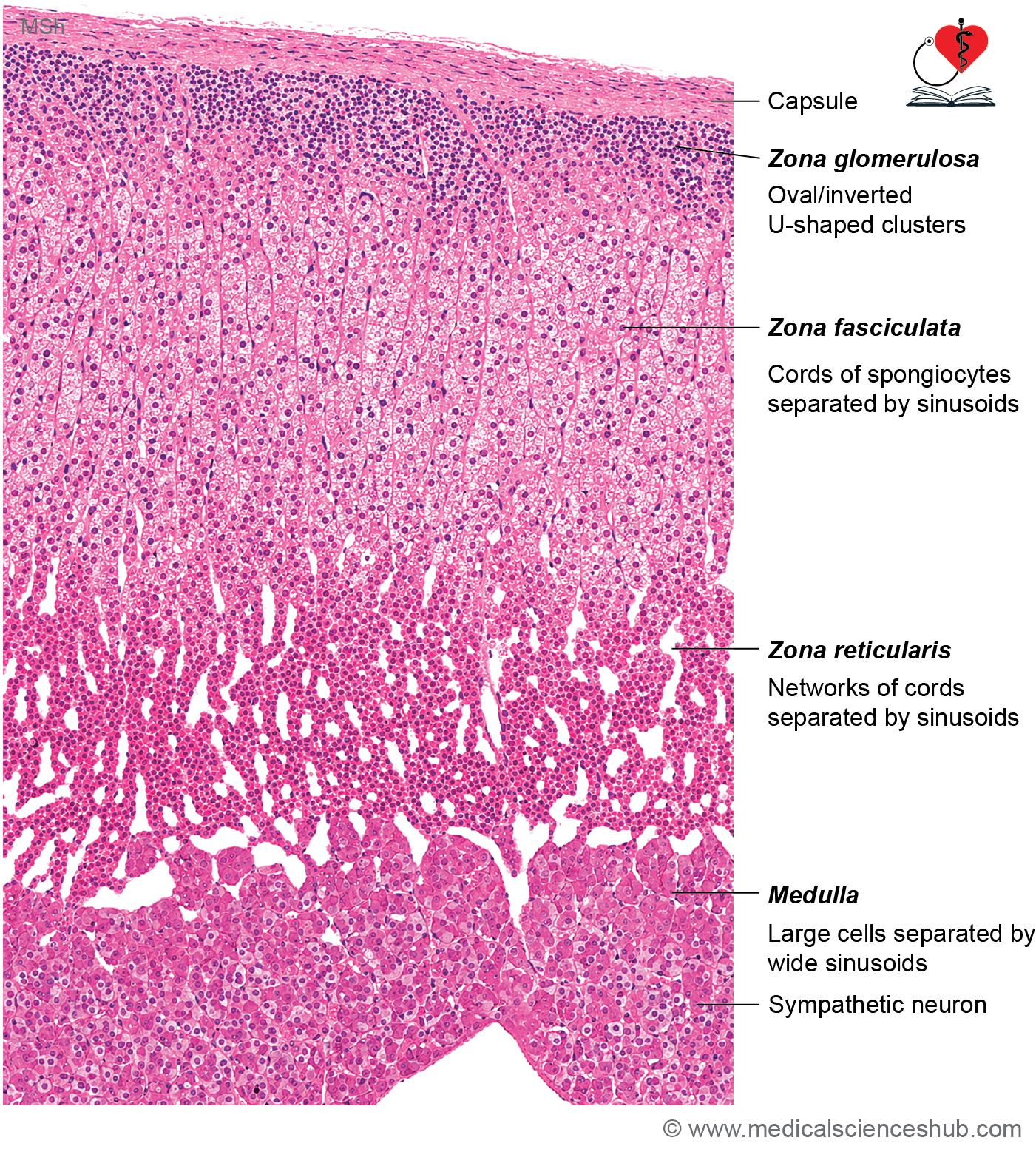
- This is the thin outer layer located immediately beneath the capsule.
- Cells are arranged in rounded clusters and curved cords resembling renal glomeruli.
- The cells are small, columnar to pyramidal in shape, with centrally placed spherical nuclei.
- Their cytoplasm is mildly eosinophilic and contains relatively few lipid droplets compared with deeper cortical cells.
Functions of Zona Glomerulosa
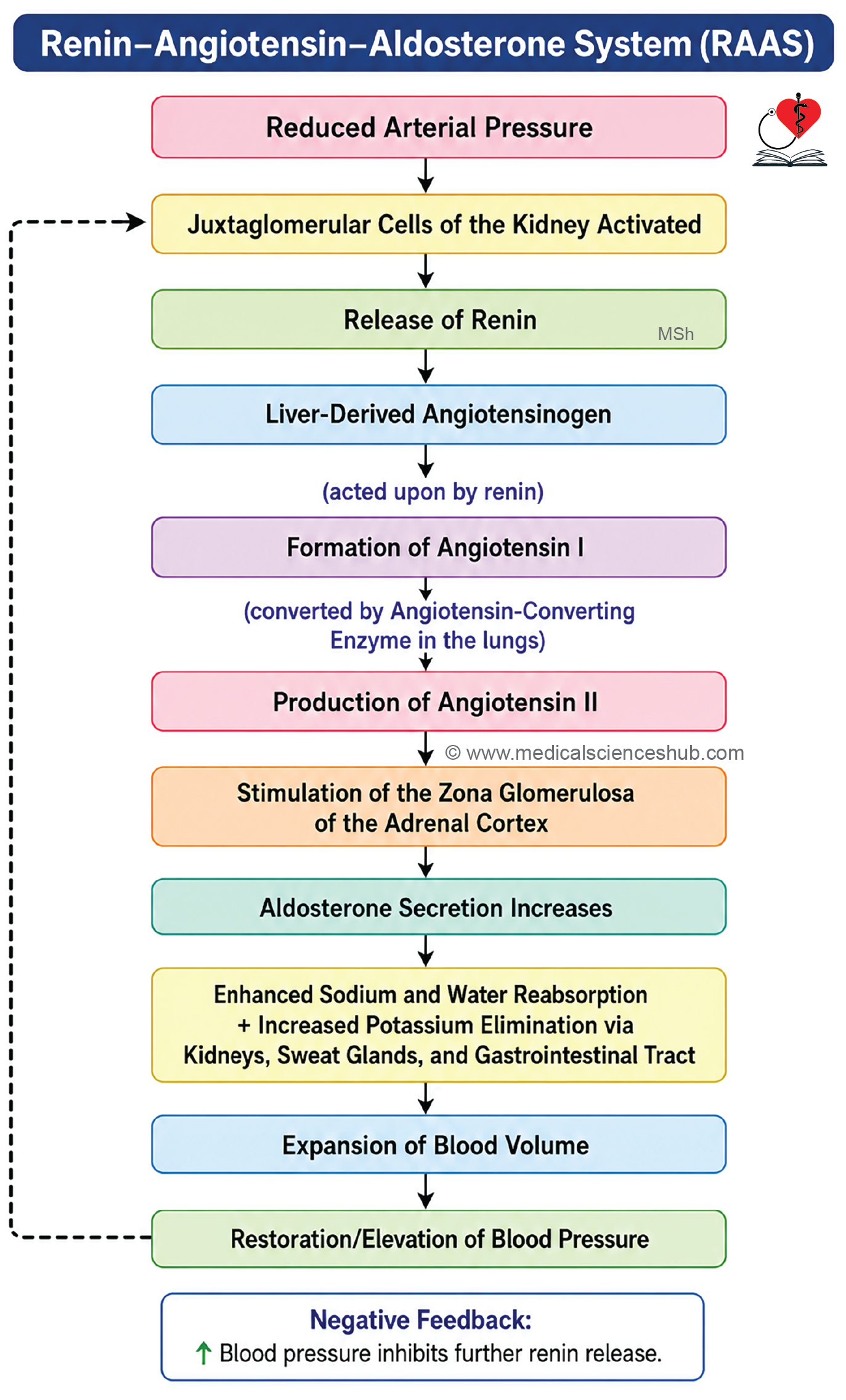
- Cells of the zona glomerulosa secrete mineralocorticoids, primarily aldosterone.
- Aldosterone regulates sodium and potassium balance, promotes water retention, and contributes to the maintenance of blood pressure and extracellular fluid volume.
- Secretion of aldosterone is mainly controlled by the renin–angiotensin–aldosterone system (RAAS) and, to a lesser extent, by blood potassium levels.
Zona Fasciculata
- The zona fasciculata is the middle and largest layer of the adrenal cortex, comprising approximately 80% of its thickness.
- Cells are arranged in long, straight cords that are typically one to two cells thick and are separated by sinusoidal capillaries.
- The cells, known as spongiocytes, are large, polyhedral, and larger than those of the zona glomerulosa.
- Their cytoplasm appears vacuolated on routine H&E staining because it contains abundant lipid droplets that are dissolved during tissue processing.
- The nuclei are large, pale, and vesicular.
- Ultrastructurally, these cells exhibit features characteristic of steroid-producing cells, including abundant smooth endoplasmic reticulum, numerous mitochondria, a prominent Golgi apparatus, and lipid droplets.
Function
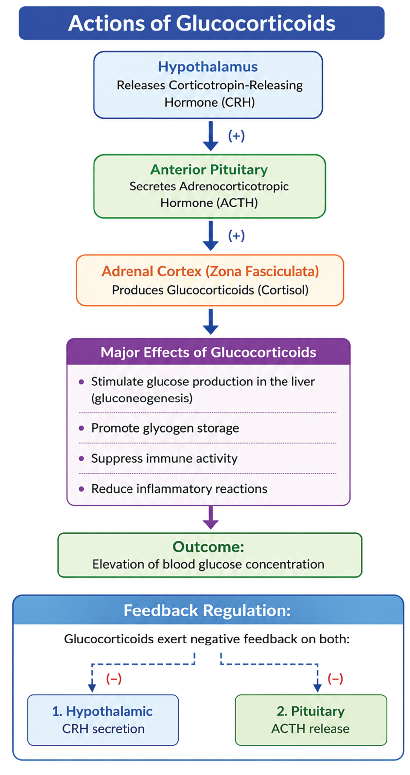
- The zona fasciculata synthesizes and secretes glucocorticoids, mainly cortisol.
- Hormone secretion is regulated primarily by adrenocorticotropic hormone (ACTH) from the anterior pituitary.
Zona Reticularis
- The zona reticularis is the innermost layer of the adrenal cortex and accounts for about 5% of cortical tissue.
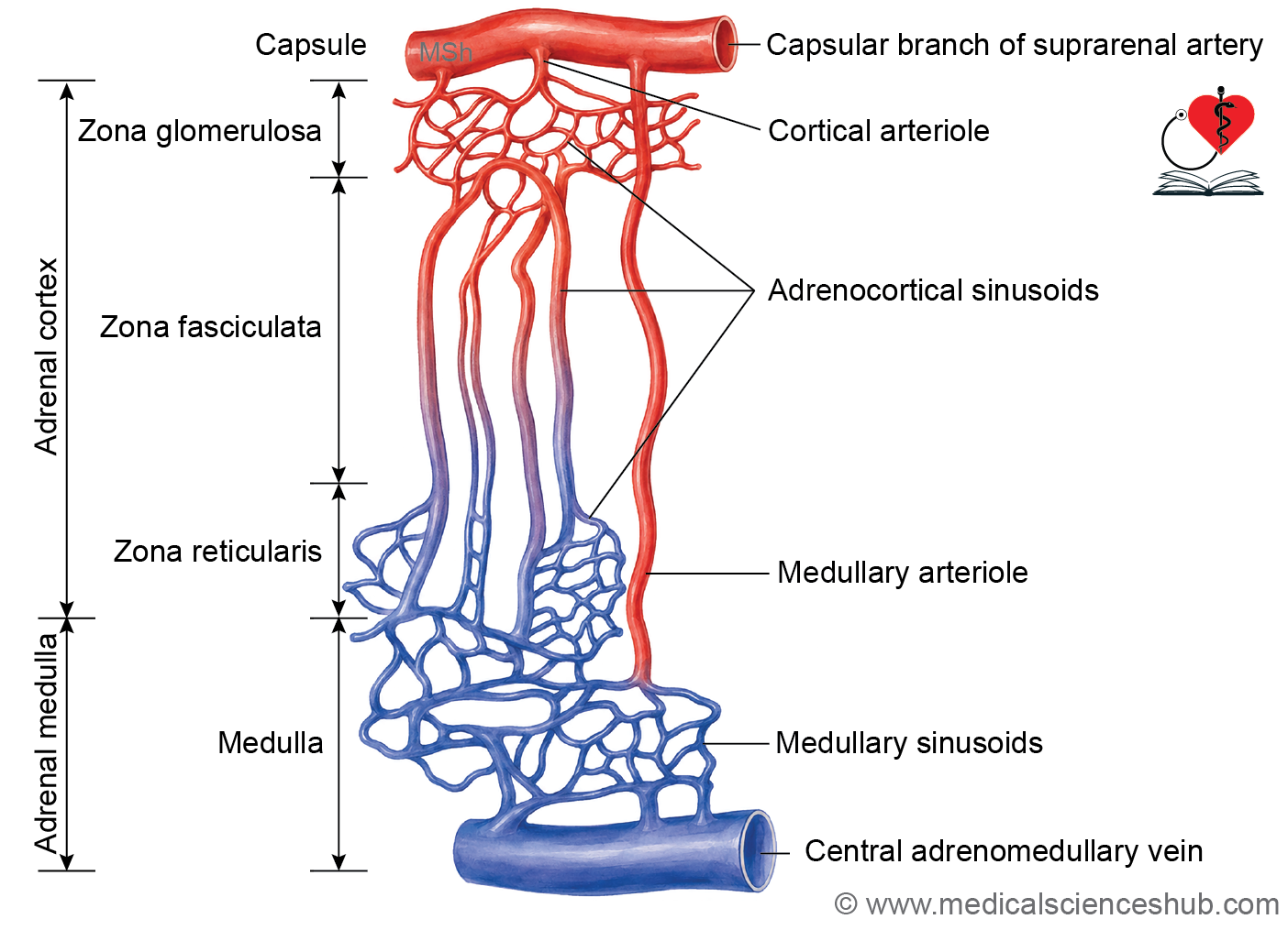
- Cells are arranged in branching and interconnected cords that form a network-like pattern, separated by fenestrated capillaries.
- These cells are smaller than spongiocytes and possess less cytoplasm, resulting in more darkly stained nuclei.
- They contain abundant lipofuscin pigment granules, which impart a yellow-brown coloration.
- Electron microscopy reveals well-developed smooth endoplasmic reticulum and relatively sparse rough endoplasmic reticulum, consistent with steroid hormone synthesis.
Function
- The zona reticularis secretes weak androgens, predominantly dehydroepiandrosterone (DHEA).
- It also produces small amounts of glucocorticoids, including cortisol.
Suprarenal Medulla
- The adrenal medulla forms the central region of the adrenal gland.
- Medullary cells are arranged in short, interconnected cords and clusters separated by numerous sinusoidal capillaries.
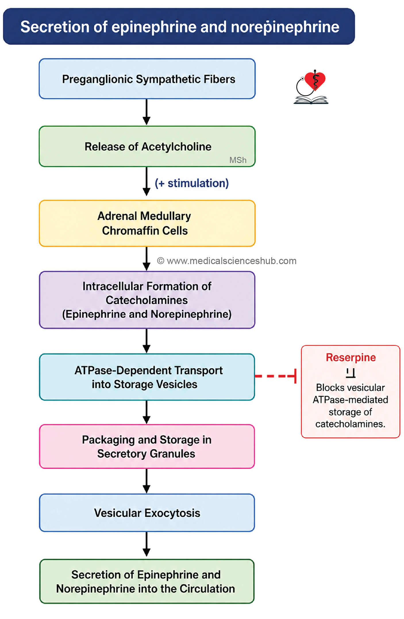
- The principal cells are pale-staining chromaffin cells (pheochromocytes), which are modified postganglionic sympathetic neurons.
- These cells are called chromaffin cells because they stain brown with chromium salts such as potassium dichromate.
- Cells of the adrenal cortex do not possess chromaffin properties.
- Ultrastructurally, chromaffin cells contain abundant rough endoplasmic reticulum and numerous membrane-bound secretory granules containing catecholamines.
- Occasional sympathetic ganglion cells may also be present within the medulla.
Functions of the Adrenal Medulla
- The adrenal medulla secretes the catecholamines epinephrine (≈80%) and norepinephrine (≈20%).
- These hormones mediate the fight-or-flight response by producing effects similar to sympathetic nervous system activation.
- Their actions include:
- Increased heart rate and cardiac output.
- Elevated blood pressure.
- Increased blood glucose concentration.
- Enhanced blood flow to the heart and skeletal muscles through vasodilation of appropriate vascular beds.
- These physiological changes prepare the body to respond rapidly to stress, physical activity, or emergencies.
CLINICAL CORRELATION
- Pheochromocytoma is a neuroendocrine tumor arising from the chromaffin cells of the adrenal medulla.
- It produces excessive catecholamines (epinephrine and norepinephrine), leading to elevated circulating hormone levels.
- Most pheochromocytomas are benign but can cause severe hypertension, tachycardia, palpitations, excessive sweating, and recurrent headaches.
- Surgical excision is the definitive treatment.
- Cushing syndrome results from excessive secretion of adrenal cortical hormones, particularly cortisol.
- Common causes include pituitary ACTH excess (Cushing disease), adrenal cortical tumors, or ectopic ACTH-producing neoplasms.
- Clinical features include central obesity, moon face, buffalo hump, thin skin, abdominal purple striae, osteoporosis, menstrual disturbances, reduced libido, and facial plethora.
- Addison disease is a form of primary adrenal insufficiency caused by inadequate production of adrenal cortical hormones.
- Typical manifestations include fatigue, muscle weakness, anorexia, postural hypotension, gastrointestinal symptoms, hyperpigmentation, and, in some cases, vitiligo.
PINEAL GLAND (EPIPHYSIS CEREBRI)
- The pineal gland is a small, cone-shaped endocrine organ situated between the two superior colliculi of the midbrain.
- It develops from the roof of the third ventricle and is a component of the epithalamus.
- The gland weighs approximately 0.1 to 0.2 g.
- Its resemblance to a pine cone gives it the name pineal gland.
Histology of Pineal Gland
- The gland is enclosed by a thin pia mater-derived capsule.
- Delicate connective tissue septa arise from the capsule and divide the gland into irregular lobules and cords of cells.
- The parenchyma contains two principal cell types:
- Pinealocytes – 95%
- Interstitial (glial) cells – 5%
Pinealocytes
- Pinealocytes are the main functional cells of the pineal gland.
- They are arranged in cords or small clusters within the lobules.
- These cells are lightly stained, polygonal, and possess a large, round, pale nucleus.
- Under the electron microscope, pinealocytes show elongated cytoplasmic processes containing secretory vesicles.
- Function: Pinealocytes synthesize and secrete melatonin.
Interstitial (Glial Cells)
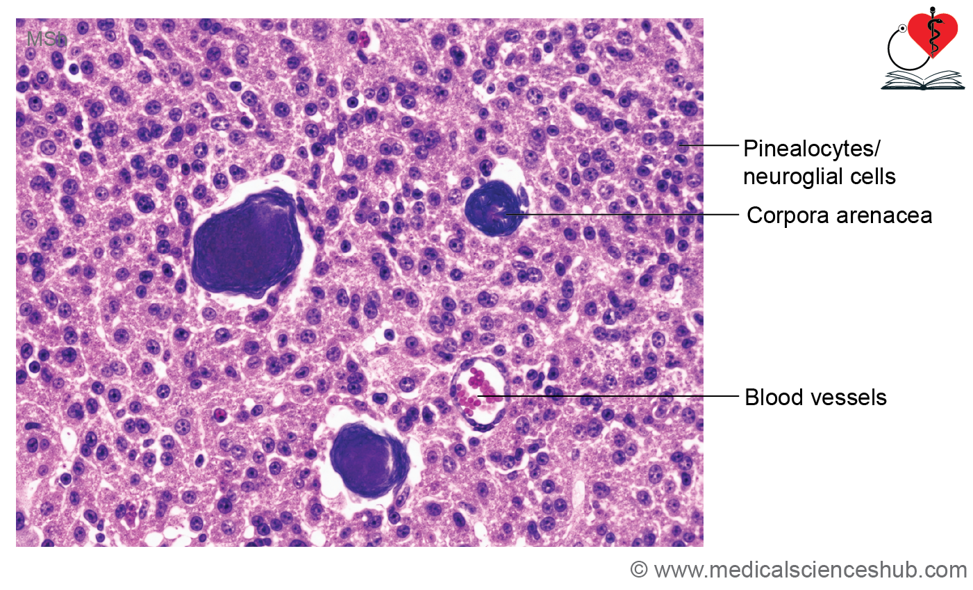
- Corpora arenacea, also known as brain sand, are the characteristic identifying feature of the pineal gland.
- They consist of calcified deposits within the gland.
- These calcifications may be present during childhood and gradually increase with advancing age.
- On Hematoxylin and Eosin (H&E) staining, they appear as dark blue, concentrically laminated structures.
- Corpora arenacea serve as an important landmark for identifying the pineal gland in both histological sections and radiological imaging.
Corpora Arenacea/Brain Sand
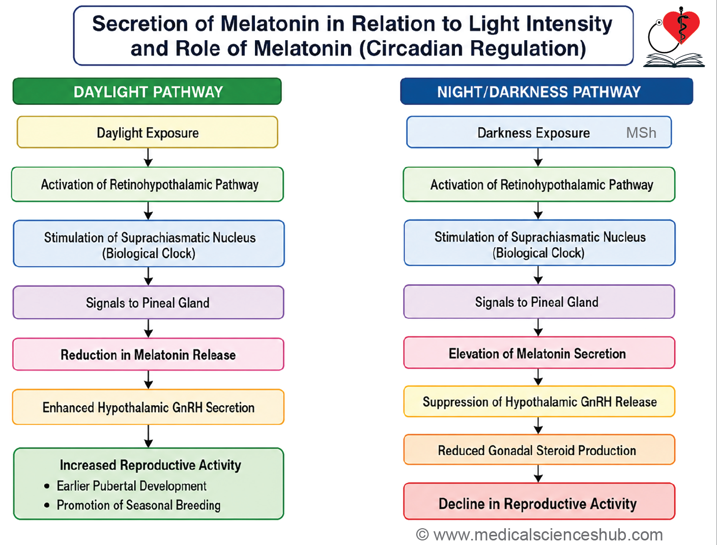
- Produces and releases melatonin.
- Melatonin secretion varies with environmental light and helps regulate the sleep-wake (circadian) rhythm.
Important Questions
- Draw a well-labeled diagram of histology of pituitary gland.
- Draw a well-labeled diagram of histology of thyroid gland.
- List the identification features of histology of thyroid gland.
- Draw a well-labeled diagram of histology of the parathyroid gland.
- Draw a well-labeled diagram of histology of the adrenal gland.
📝 Test Your Knowledge – Practice MCQs
Attempt the chapter MCQ quiz and assess your understanding of key concepts.
Click to see: Medical Disclaimer and Copyright Notice