
Competency
- AN64.1 Describe and identify the microanatomical features of spinal cord, cerebellum and cerebrum.
Introduction
- The nervous system is divided into two major components:
- Central Nervous System (CNS): Comprises the brain and spinal cord.
- Peripheral Nervous System (PNS): Includes peripheral nerves and autonomic ganglia.
- The CNS contains gray matter and white matter.
- Gray matter consists mainly of neuronal cell bodies, dendrites, and neuroglial cells.
- White matter is composed predominantly of myelinated and unmyelinated axons.
- Nuclei are localized collections of neuronal cell bodies embedded within the white matter of the CNS.
- The CNS is protected by three connective tissue coverings called the meninges: dura mater, arachnoid mater, and pia mater.
Spinal Cord
- The spinal cord is a cylindrical structure of the central nervous system that gives rise to 31 pairs of spinal nerves.
- It measures approximately 45 cm in length in adults and extends from the upper border of the atlas to the level of the L1–L2 vertebrae.
- The spinal cord is enclosed by three meninges:
- Dura mater (outer layer)
- Arachnoid mater (middle layer)
- Pia mater (inner layer)
- Two enlargements are present:
- Cervical enlargement (C4–T1 segments) supplying the upper limbs.
- Lumbosacral enlargement (L2–S3 segments) supplying the lower limbs.
- Surface features include:
- Anterior median fissure
- Posterior median sulcus
- Ventrolateral and dorsolateral sulci
- Ventral (motor) and dorsal (sensory) nerve roots
- The tapered lower end of the spinal cord is called the conus medullaris.
- The filum terminale, a fibrous extension of pia mater, continues inferiorly from the conus medullaris and helps anchor the spinal cord.
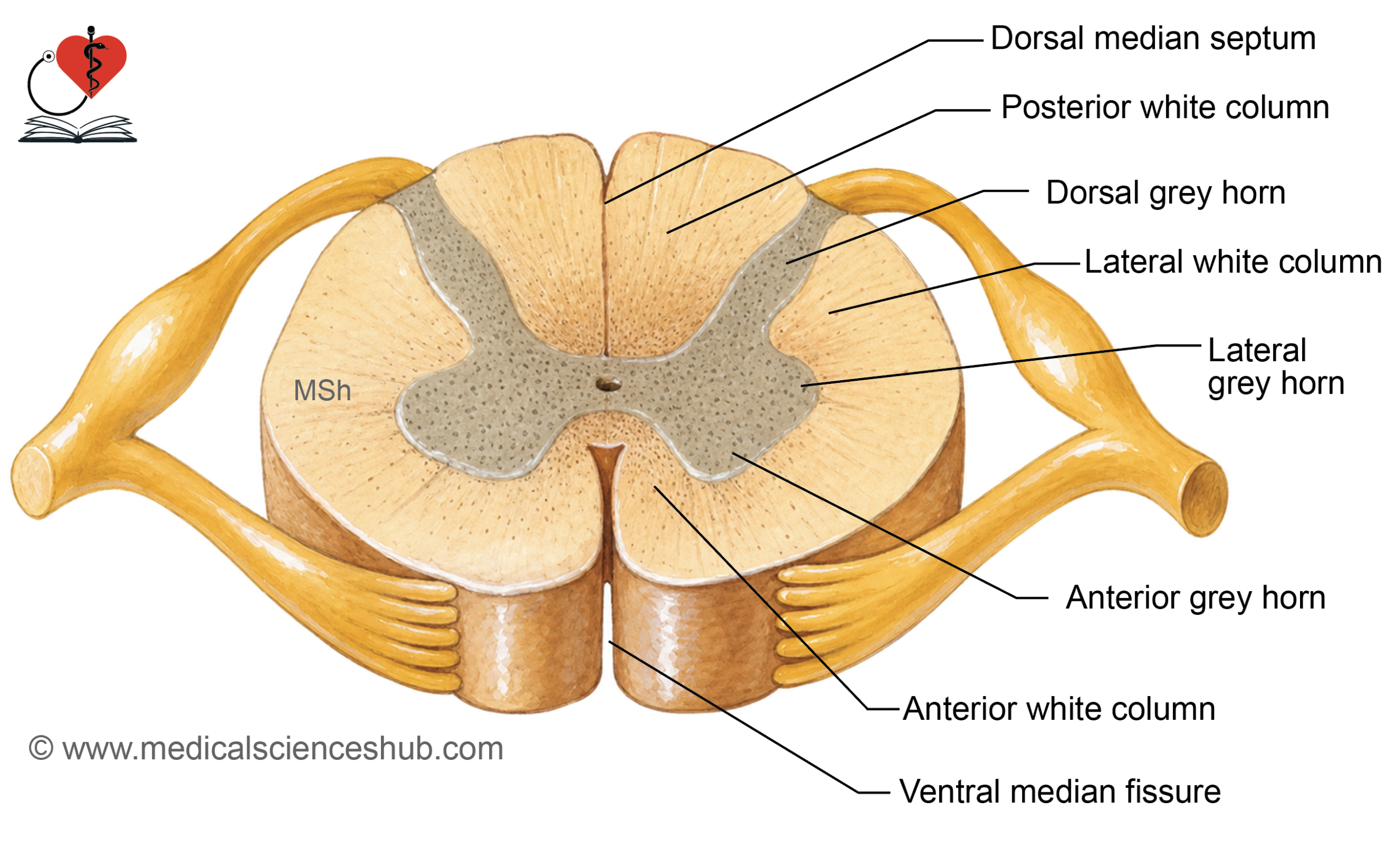
Histology of Spinal Cord
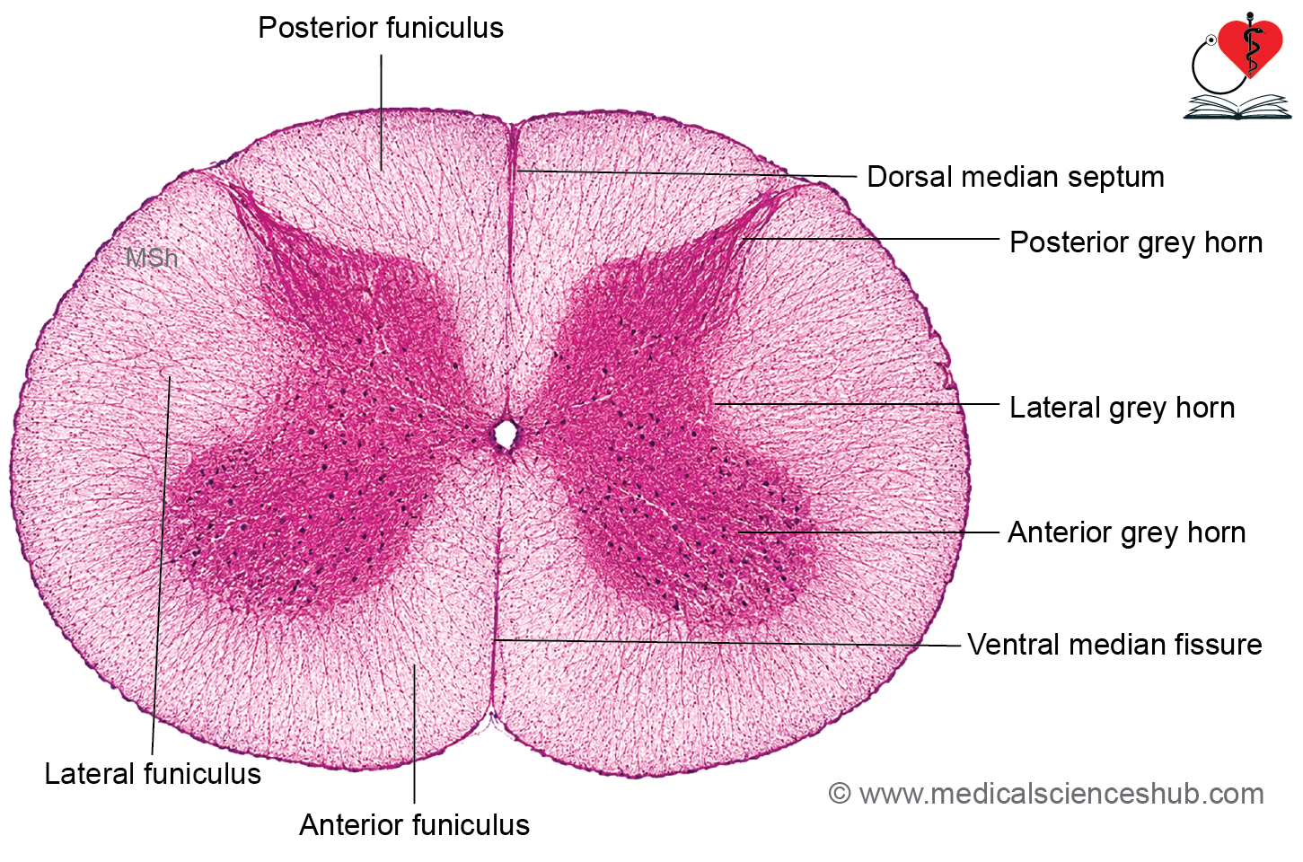
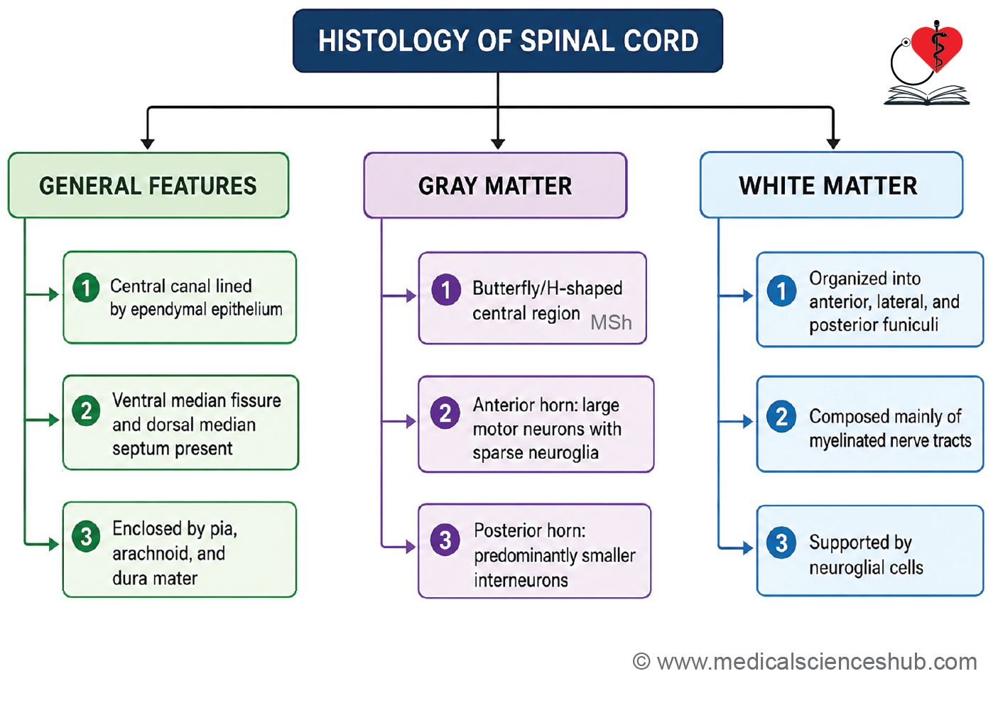
- A transverse section of the spinal cord shows a central central canal, which is lined by ependymal cells forming a simple ciliated cuboidal to low columnar epithelium.
- The central canal is surrounded by gray matter, while white matter occupies the peripheral region.
Gray Matter
- Gray matter has a characteristic H-shaped (butterfly-shaped) appearance.
- It consists of anterior (ventral) horns and posterior (dorsal) horns.
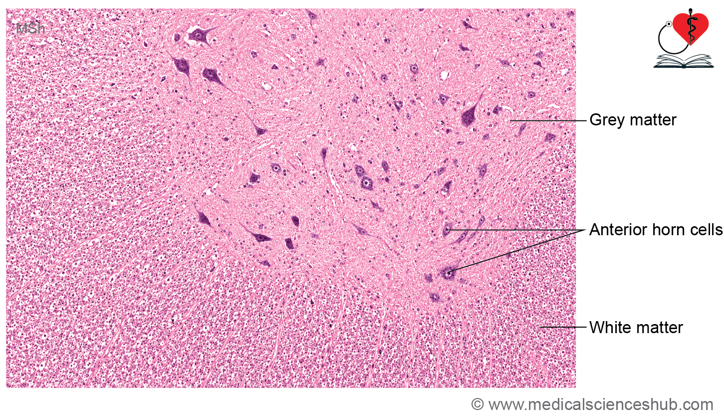
- The anterior horns contain large multipolar motor neurons known as anterior horn cells.
- These neurons possess large vesicular nuclei, prominent nucleoli, and abundant Nissl substance.
- Axons of anterior horn cells form the ventral roots of spinal nerves.
- Neurons in the posterior horns are generally smaller and are involved primarily in sensory processing.
- Gray matter also contains numerous neuroglial cells, particularly microglia, and a rich vascular supply.
White Matter
- White matter is composed predominantly of myelinated nerve fibers and supporting neuroglial cells.
- During routine H&E staining, myelin is dissolved, producing clear spaces around axons.
- White matter is organized into three regions:
- Anterior funiculus
- Lateral funiculus
- Posterior funiculus
- The entire spinal cord is externally covered by pia mater, a delicate vascular connective tissue layer closely adherent to its surface.
CLINICAL CORRELATION
- Multiple sclerosis (MS) is a chronic demyelinating disorder of the central nervous system, affecting the brain and spinal cord. It results from progressive destruction of myelin sheaths, leading to impaired nerve conduction. Clinical manifestations may include visual disturbances, muscle weakness, sensory deficits, impaired coordination, and other neurological symptoms.
- MS is considered an autoimmune disease, although its exact cause remains uncertain.
- Guillain–Barré syndrome (GBS) is an acute autoimmune disorder characterized by demyelination of peripheral nerves. It typically presents with rapidly progressive numbness, weakness, and ascending paralysis.
- Severe cases may involve respiratory muscles, causing respiratory failure that requires urgent medical management.
- Early diagnosis and treatment are important to improve recovery and reduce complications.
Regional Characteristics of Spinal Cord Enlargements of Spinal Cord
- The spinal cord exhibits two prominent enlargements due to an increased number of motor neurons required for innervation of the limbs.
- Cervical enlargement:
- Supplies the upper limbs through the brachial plexus.
- Extends approximately from C3 to T2 spinal cord segments.
- Reaches its maximum diameter near the C6 spinal cord segment.
- Lumbosacral enlargement:
- Supplies the lower limbs through the lumbosacral plexus.
- Extends from approximately L1 to S3 spinal cord segments.
- Attains its greatest diameter near the level corresponding to the T12 vertebra.
- These enlargements occur because limb innervation requires a larger population of sensory and motor neurons.
- The amount and configuration of grey matter vary among spinal cord segments according to the volume of tissue they innervate.
- Consequently, gray matter is most abundant in the cervical and lumbosacral regions, producing the characteristic enlargements of the spinal cord.
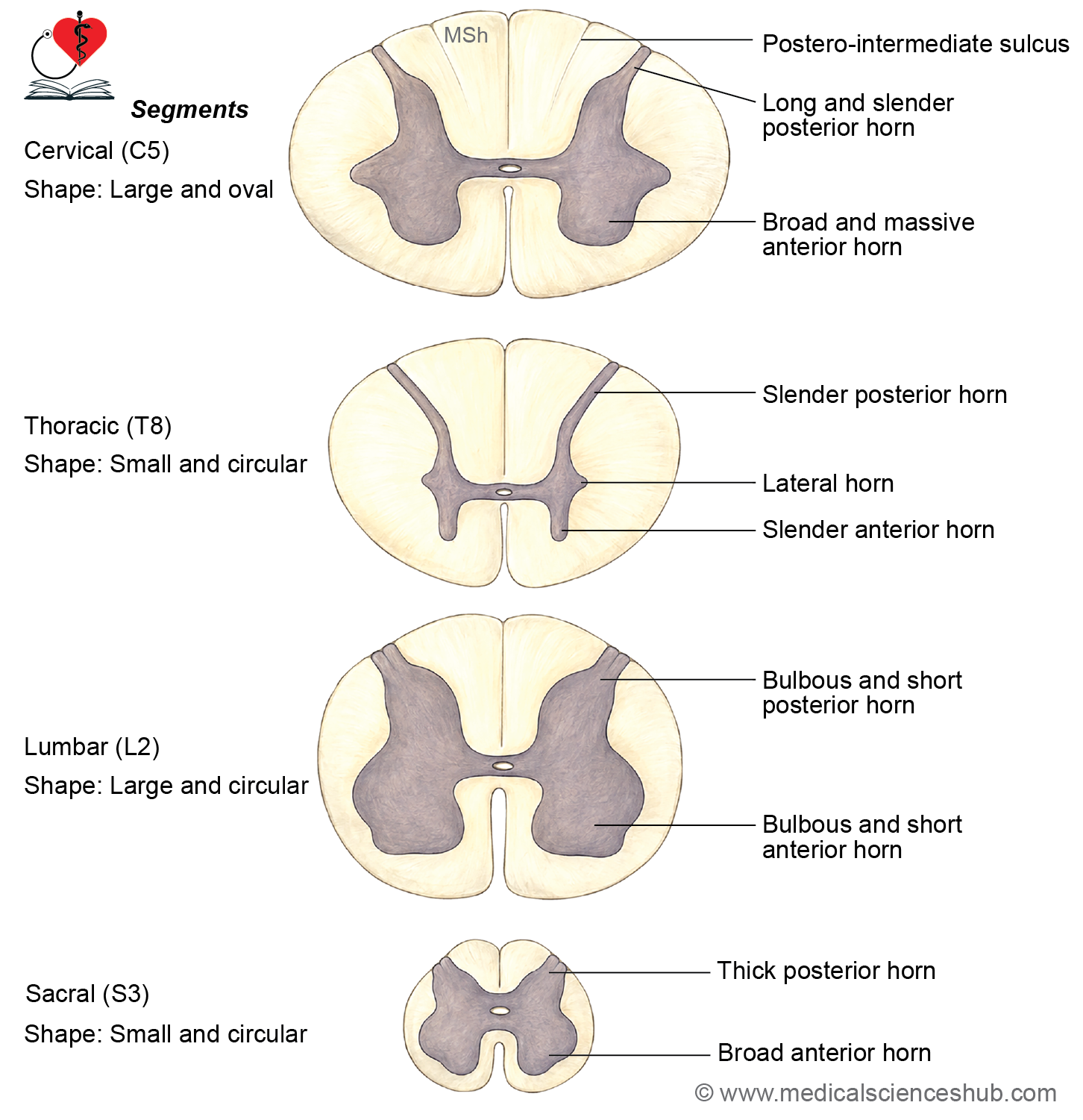
Table 23.1: Regional characteristics of spinal gray matter
| Spinal Cord Region | Shape of Transverse Section | Anterior (Ventral) Horn | Lateral Horn | Posterior (Dorsal) Horn | Amount of White Matter | Posterior Intermediate Sulcus |
|---|---|---|---|---|---|---|
| Cervical | Oval | Narrow in upper cervical segments (C1–C3); enlarged in lower cervical segments (C4–C8) due to the cervical enlargement | Absent | Long and slender | Greatest amount | Present |
| Thoracic | Small and nearly circular | Relatively slender, except at T1 where it is more prominent | Present, especially from T1 to L2 | Long and slender | Abundant | Present |
| Lumbar | Large and circular | Broad and well developed due to the lumbar enlargement | Present mainly in L1–L2 segments | Broad | Reduced compared with cervical and thoracic regions | Absent |
| Sacral | Small and circular | Broad | Present in parasympathetic segments (S2–S4) | Thick and prominent | Minimal | Absent |
Cerebellum
- The cerebellum (“small brain”) is extensively folded, allowing a large number of neurons to be packed into a limited space.
- Its surface folds are called folia, which are separated by deep fissures.
- The internal white matter exhibits a characteristic branching pattern known as the arbor vitae (“tree of life”).
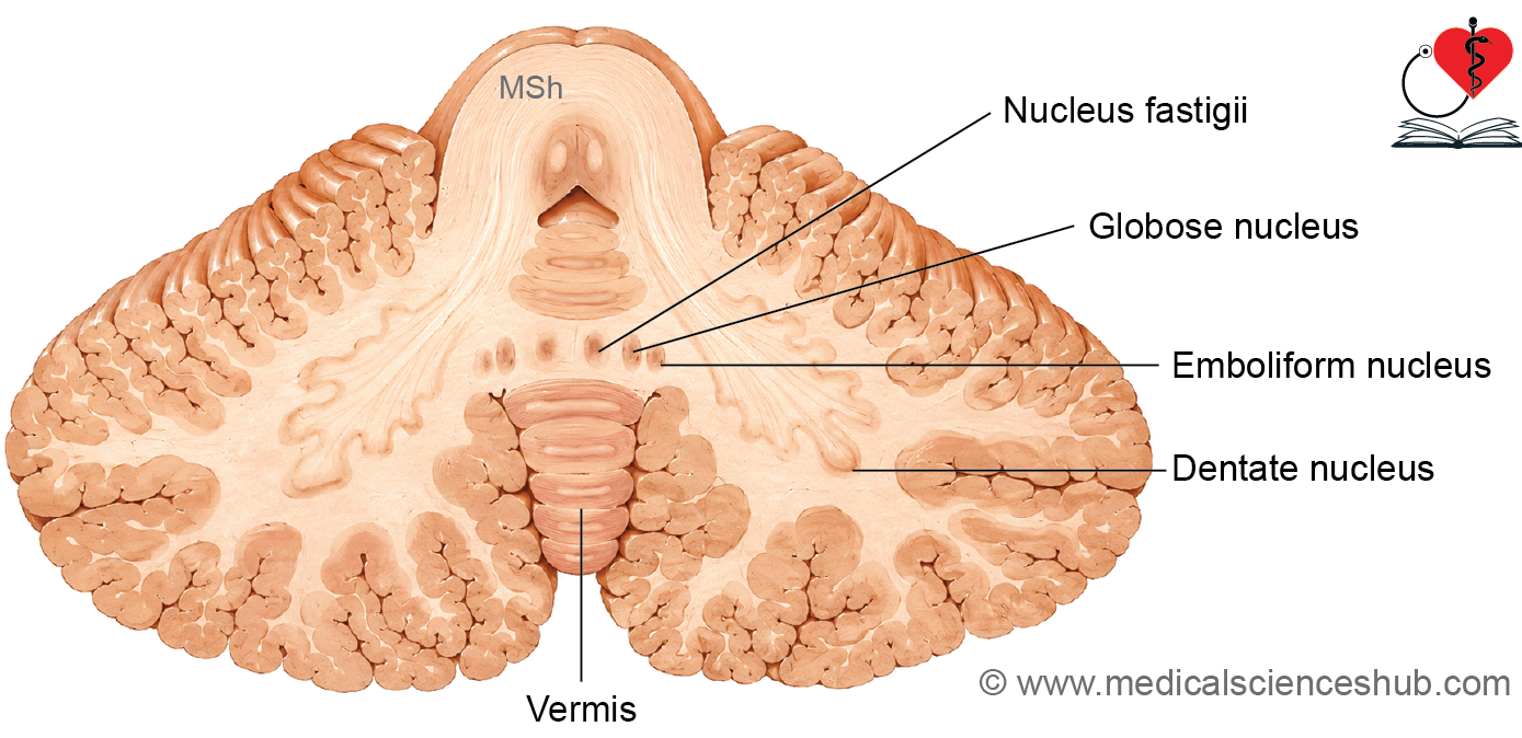
- The cerebellum consists of two cerebellar hemispheres connected in the midline by the vermis.
- Deep fissures subdivide the cerebellum into several lobules.
Histology of Cerebellum
- The cerebellar structure is remarkably uniform throughout and is described as homotypical.
- It is composed of:
- An outer cerebellar cortex (gray matter)
- An inner white matter core
- Embedded within the white matter are four pairs of deep cerebellar nuclei:
- Dentate nucleus
- Globose nucleus
- Emboliform nucleus
- Fastigial nucleus
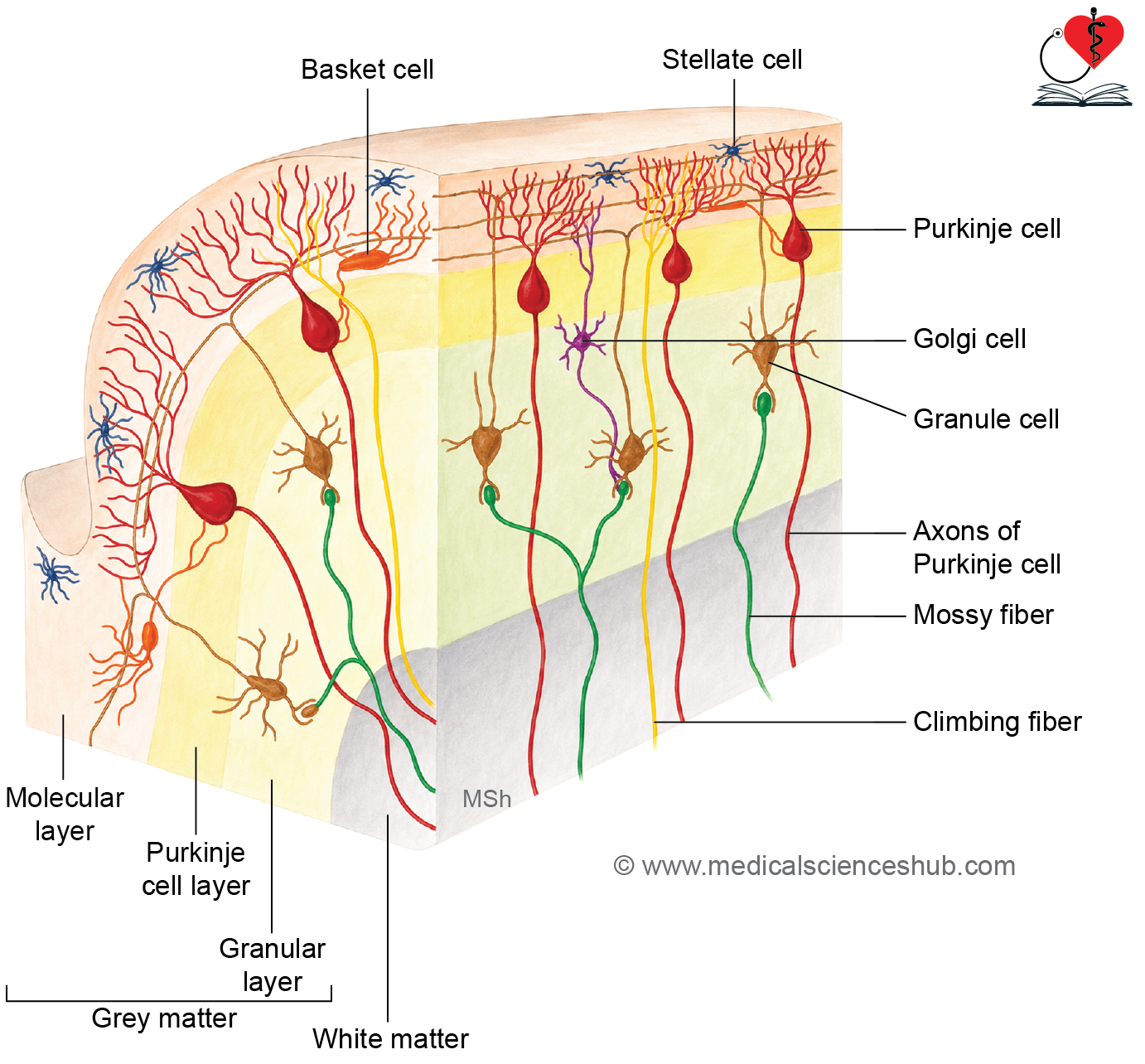
- The cerebellar cortex has three distinct layers:
- Molecular layer (outer)
- Purkinje cell layer (middle)
- Granular layer (inner)
- The presence of these three cortical layers is a key histological feature of the cerebellum.
Molecular Layer
- The molecular layer lies immediately beneath the pia mater and appears lightly stained on H&E sections.
- It contains relatively few cell bodies but numerous nerve fibers.
- The principal cells are stellate cells in the superficial region and basket cells in the deeper region.
- This layer contains extensive dendritic branches of Purkinje cells, parallel fibers derived from granule cell axons, and climbing fibers.
Purkinje Cell Layer
- The Purkinje cell layer consists of a single row of large, flask-shaped Purkinje neurons situated between the molecular and granular layers.
- Dendrites arise from the apex of each cell and branch extensively within the molecular layer.
- A single axon emerges from the base, traverses the granular layer and white matter, and terminates in the deep cerebellar nuclei.
- Purkinje cells constitute the sole output neurons of the cerebellar cortex.
Granular Layer
- The granular layer lies between the Purkinje cell layer and white matter and stains deeply because of its densely packed cells.
- It contains numerous granule cells and larger Golgi cells.
- Granule cell axons ascend into the molecular layer and form parallel fibers.
- Mossy fibers terminate within this layer and synapse with granule cells.
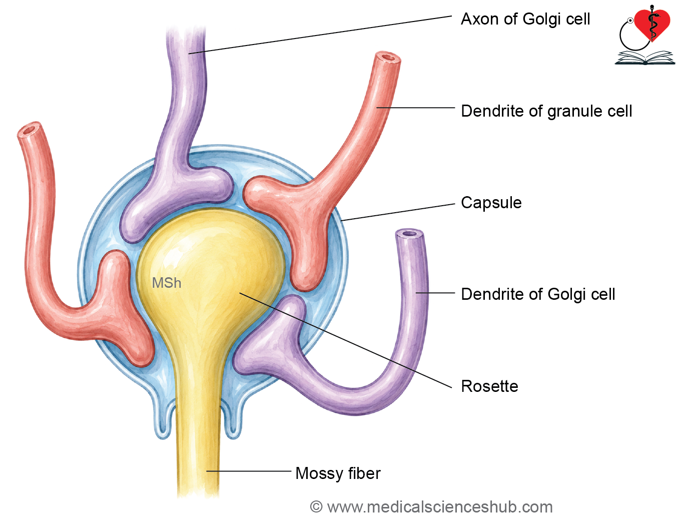
- Characteristic pale areas called cerebellar glomeruli are formed by mossy fiber terminals surrounded by granule cell dendrites and Golgi cell processes.
White Matter
- The cerebellar white matter consists mainly of myelinated nerve fibers and supporting neuroglial cells.
- It forms the central core of the cerebellum and continues into the deep cerebellar nuclei.
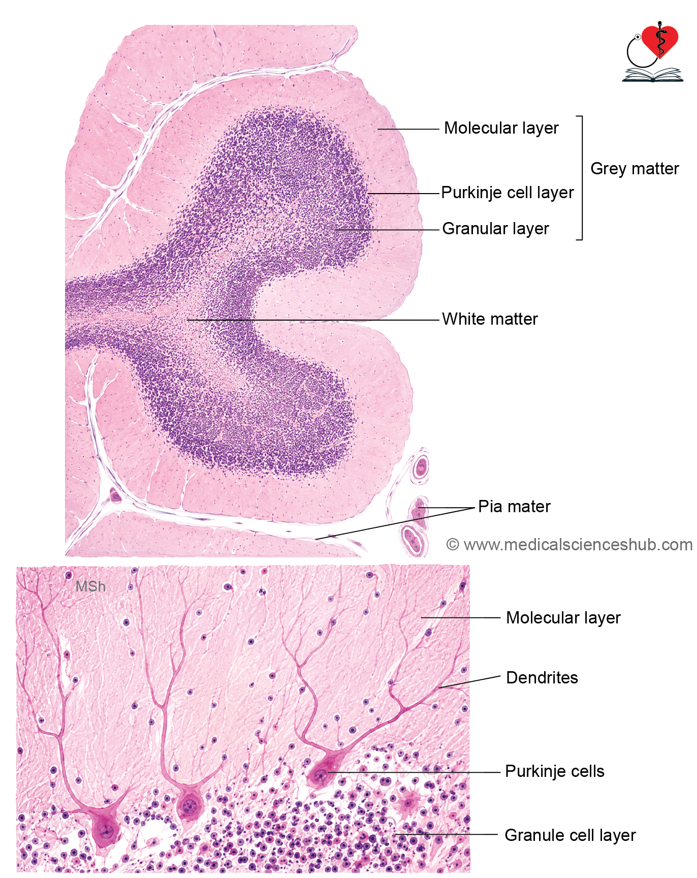
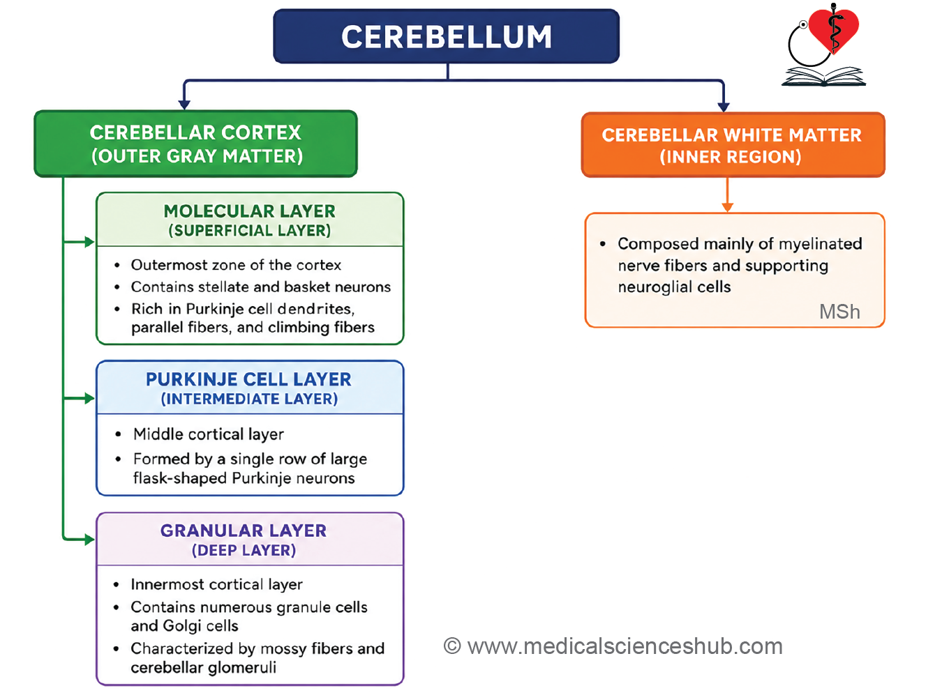
Table 23.2: Cells of cerebellum
| Cell Type | Location | Morphological Features | Dendrites | Axon |
|---|---|---|---|---|
| Cell Type | Superficial part of the molecular layer | Small interneurons | Remain within the molecular layer and receive input from parallel fibers of granule cells | Confined to the molecular layer; form inhibitory synapses on the dendrites of Purkinje cells |
| Stellate Cells | Deeper region of the molecular layer | Small neurons with a relatively small nucleus | Receive synaptic input from parallel fibers; interact with Purkinje cells and other cerebellar afferent fibers | Axons form basket-like networks around Purkinje cell bodies and exert inhibitory effects |
| Basket Cells | Purkinje cell layer, between the molecular and granular layers | Large flask-shaped neurons; principal output cells of the cerebellar cortex | Extensively branched dendritic tree projecting into the molecular layer | Single axon projects to the deep cerebellar nuclei |
| Purkinje Cells | Granular layer | Small, densely packed spherical neurons; most numerous neurons in the cerebellum | Short dendrites participate in cerebellar glomeruli and receive input from mossy fibers | Axons ascend to the molecular layer and bifurcate into parallel fibers |
| Granule Cells | Superficial portion of the granular layer | Large stellate inhibitory neurons; predominantly GABAergic | Extend into the molecular layer and receive synaptic input from parallel fibers | Axons terminate within cerebellar glomeruli of the granular layer |
Afferent Fibers of Cerebellum
- The cerebellum receives sensory and motor information through two major afferent fiber systems: climbing fibers and mossy fibers.
Climbing Fibers
- Climbing fibers originate from the inferior olivary nucleus of the medulla.
- They enter the cerebellar cortex and form powerful synaptic connections with the dendrites of Purkinje cells in the molecular layer.
- Each Purkinje cell is typically innervated by a single climbing fiber.
Mossy Fibers
- Mossy fibers include all cerebellar afferents other than olivocerebellar fibers.
- They arise from sources such as the vestibulocerebellar, pontocerebellar, and spinocerebellar pathways.
- Mossy fibers terminate in the granular layer, where they form enlarged endings called rosettes.
- These terminals synapse with dendrites of granule cells and processes of Golgi cells.
- The synaptic complex formed by mossy fiber rosettes, granule cell dendrites, and Golgi cell processes constitutes a cerebellar glomerulus, which appears as a pale-staining area in the granular layer.
Cerebrum
- The cerebrum consists of an outer gray matter layer, known as the cerebral cortex, and an inner white matter core.
- The cerebral cortex contains neuronal cell bodies, neuroglial cells, nerve processes, blood vessels, and numerous synapses that facilitate information processing.
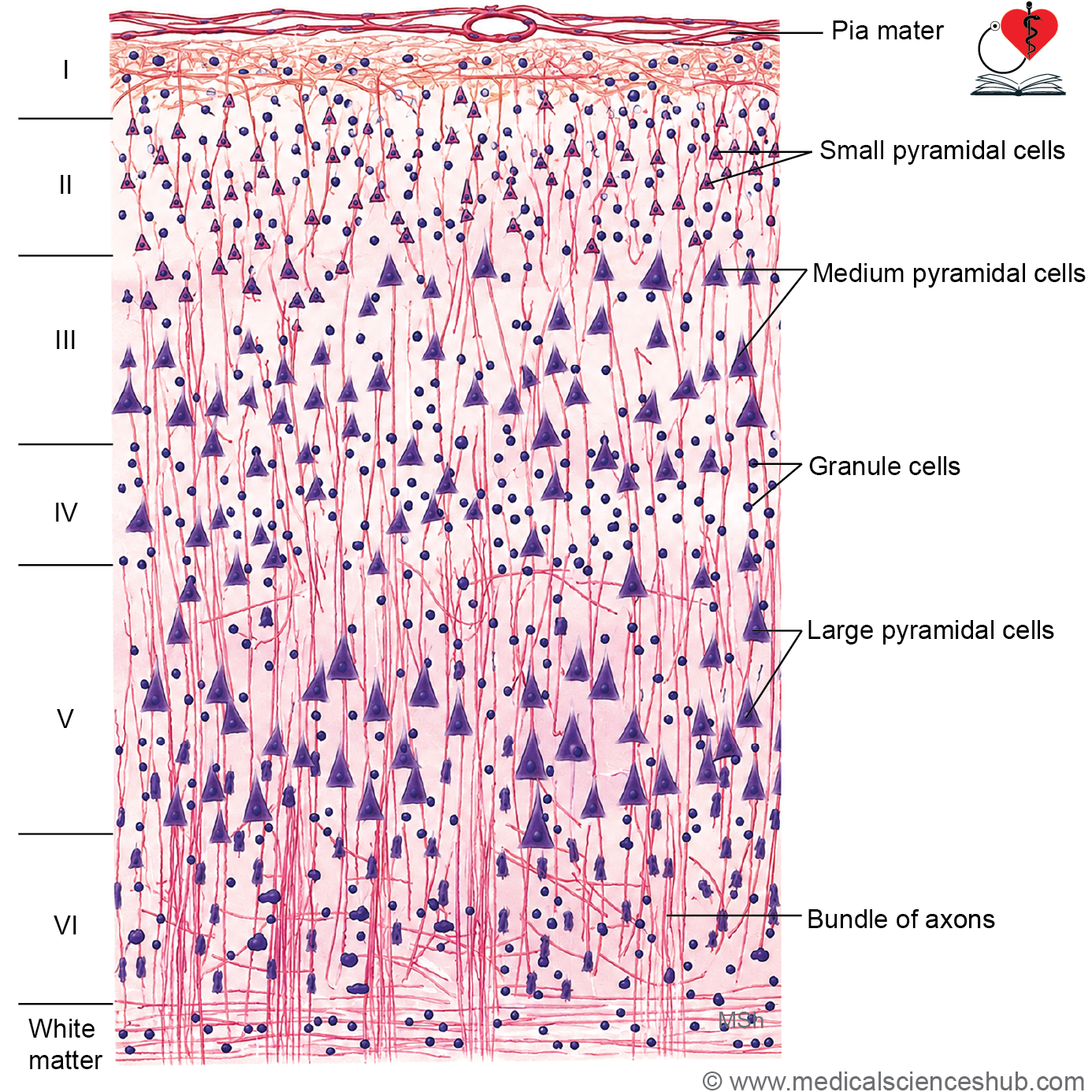
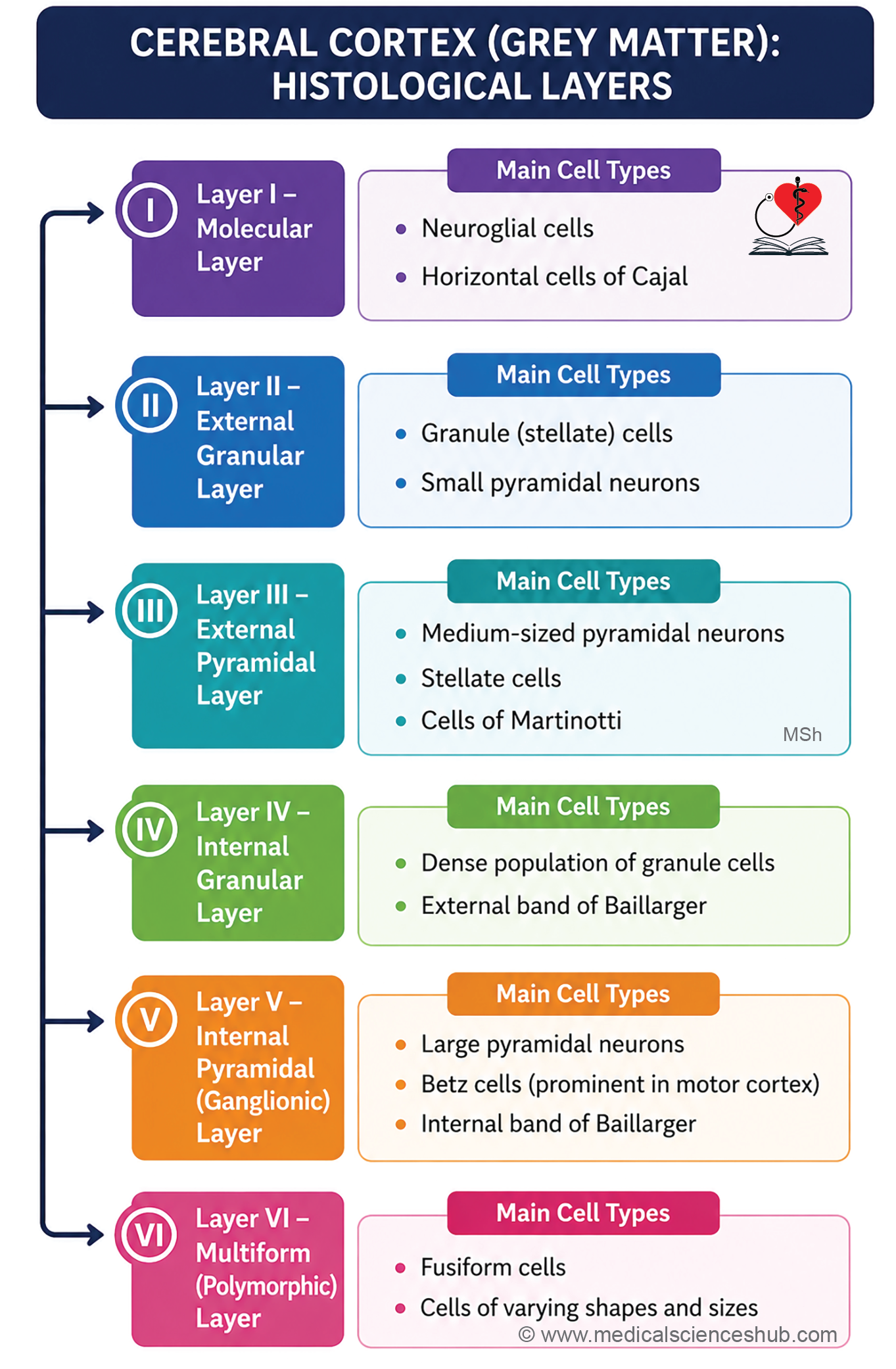
- Histologically, the cerebral cortex is organized into six distinct layers.
Neurons of Cerebral Cortex
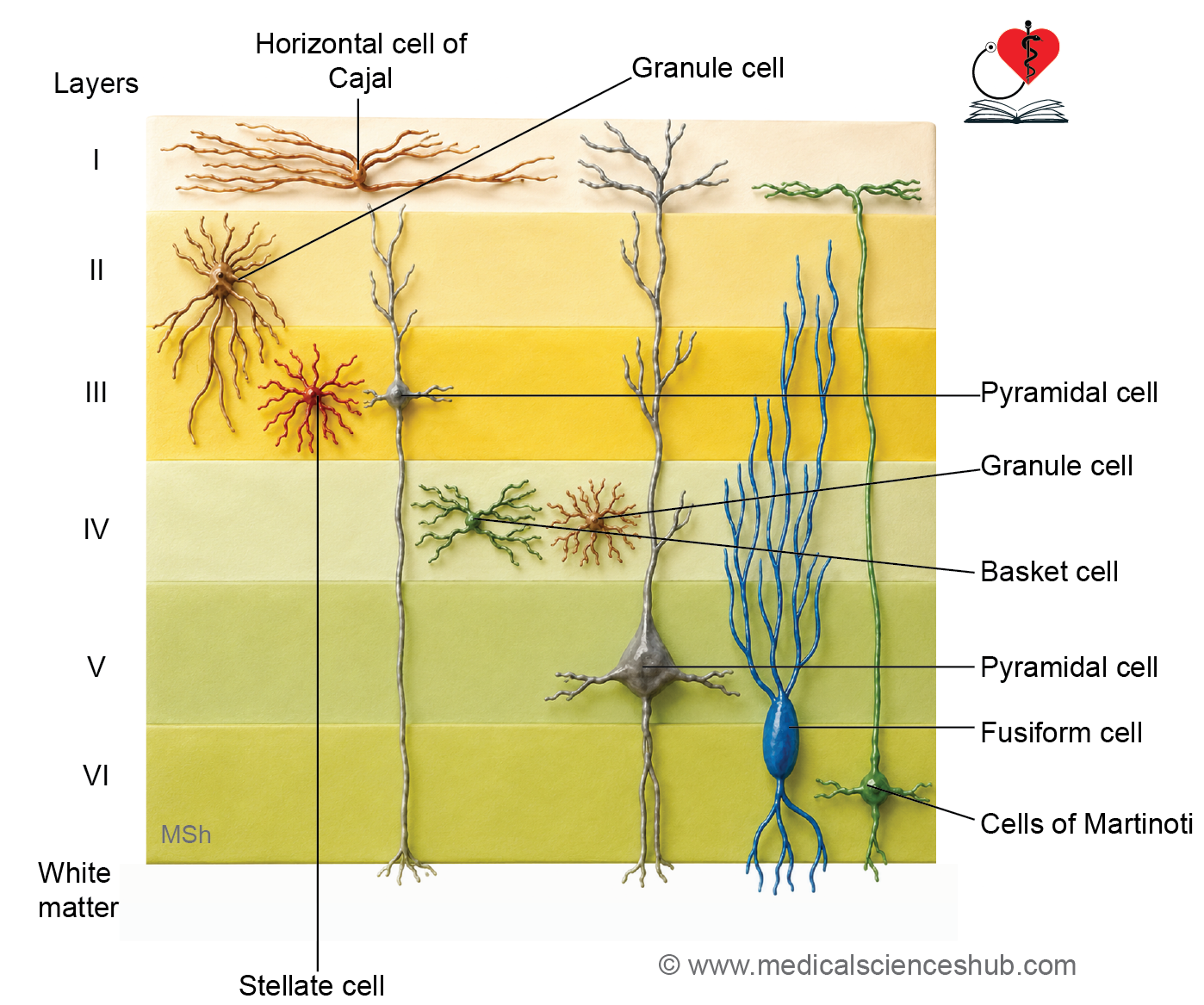
The cortex contains several neuronal types, including pyramidal cells, stellate cells, Martinotti cells, basket cells, fusiform cells, horizontal cells of Cajal, double bouquet cells, and chandelier cells.
Pyramidal CellsIdentification feature
- Pyramidal cells are the most abundant cortical neurons, constituting nearly two-thirds of the neuronal population.
- They are multipolar, triangular-shaped neurons with the apex directed toward the cortical surface.
- Their processes include:
- An apical dendrite extending toward the cortical surface.
- Multiple basal dendrites arising from the base of the cell body.
- A single axon originating from the basal region.
- These neurons serve as the principal output cells of the cerebral cortex.
Martinotti Cells
- Martinotti cells are small triangular or polygonal interneurons.
- They are commonly located in the external pyramidal layer.
- Their axons ascend toward the superficial layers of the cortex.
Stellate Neurons (Granule Cells)
- Stellate cells are small, star-shaped multipolar neurons.
- Because of their granular appearance, they are also referred to as granule cells.
- They function mainly as local interneurons within the cerebral cortex.
Layer of Cerebral Cortex
The cerebral cortex is organized into six distinct layers, each containing characteristic neuronal populations and fiber arrangements.
I. Molecular or Plexiform Layer
- This superficial layer lies immediately beneath the pia mater.
- It contains numerous nerve fibers, neuroglial processes, and a few capillaries.
- Horizontal cells of Cajal are present in this layer.
- Cell bodies are sparse, making the layer appear relatively cell-poor.
II. External Granular Layer
- This layer contains mainly stellate (granule) cells and small pyramidal neurons.
- It participates in local cortical processing and interconnections.
III. External Pyramidal Layer
- This layer is composed predominantly of medium-sized pyramidal cells, along with stellate and Martinotti cells.
- Axons from pyramidal neurons contribute to association and commissural fibers, connecting different cortical regions.
IV. Internal Granular Layer
- This densely packed layer contains numerous small stellate cells.
- It receives significant sensory input from the thalamus.
- A horizontal band of myelinated fibers, known as the external band of Baillarger, is present within this layer.
V. Internal Pyramidal Layer
- This layer contains large pyramidal neurons.
- In the motor cortex, it houses giant Betz cells, which are among the largest neurons in the central nervous system.
- It also contains Martinotti, fusiform, and stellate cells.
- The internal band of Baillarger is formed by horizontally arranged fibers within this layer.
VI. Multiform Layer/Layer of Polymorphic/ Fusiform Cells
- This deepest cortical layer contains fusiform cells, Martinotti cells, and a few stellate neurons.
- Fusiform cells are spindle-shaped and oriented perpendicular to the cortical surface, with dendrites extending from both ends.
White Matter
- Deep to the sixth cortical layer lies the white matter.
- It consists predominantly of myelinated nerve fibers, neuroglial cells, and axonal processes that connect cortical and subcortical regions.
Important Questions
- Draw a well-labelled diagram of histology of spinal cord.
- Draw a well-labelled diagram of histology of cerebellum.
- List the cells of cerebellar cortex.
- Draw a well-labelled diagram of histology of cerebral cortex.
📝 Test Your Knowledge – Practice MCQs
Attempt the chapter MCQ quiz and assess your understanding of key concepts.
Click to see: Medical Disclaimer and Copyright Notice