
Competency
- AN43.3 Identify, describe, and draw microanatomy of cochlea– organ of Corti.
Introduction
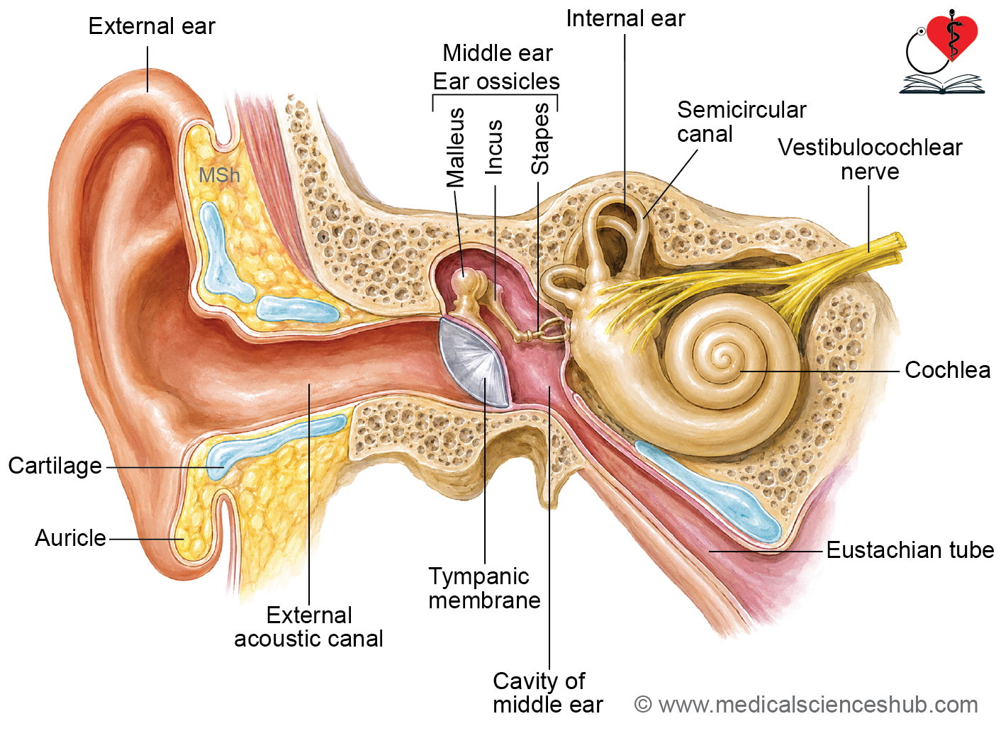
- The ear is the sensory organ responsible for hearing and maintenance of equilibrium (balance).
- It is divided into three parts: external ear, middle ear, and internal ear.
External Ear
- The external ear consists of the auricle (pinna) and the external acoustic meatus.
- The auricle contains irregularly shaped elastic cartilage covered by thin, tightly adherent skin.
- Its skin contains hair follicles, sebaceous glands, and sweat glands.
- The auricle collects and directs sound waves toward the auditory canal.
External Acoustic Meatus
- The external acoustic meatus is a tubular passage extending from the concha to the tympanic membrane.
- It has two parts:
- Outer one-third: cartilaginous portion.
- Inner two-thirds: bony portion.
- The canal is lined by thin skin.
- It contains ceruminous glands, which are modified apocrine sweat glands that secrete cerumen (earwax).
- Cerumen helps protect the canal by trapping dust and foreign particles.
- The tympanic membrane (eardrum) separates the external ear from the middle ear and transmits sound vibrations to the auditory ossicles.
Middle Ear
- The middle ear is an air-filled cavity located within the petrous part of the temporal bone.
- It is lined mainly by simple cuboidal to ciliated columnar epithelium.
- The middle ear contains three auditory ossicles:
- Malleus
- Incus
- Stapes
- These ossicles transmit sound vibrations from the tympanic membrane to the inner ear.
- Two small skeletal muscles, the tensor tympani and stapedius, regulate ossicular movement and help protect the inner ear from excessive sound vibrations.
- The auditory (Eustachian) tube connects the middle ear to the nasopharynx and helps equalize air pressure across the tympanic membrane.
Internal Ear
- The internal ear is situated within the petrous temporal bone and contains the organs of hearing and balance.
- It is composed of:
- Bony labyrinth
- Membranous labyrinth
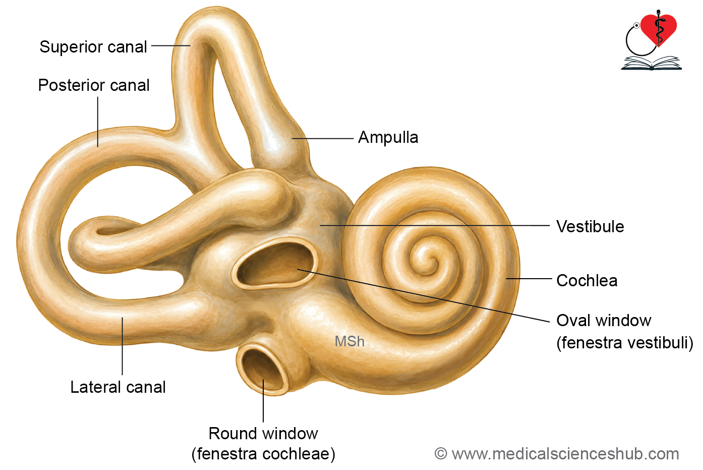
- The bony labyrinth consists of three regions:
- Cochlea
- Vestibule
- Semicircular canals
- The membranous labyrinth is a system of interconnected ducts and sacs located within the bony labyrinth and contains the sensory receptors for hearing and equilibrium.
Tympanic membrane
- The tympanic membrane (eardrum) is a thin structure that separates the external acoustic meatus from the middle ear cavity.
- It is oval in shape and measures approximately 9–10 mm in diameter.
- The membrane has an outer and an inner surface.
Histological Layers
- Outer cutaneous layer: Continuous with the skin of the external auditory canal.
- Middle fibrous layer: Provides strength and elasticity; contains superficial radial and deep circular collagen fibers.
- Inner mucosal layer: Lined by ciliated simple columnar epithelium, continuous with the mucosa of the middle ear.
- The tympanic membrane transmits sound vibrations from the external ear to the auditory ossicles of the middle ear.
Internal Ear
- The internal ear is located within the petrous part of the temporal bone and contains the sensory organs for hearing and balance.
- It consists of two major components:
- Bony labyrinth
- Membranous labyrinth
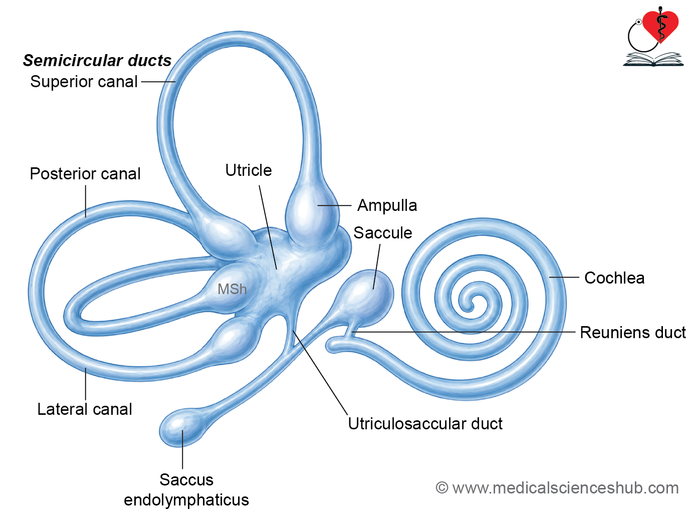
- The membranous labyrinth includes the cochlear duct, semicircular ducts, utricle, saccule, ductus reuniens, and the endolymphatic duct and sac.
- It contains endolymph, while the space between the membranous and bony labyrinths contains perilymph.
Bony Labyrinth
The bony labyrinth is composed of three parts:
- Cochlea (anterior)
- Vestibule (central)
- Semicircular canals (posterior)
Cochlea
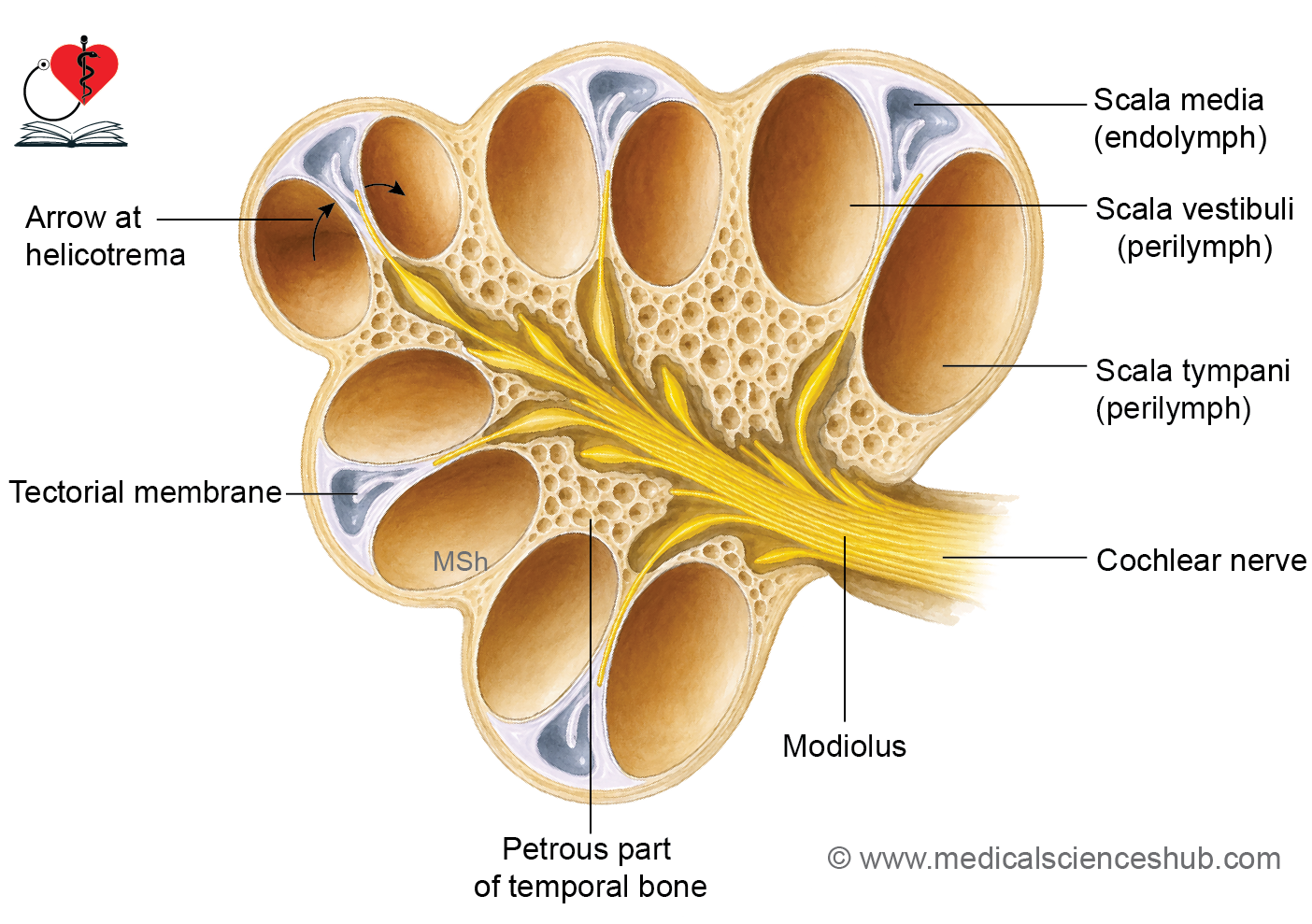
- The cochlea is a cone-shaped, spiral bony canal resembling a snail shell.
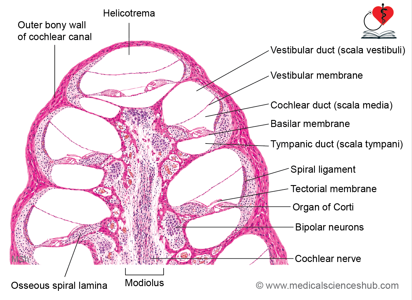
- It winds around a central bony axis called the modiolus, completing approximately 2¾ turns.
- A bony projection, the spiral lamina, extends from the modiolus and partially divides the cochlear canal.
- This division forms the scala vestibuli above and the scala tympani below, both filled with perilymph.
- The basilar membrane completes the separation between these chambers.
- At the apex of the cochlea, the scala vestibuli and scala tympani communicate through the helicotrema.
- The cochlear nerve emerges from the base of the cochlea and carries auditory information to the brain.
Vestibule
- The vestibule is the central chamber of the bony labyrinth and connects the cochlea with the semicircular canals.
- It communicates with the middle ear through two openings:
- Oval window (fenestra vestibuli), occupied by the footplate of the stapes.
- Round window (fenestra cochleae), closed by the secondary tympanic membrane.
- The vestibule also contains the opening of the vestibular aqueduct, through which the endolymphatic duct passes.
Semicircular Canals
- The bony labyrinth contains three semicircular canals:
- Superior (anterior)
- Posterior
- Lateral (horizontal)
- Each canal has an expanded end called the ampulla, which contains sensory structures for balance.
- The canals open into the vestibule through five openings.
- They are arranged approximately at right angles to one another, allowing detection of rotational movements of the head in different planes.
Membranous Labyrinth
- The membranous labyrinth is a system of membrane-lined chambers and ducts filled with endolymph.
- It contains the sensory receptors responsible for hearing and balance:
- Organ of Corti for hearing.
- Maculae for static equilibrium and linear acceleration.
- Cristae ampullares for dynamic equilibrium and rotational movements.
Components of the Membranous Labyrinth
- Cochlear duct (scala media) within the cochlea.
- Utricle and saccule, which contain maculae and lie within the vestibule.
- Semicircular ducts, which contain cristae and lie within the bony semicircular canals.
Duct of Cochlea/Scala Media
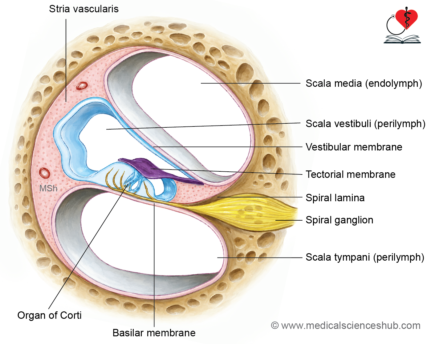
- The cochlear duct occupies the space between the scala vestibuli and scala tympani.
- In cross-section, it is triangular and contains endolymph.
- Its walls are:
- Floor: Basilar membrane.
- Roof: Vestibular (Reissner’s) membrane.
- Lateral wall: Cochlear wall containing the stria vascularis.
- The basilar membrane supports the Organ of Corti, the receptor organ for hearing.
- Sensory cells of the Organ of Corti are innervated by peripheral processes of bipolar neurons located in the spiral ganglion. Their central processes form the cochlear nerve.
- The vestibular membrane separates the cochlear duct from the scala vestibuli.
- The basilar membrane extends from the spiral lamina to the spiral ligament, a thickened connective tissue structure of the lateral cochlear wall.
- The cochlear duct communicates with the saccule through the narrow ductus reuniens.
- The stria vascularis is a highly vascular epithelium that produces and maintains the ionic composition of endolymph.
Saccule and Utricle
- The saccule and utricle are components of the membranous labyrinth and are involved in maintaining balance.
- Their walls consist of a thin connective tissue layer lined by simple squamous or simple cuboidal epithelium.
- Both structures are located within the vestibule of the inner ear.
- The saccule communicates with the cochlear duct through the ductus reuniens.
- The utricle connects with the semicircular ducts through five openings.
- The ducts of the saccule and utricle unite to form the endolymphatic duct, which expands distally as the endolymphatic sac.
- The medial walls of the saccule and utricle contain specialized sensory areas called maculae. Maculae are innervated by fibers from the vestibular ganglion. They detect linear acceleration and changes in head position relative to gravity.
Semicircular Ducts
- Three semicircular ducts lie within the superior, posterior, and lateral semicircular canals.
- Each duct has a dilated end called an ampulla, which contains a sensory structure known as the crista ampullaris.
- The ducts are oriented approximately at right angles to one another.
- This arrangement enables detection of angular (rotational) acceleration of the head, helping maintain dynamic equilibrium.
Organ of Corti
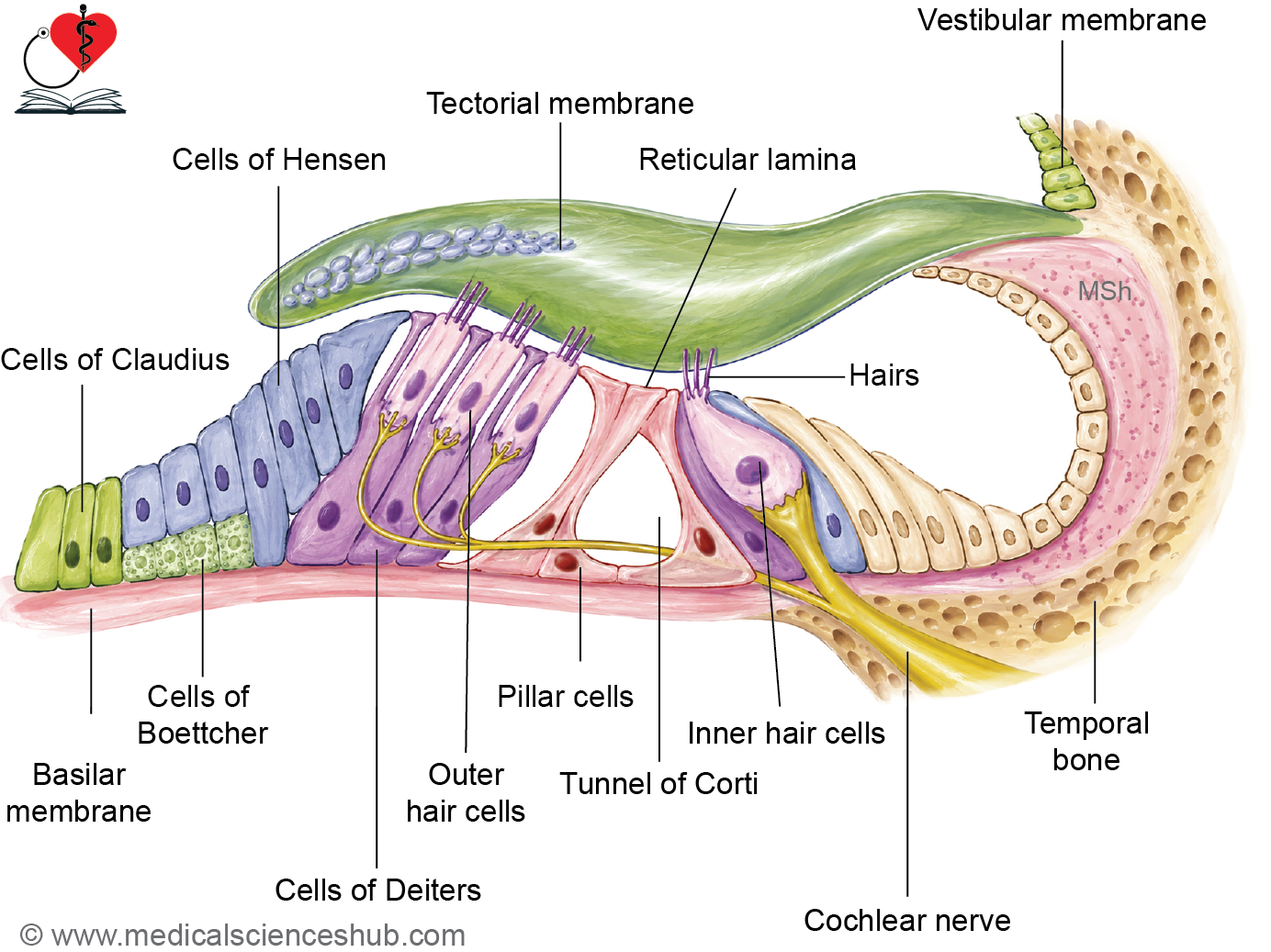
- The Organ of Corti is the specialized sensory organ responsible for hearing.
- It is located on the basilar membrane within the cochlear duct and extends throughout the spiral turns of the cochlea.
- Its main components are supporting cells, hair cells, and the tectorial membrane.
Tectorial Membrane
- The tectorial membrane is a gelatinous, glycoprotein-rich structure that overlies the Organ of Corti.
- The tips of the stereocilia of hair cells project into or contact this membrane, allowing mechanical stimulation during sound transmission.
Supporting Cells
- Supporting cells provide structural stability and maintain the architecture of the Organ of Corti.
- They include:
- Inner and outer pillar (rod) cells
- Deiters’ (phalangeal) cells
- Hensen’s cells
- Claudius cells
- Boettcher cells
- Inner and outer pillar cells form the Tunnel of Corti, a fluid-filled space containing cortilymph.
- The apical processes of pillar cells contribute to the formation of the reticular lamina.
- Deiters’ cells support the outer hair cells, while Hensen’s and Claudius cells provide additional lateral support.
Hair Cells
- Hair cells are the sensory receptor cells of hearing.
- They are columnar to flask-shaped cells supported by adjacent supporting cells and do not directly contact the basilar membrane.
- Their apical surfaces contain a cuticular plate and bundles of stereocilia, which are essential for mechanotransduction.
- Each hair cell possesses approximately 30–100 stereocilia arranged in rows of increasing height.
- Hair cells are organized into:
- One row of inner hair cells
- Three to four rows of outer hair cells
- Sensory nerve endings from bipolar neurons of the spiral ganglion terminate on these hair cells and transmit auditory information to the brain.
Spiral Limbus
- The spiral limbus is a connective tissue ridge projecting from the modiolus into the cochlear duct.
- It serves as an attachment site for the tectorial membrane, helping maintain the structural organization necessary for sound perception.
Macula
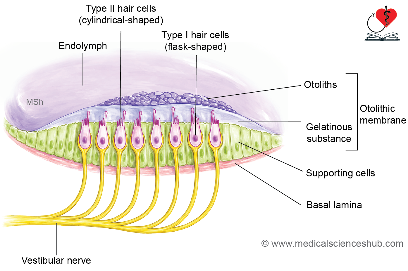
- Maculae are specialized sensory regions located in the utricle and saccule, collectively known as the otolith organs.
- The sensory areas are termed macula utriculi and macula sacculi.
- A curved central region called the striola divides each macula into two functional zones.
- Each macula consists of:
- Sensory neuroepithelium
- Otolithic membrane
- Each macula consists of:
Sensory Neuroepithelium
- It contains hair cells and supporting cells.
- Supporting cells are tall columnar cells that secrete the gelatinous otolithic membrane.
- Hair cells possess numerous stereocilia and a single longer kinocilium on their apical surface.
- Both stereocilia and kinocilium project into the overlying otolithic membrane.
- Two types of hair cells are present:
- Type I hair cells: Flask-shaped.
- Type II hair cells: Columnar in shape.
Otolithic Membrane
- The otolithic membrane is a gelatinous layer covering the sensory epithelium.
- It contains otoliths (otoconia), which are microscopic crystals of calcium carbonate embedded in a protein-rich matrix.
- These crystals increase the membrane’s weight and sensitivity to gravitational forces and linear movements.
Functions of Maculae
- Maculae are receptors for static equilibrium.
- They detect the position of the head relative to gravity and sense linear acceleration, such as forward, backward, or vertical movements.
- Through connections with the vestibular system, they contribute to the maintenance of posture and balance.
- The saccular macula may also have a limited role in detecting low-frequency sound vibrations.
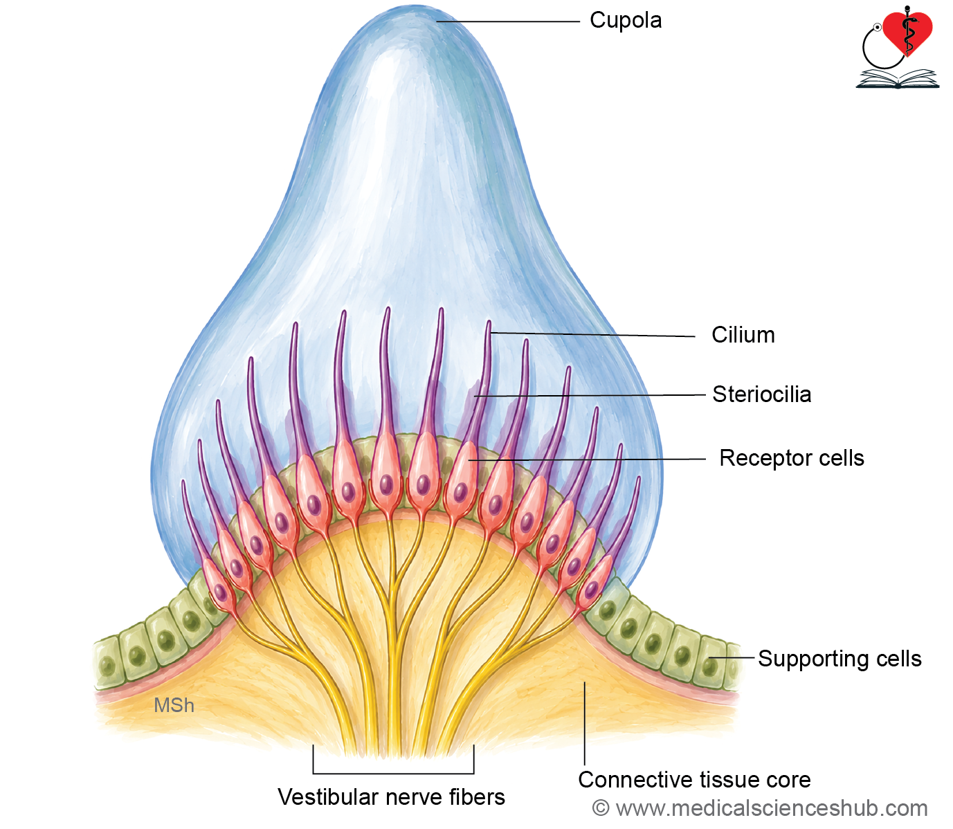
Ampullary crests
- Ampullary crests (cristae ampullares) are specialized sensory structures located within the ampulla of each semicircular duct.
- The long axis of each crest is oriented perpendicular to the long axis of the corresponding semicircular duct.
- Each crest is covered by sensory epithelium containing hair cells and supporting cells.
Hair Cells
- Hair cells are the sensory receptors for detecting rotational movements of the head.
- Their apical surface bears:
- A single kinocilium
- Multiple stereocilia
- The stereocilia and kinocilium project into a gelatinous structure called the cupula.
- Two types of hair cells are present:
- Type I hair cells: Flask-shaped.
- Type II hair cells: Columnar.
- Each hair cell receives afferent nerve endings from the vestibular nerve.
Supporting Cells
- Supporting (sustentacular) cells are elongated cells that surround and support the hair cells.
- They provide structural support, metabolic nourishment, and help maintain the composition of endolymph.
Functions of Ampullary Crests
- Ampullary crests are receptors for dynamic equilibrium.
- During head rotation, inertia causes movement of endolymph within the semicircular ducts.
- This fluid movement deflects the cupula, bending the stereocilia and kinocilium of hair cells.
- Mechanical deformation of hair cells generates nerve impulses that inform the brain about angular acceleration and rotational movements of the head.
Important Questions
- Write a short note on organ of Corti.
- List the names of cells of organ of Corti.
📝 Test Your Knowledge – Practice MCQs
Attempt the chapter MCQ quiz and assess your understanding of key concepts.
Click to see: Medical Disclaimer and Copyright Notice