
Competencies
- AN21.1: Identify and describe the salient features of sternum, typical rib, and typical thoracic vertebra.
- AN21.2: Identify and describe the features of 1st, 2nd, 11th, and 12th ribs, and 1st, 11th, and 12th thoracic vertebrae.
This chapter includes features of typical and atypical thoracic vertebrae.
Introduction
- The human vertebral column is also known as the backbone or spine.
- It forms the central supporting axis of the body and provides attachment and support for bones and muscles.
- The thoracic vertebrae include typical vertebrae as well as atypical vertebrae, such as T1, T11, and T12.
Structure
- The vertebral column consists of 33 vertebrae.
- Based on their location, the vertebrae are classified into five regions:
- The cervical region consists of 7 vertebrae.
- The thoracic region consists of 12 vertebrae.
- The lumbar region consists of 5 vertebrae.
- The sacral region consists of 5 fused vertebrae, which together form the sacrum.
- The coccygeal region usually consists of 4 fused vertebrae, which together form the coccyx.
- The upper 24 vertebrae, comprising 7 cervical, 12 thoracic, and 5 lumbar vertebrae, remain separate and are referred to as movable or true vertebrae.
- The lower 9 vertebrae, comprising 5 sacral and 4 coccygeal vertebrae, are fused and are referred to as fixed or false vertebrae.
Length of the Vertebral Column
- The vertebral column measures approximately 70 cm in adult males. It measures approximately 60 cm in adult females. Intervertebral discs contribute approximately one-fifth of the total length of the vertebral column.
Curvatures
- The vertebral column has primary and secondary curvatures in the sagittal plane.
Primary Curvatures
- Primary curvatures are present at birth. They include the thoracic and sacral curvatures. Both primary curvatures are concave anteriorly.
Secondary Curvatures
- Secondary curvatures develop after birth as the child acquires postural control. They include the cervical and lumbar curvatures. Both secondary curvatures are convex anteriorly.
- The cervical curvature develops at about 4–5 months of age, when the infant begins to hold the head upright. The lumbar curvature develops as the child begins to stand and walk, usually between 12 and 18 months of age.
Lateral Curvature
- A slight lateral curvature may be present in the thoracic region. It is commonly described as being convex toward the right, which may be related to greater use of the right upper limb.
Parts of Typical Vertebra
A typical vertebra consists of two main parts:
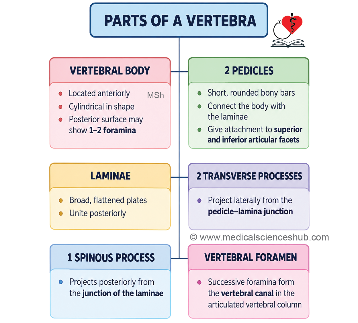
- The body is situated anteriorly.
- The vertebral arch lies posteriorly and consists of two pedicles and two laminae.
Body
- The vertebral body is cylindrical in shape. Its superior and inferior surfaces are rough and are connected to the intervertebral discs.
- The anterior surface is convex from side to side and concave vertically.
- The posterior surface is concave and forms the anterior boundary of the vertebral foramen. The posterior surface contains one or more openings for the basivertebral veins and nutrient vessels.
Vertebral Arch
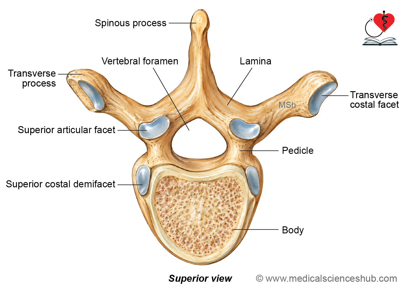
- The vertebral arch is formed by two pedicles and two laminae. It gives rise to the transverse, articular, and spinous processes.
Pedicles
- Each pedicle is a short, rounded bony bar that extends posteriorly from the vertebral body. The pedicles connect the vertebral body with the laminae.
Laminae
- Each lamina is a flat plate of bone extending posteriorly and medially from the pedicle. The right and left laminae unite in the midline posteriorly.
Vertebral Foramen
- The space enclosed by the vertebral body anteriorly and the vertebral arch posteriorly is called the vertebral foramen.
Vertebral Canal
- When the vertebrae are articulated, successive vertebral foramina collectively form the vertebral canal. The vertebral canal contains the spinal cord and its coverings, blood vessels, nerve roots, and associated structures.
Transverse Processes
- Two transverse processes project laterally, one on each side, from the junction of the pedicle and lamina.
Articular Processes
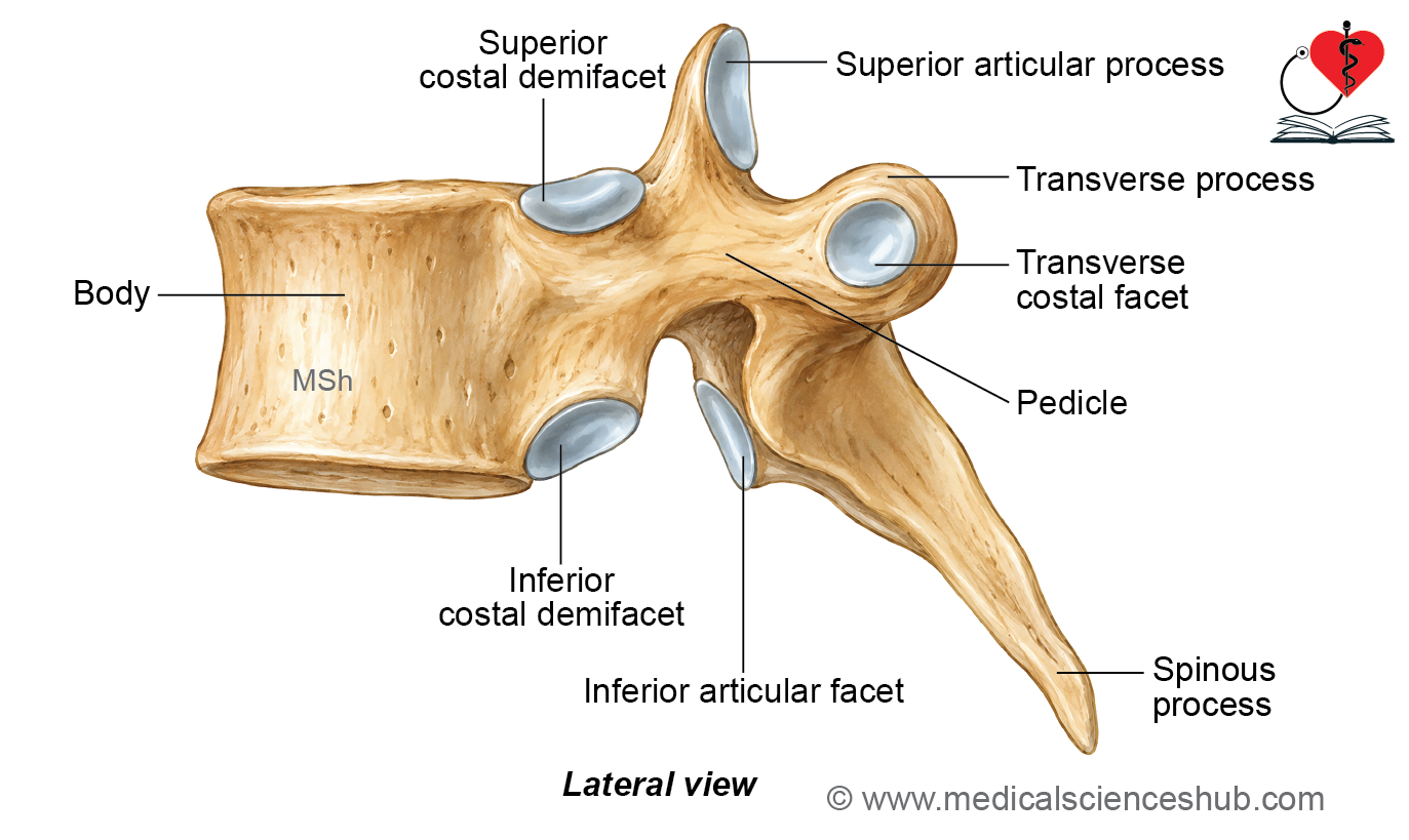
- Four articular processes arise from the junctions of the pedicles and laminae. The two superior articular processes project superiorly and articulate with the vertebra above. The two inferior articular processes project inferiorly and articulate with the vertebra below.
Spinous Process
- The two laminae unite posteriorly in the midline and continue as the spinous process. The spinous process projects posteriorly and provides attachment to muscles and ligaments.
Vertebral Notches
- The superior and inferior borders of each pedicle are notched. These depressions are called the superior and inferior vertebral notches, respectively. The inferior vertebral notch is usually deeper than the superior vertebral notch.
Intervertebral Foramen
In the articulated vertebral column, the inferior vertebral notch of one vertebra and the superior vertebral notch of the vertebra below form an intervertebral foramen. Each intervertebral foramen transmits a spinal nerve and associated blood vessels.
Thoracic Vertebrae
- The thoracic region of the vertebral column consists of 12 vertebrae, designated T1 to T12.
Identification Feature
- The characteristic feature of thoracic vertebrae is the presence of costal facets for articulation with the ribs.
- Costal facets are present on the sides of the vertebral bodies, with additional facets on the transverse processes in most thoracic vertebrae.
Classification
- Thoracic vertebrae are classified into typical and atypical vertebrae.
- Typical thoracic vertebrae include T2–T8.
- Atypical thoracic vertebrae include T1 and T9–T12.
Typical Thoracic Vertebra
- The second to eighth thoracic vertebrae (T2–T8) are considered typical thoracic vertebrae.
Identification Features
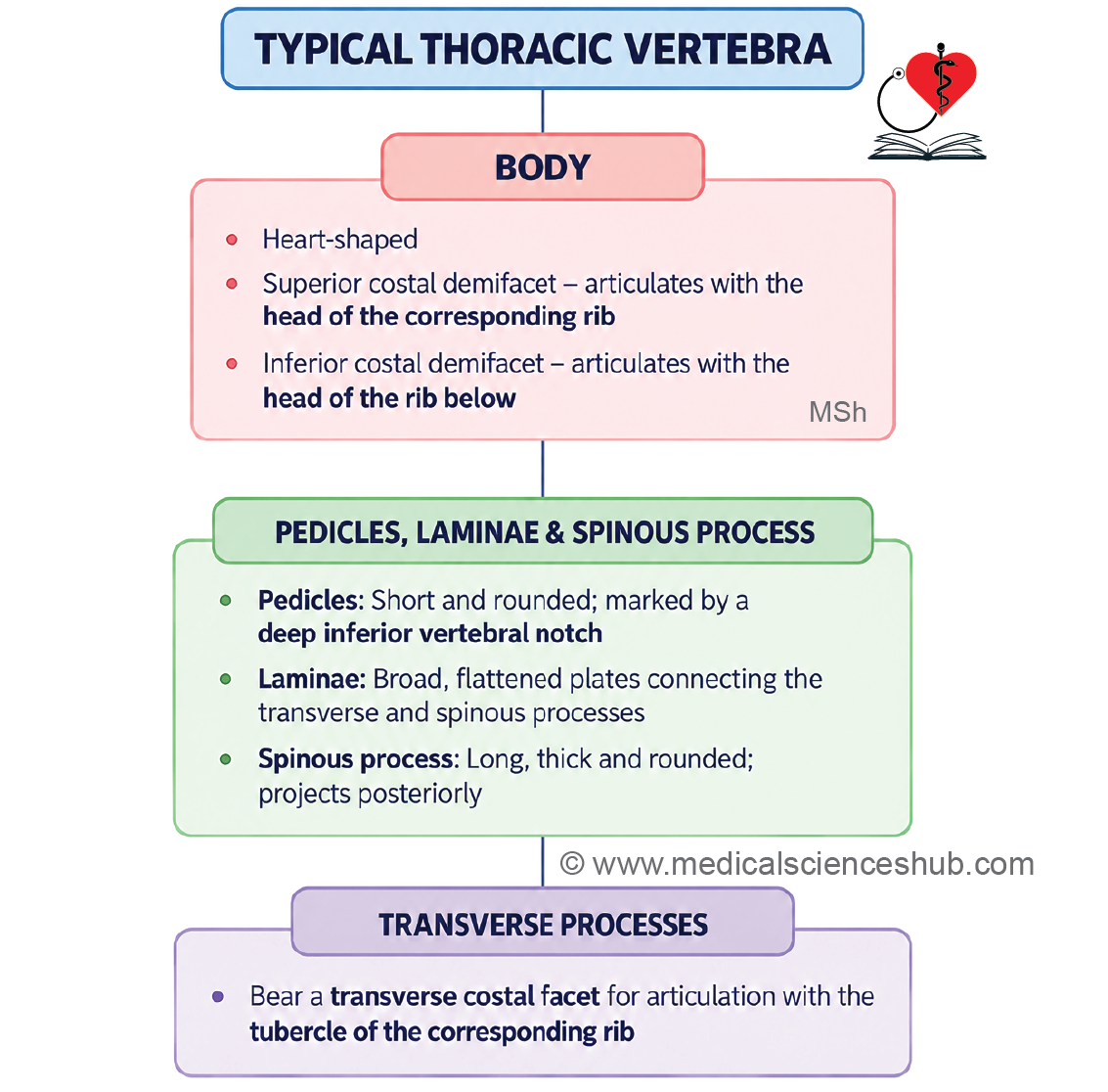
- The body is heart-shaped and bears costal facets for articulation with the ribs.
- The vertebral foramen is small and circular.
- The spinous process is long and slopes downward and backward.
- The pedicles are attached to the upper part of the vertebral body, resulting in a deep inferior vertebral notch.
Features
• The typical thoracic vertebra has body, pedicle, transverse process, vertebral foramen, and spine.
Body
- The body appears heart-shaped when viewed from above. Each side of the vertebral body bears a superior and an inferior costal demifacet.
Costal Facets
- The superior costal demifacet is larger and lies near the upper border of the body, close to the pedicle. It articulates with the head of the numerically corresponding rib.
- The inferior costal demifacet is smaller and lies near the lower border of the body, anterior to the inferior vertebral notch. It articulates with the head of the rib immediately below.
Attachments to the Body
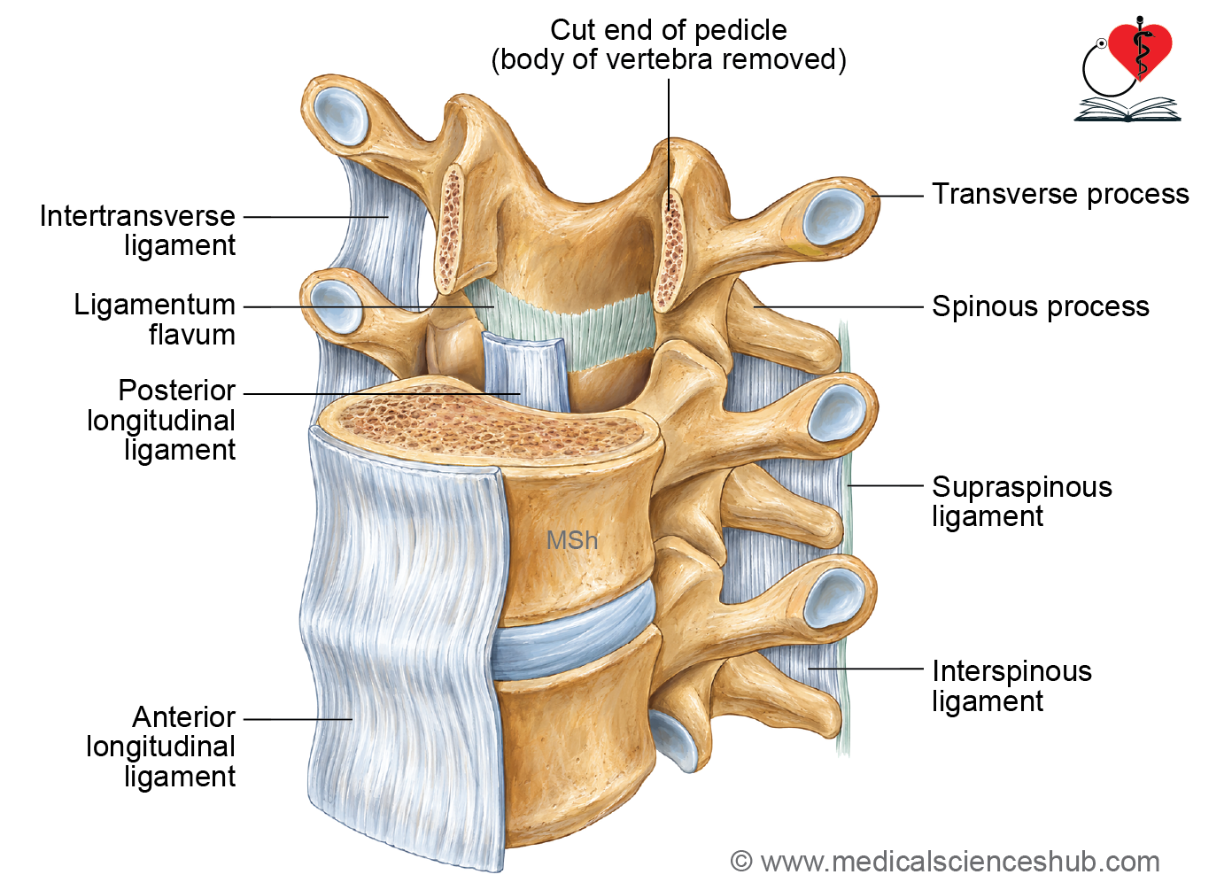
- The anterior longitudinal ligament is attached to the anterior aspect of the vertebral bodies and intervertebral discs.
- The posterior longitudinal ligament is attached along the posterior aspect of the vertebral bodies within the vertebral canal.
- Basivertebral veins pass through vascular foramina on the posterior surface of the vertebral body.
Pedicles
- The right and left pedicles are short, rounded bony processes that connect the vertebral body with the posterior elements. Each pedicle arises from the upper part of the vertebral body. As a result, the superior vertebral notch is shallow, while the inferior vertebral notch is deep.
Laminae
- The right and left laminae are thick bony plates that extend posteriorly from the pedicles. The two laminae unite posteriorly in the midline.
- The ligamenta flava are attached to the laminae and connect the laminae of adjacent vertebrae.
Spinous Process
- The spinous process is long and directed downward and backward. The spinous processes of the middle thoracic vertebrae overlap the vertebrae below. The spinous process of T1 is nearly horizontal, while that of T8 is particularly long.
Attachments
- The spinous processes provide attachment to the supraspinous and interspinous ligaments. They also provide attachment to several back muscles, depending on the vertebral level, including trapezius, rhomboids, latissimus dorsi, serratus posterior superior, and serratus posterior inferior.
Transverse Processes
- Two transverse processes project laterally and backward from the vertebral arch. Each transverse process bears a costal facet on its anterior surface. This facet articulates with the tubercle of the numerically corresponding rib.
Attachments
- The lateral costotransverse ligament is attached near the tip of the transverse process.
- The superior costotransverse ligament connects the rib to the transverse process of the vertebra above.
- The intertransverse ligaments connect the transverse processes of adjacent vertebrae.
Superior Articular Processes
- The superior articular processes arise near the junction of the pedicles and laminae.
- Their articular facets are flat and face mainly backward.
Inferior Articular Processes
- The inferior articular processes arise from the laminae. Their articular facets are flat and face mainly forward.
Vertebral Foramen
The vertebral foramen is relatively small and circular in outline.
Atypical Thoracic Vertebrae
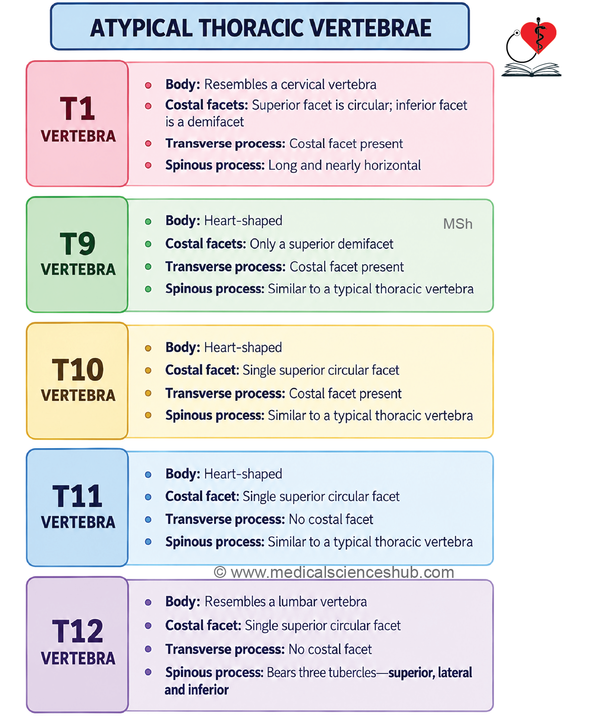
- The atypical thoracic vertebrae include T1 and T9–T12. They differ from typical thoracic vertebrae mainly in the shape of the body, arrangement of costal facets, and features of the transverse and spinous processes.
First Thoracic Vertebra
Identification features
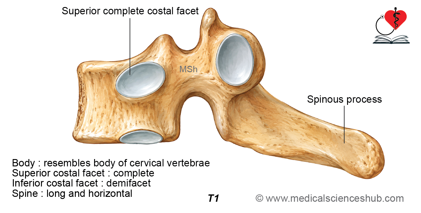
- The first thoracic vertebra resembles the seventh cervical vertebra in several features.
- Its body is broad from side to side and resembles that of a cervical vertebra rather than the heart-shaped body of a typical thoracic vertebra.
- Each side of the body bears a complete superior costal facet for articulation with the head of the first rib.
- An inferior costal demifacet is present for articulation with part of the head of the second rib.
- The spinous process is thick, long, and nearly horizontal rather than being directed downward.
- The superior vertebral notches are relatively deep.
- The transverse processes do not contain foramina transversaria.
Ninth Thoracic Vertebra
Identification features
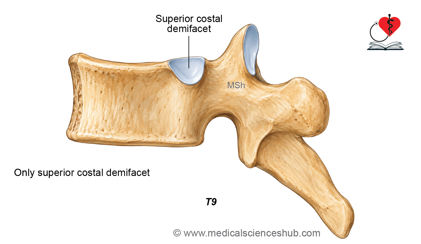
- The ninth thoracic vertebra generally resembles a typical thoracic vertebra.
- Each side of its body usually bears a superior costal demifacet for articulation with the head of the ninth rib.
- The inferior costal demifacet is usually absent.
Tenth Thoracic Vertebra
Identification features
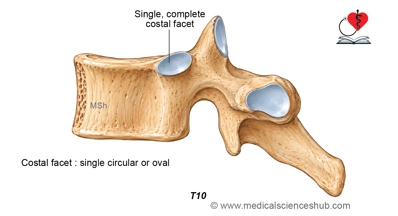
- Each side of the body bears a single complete costal facet for articulation with the head of the tenth rib.
- The costal facet is circular or oval and may extend onto the upper part of the pedicle.
- An inferior costal facet is absent.
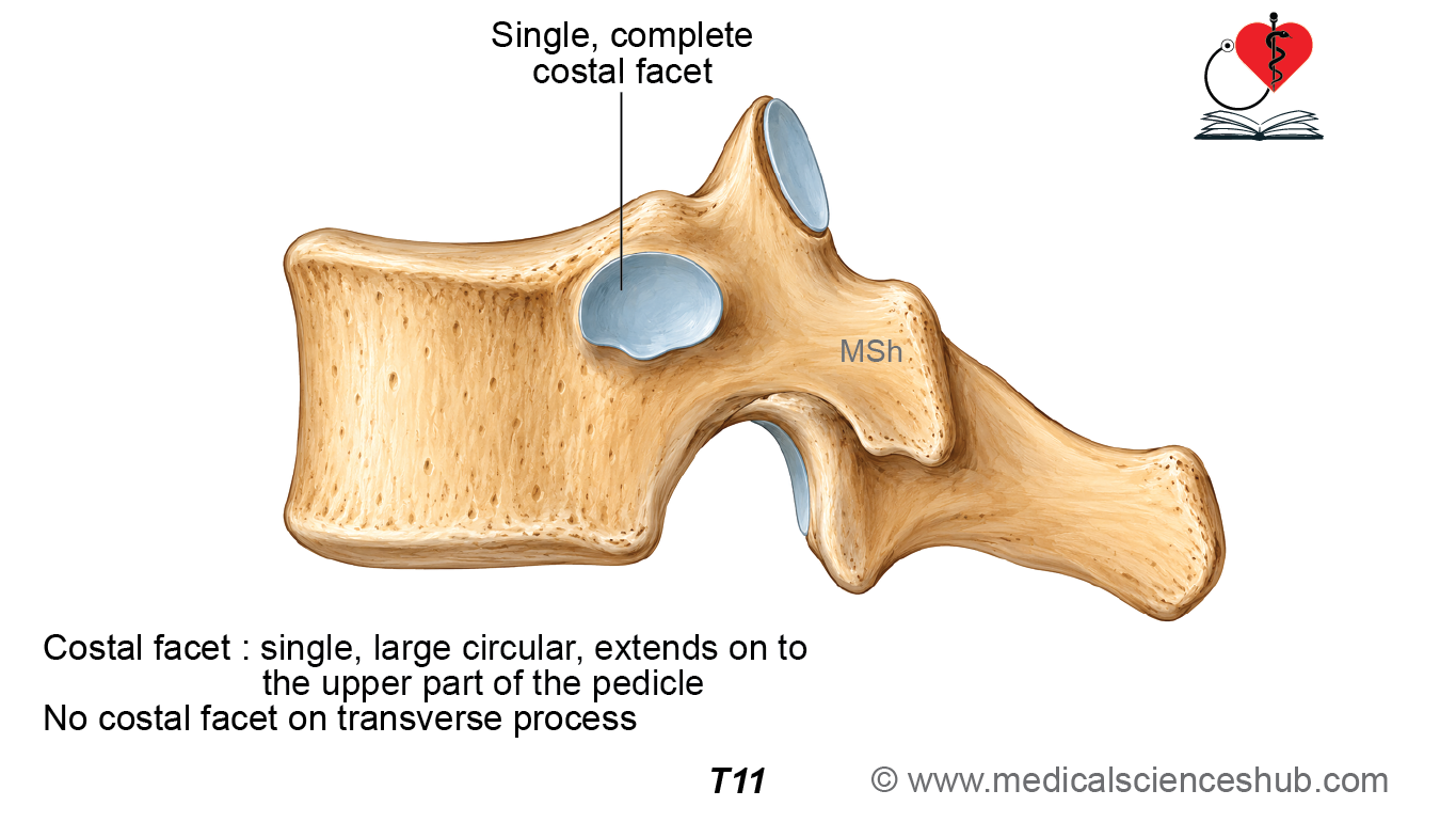
Eleventh Thoracic Vertebra
Identification features
- Each side of the body bears a single large, complete costal facet for articulation with the head of the eleventh rib.
- The costal facet extends onto the upper part of the pedicle.
- The transverse process does not bear a costal facet because the eleventh rib does not articulate with it.
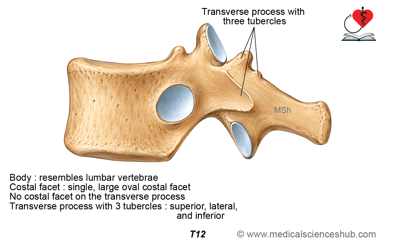
Twelfth Thoracic Vertebra
Identification features
- Each side of the body bears a single large, complete oval costal facet for articulation with the head of the twelfth rib.
- The body is broad from side to side and resembles that of a lumbar vertebra.
- The transverse process is short and does not bear a costal facet.
- The transverse process has three tubercles: superior, lateral, and inferior.
- The superior tubercle corresponds to the mammillary process of a lumbar vertebra.
- The inferior tubercle corresponds to the accessory process of a lumbar vertebra.
- The lateral tubercle represents the true transverse process.
Ossification of Typical Vertebra
- A typical thoracic vertebra develops by endochondral ossification.
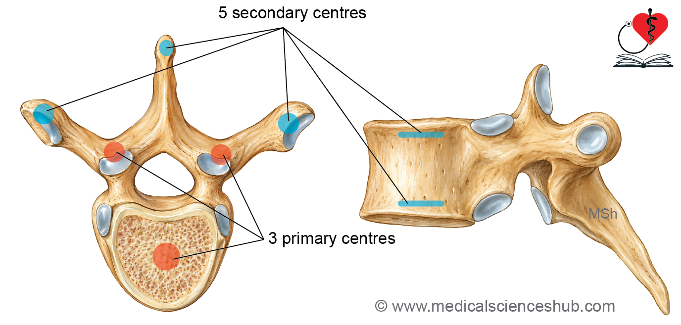
- It usually ossifies from three primary and five secondary ossification centers.
Primary Ossification Centers
- Three primary ossification centers are present:
- One center develops in the centrum, which forms the vertebral body.
- One center develops in each half of the vertebral arch.
- The primary ossification centers appear during the 8th–9th week of intrauterine life.
- At birth, each vertebra consists of three bony parts: the body and two halves of the vertebral arch.
- These three parts remain connected by cartilage at birth.
- The two halves of the vertebral arch unite posteriorly during early childhood.
- The vertebral arch subsequently fuses with the centrum, usually between 3 and 6 years of age.
Secondary Ossification Centers
- Five secondary ossification centers appear around puberty:
- One center appears at the tip of each transverse process.
- One center appears at the tip of the spinous process.
- One annular epiphysis develops along the superior surface of the vertebral body.
- One annular epiphysis develops along the inferior surface of the vertebral body.
- The secondary ossification centers usually fuse with the rest of the vertebra by about 25 years of age.
Clinical Anatomy
Spina Bifida
- Spina bifida results from failure of fusion of the two halves of the vertebral arch. It is associated with defective development of the neural tube.
Hemivertebra
- The vertebral body develops from ossification centers associated with the two sides of the centrum. Failure of development on one side may result in formation of only half of the vertebral body, producing a hemivertebra. This congenital anomaly can produce lateral curvature of the vertebral column. An associated rib may also be absent or malformed.
Spondylolisthesis
- Spondylolisthesis is the displacement of one vertebra relative to the vertebra below it. The displacement most commonly occurs in the lumbar region.
Intervertebral Disc Prolapse
- Degenerative changes in the annulus fibrosus may weaken it and lead to tearing or rupture. The nucleus pulposus may protrude or herniate through the weakened annulus fibrosus, producing an intervertebral disc prolapse.
- Disc prolapse commonly occurs in a posterolateral direction. The herniated disc may compress an adjacent spinal nerve root. Nerve root compression can produce pain that radiates along the distribution of the affected nerve.
📝 Test Your Knowledge – Practice MCQs
Attempt the chapter MCQ quiz and assess your understanding of key concepts.
Click to see: Medical Disclaimer and Copyright Notice