
Competencies
- AN21.8: Describe and demonstrate type, articular surfaces, and movements of manubriosternal, costovertebral, costotransverse, and xiphisternal joints.
- AN21.10: Describe costochondral and interchondral joints.
Introduction
- The thoracic cage includes the following joints:
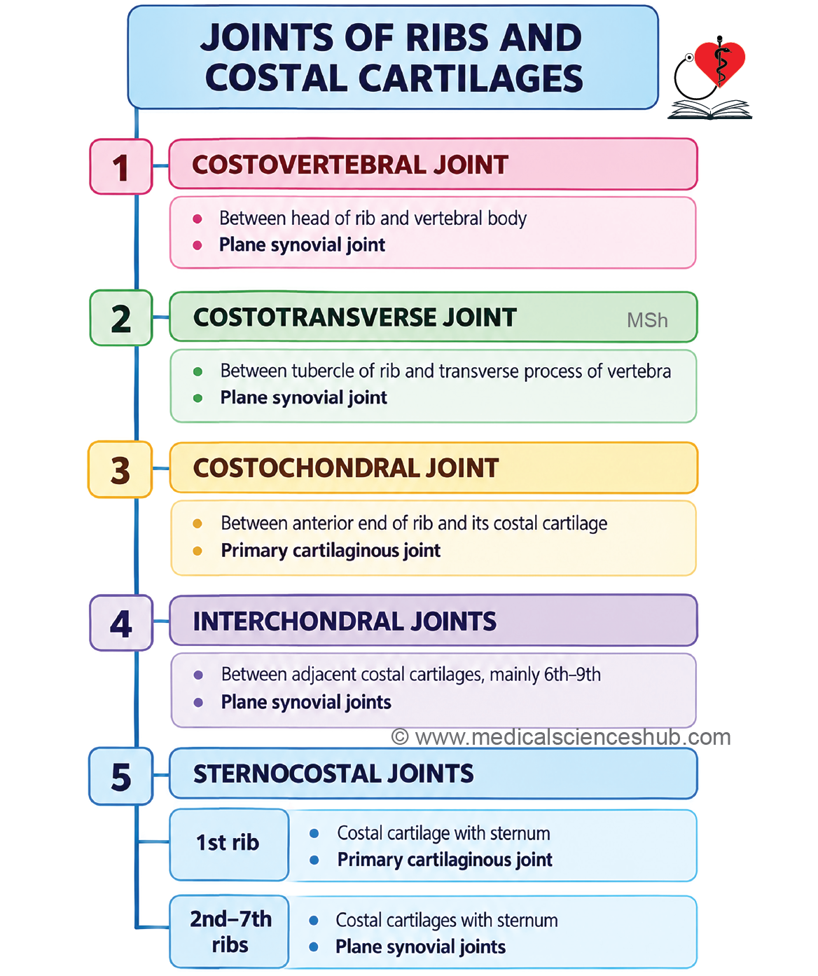
- Costovertebral joints connect the heads of the ribs with the thoracic vertebrae.
- Costotransverse joints connect the tubercles of the ribs with the transverse processes of the thoracic vertebrae.
- Costochondral joints connect the ribs with their respective costal cartilages.
- Interchondral joints are formed between adjacent costal cartilages.
- Sternochondral joints connect the costal cartilages with the sternum.
- The manubriosternal joint is formed between the manubrium and body of the sternum.
- The xiphisternal joint is formed between the body of the sternum and the xiphoid process.
- Intervertebral joints are present between adjacent vertebrae.
Movements
- Most joints of the thoracic cage permit only a limited range of movement.
- These small movements collectively allow expansion and contraction of the thoracic cage during respiration.
Costovertebral Joint
Articulation
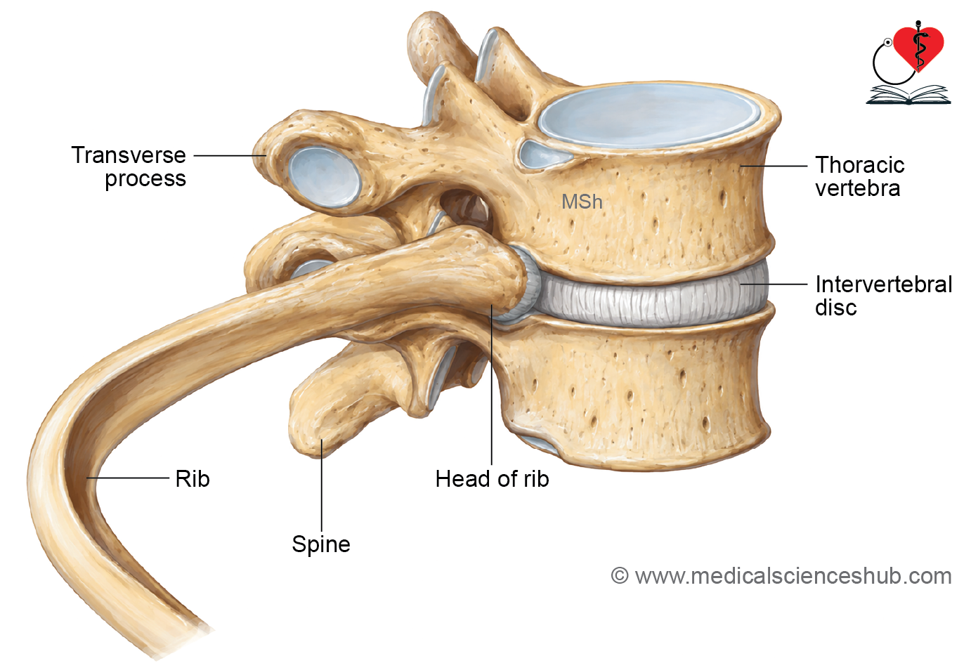
- The costovertebral joint is formed between the head of a rib and the costal facets on the bodies of thoracic vertebrae.
- The head of a typical rib articulates with:
- The body of the numerically corresponding thoracic vertebra.
- The body of the thoracic vertebra immediately above it.
- The intervening intervertebral disc.
Type
The costovertebral joint is a plane synovial joint.
Ligaments
Capsular Ligament
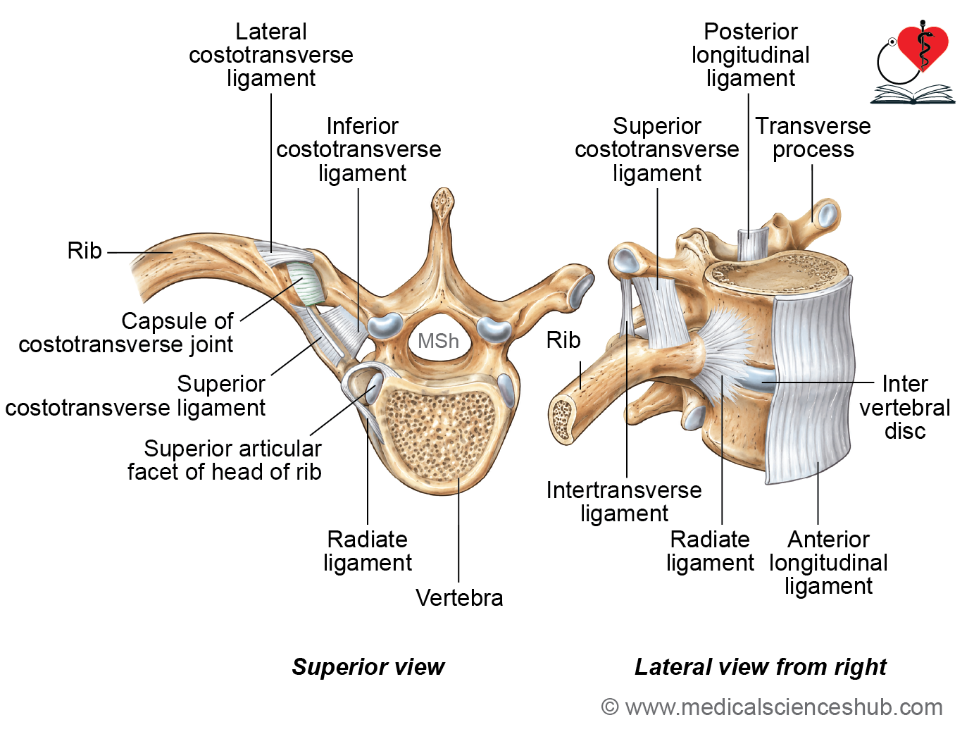
- A fibrous capsule surrounds the joint and is attached to the margins of the articular surfaces.
Radiate Ligament
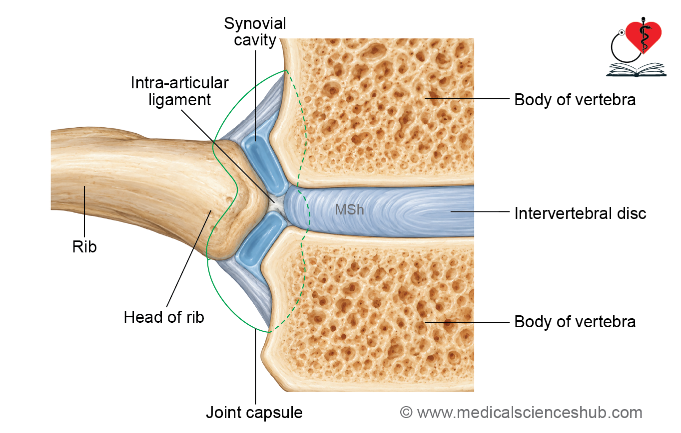
- The radiate ligament connects the anterior aspect of the head of the rib to the adjacent vertebral bodies and intervertebral disc.
- It consists of three bands:
- The upper band passes to the body of the vertebra above.
- The middle band is attached to the intervertebral disc.
- The lower band passes to the body of the numerically corresponding vertebra.
Intra-articular Ligament
- The intra-articular ligament extends from the crest on the head of the rib to the intervertebral disc. It separates the joint cavity into two synovial compartments.
Special Features
- The 1st, 10th, 11th, and 12th ribs usually articulate only with their numerically corresponding vertebral bodies. Therefore, their costovertebral joints have a single joint cavity.
Costotransverse Joints
Articulation
- A costotransverse joint is formed between:
- The articular facet on the tubercle of a rib.
- The costal facet on the transverse process of the numerically corresponding thoracic vertebra.
- Costotransverse joints are absent for the 11th and 12th ribs because these ribs do not articulate with the transverse processes.
Type
The costotransverse joint is a plane synovial joint.
Ligaments
Capsular Ligament
- The fibrous capsule surrounds the joint and is attached to the margins of the articulating facets.
Superior Costotransverse Ligament
- The superior costotransverse ligament extends from the neck of the rib to the transverse process of the vertebra above. It consists of anterior and posterior laminae.
- The anterior lamina extends from the crest of the neck of the rib to the inferior border of the transverse process of the vertebra above. The posterior lamina extends from the posterior surface of the neck of the rib to the inferior border of the transverse process of the vertebra above.
Inferior Costotransverse Ligament
- The inferior costotransverse ligament extends from the rough posterior surface of the neck of the rib to the anterior surface of the transverse process of the corresponding vertebra.
Lateral Costotransverse Ligament
- The lateral costotransverse ligament extends from the non-articular part of the tubercle of the rib to the tip of the transverse process of the corresponding vertebra.
Joints of Costal Cartilages
Costochondral joints
Articulation
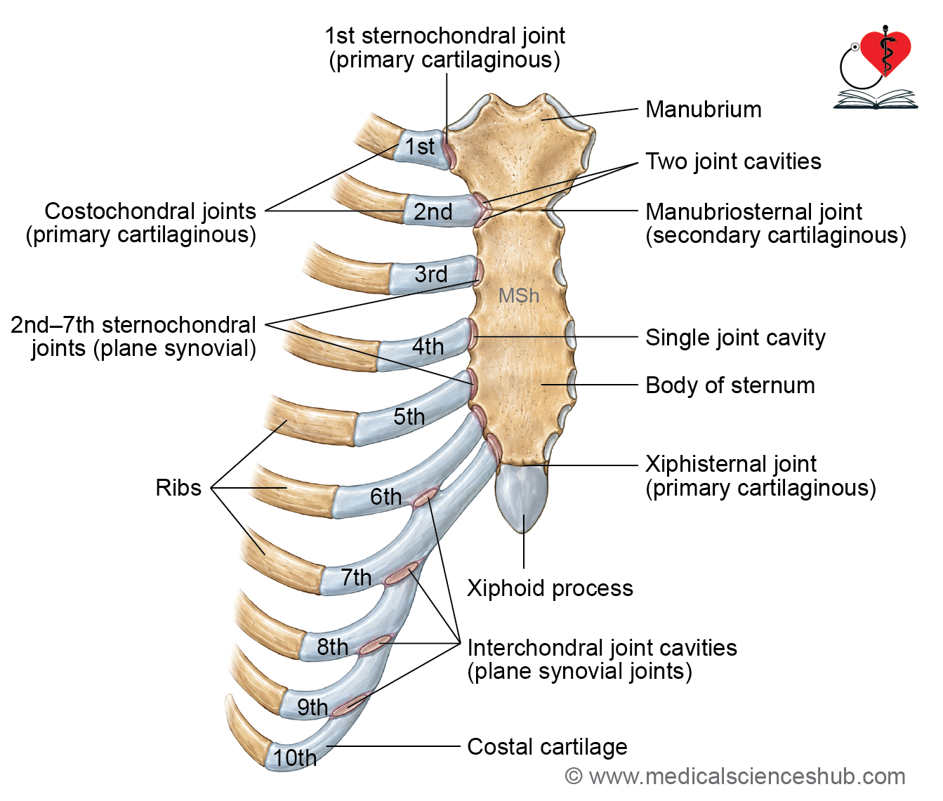
- The costochondral joints are formed between the sternal ends of the ribs and the lateral ends of their corresponding costal cartilages.
Type
- These joints are primary cartilaginous joints (synchondroses).
Movements
- No movement occurs at the costochondral joints.
- However, the flexibility of the costal cartilages contributes to movements of the thoracic cage during respiration.
Interchondral joints
Articulation
- Interchondral joints are present between the adjacent costal cartilages.
- Synovial interchondral joints are commonly present between the 6th and 7th, 7th and 8th, and 8th and 9th costal cartilages.
Type
- These joints are plane synovial joints.
Special Feature
- The connection between the 9th and 10th costal cartilages is usually fibrous rather than synovial.
Sternochondral joint
Articulation
- The sternochondral joints are formed between the medial ends of the costal cartilages and the lateral borders of the sternum.
Type
- The first sternochondral joint is a primary cartilaginous joint or synchondrosis.
- The 2nd–7th sternochondral joints are plane synovial joints.
Special Feature
- The cavity of the second sternochondral joint is commonly divided into two compartments by an intra-articular ligament.
Manubriosternal Joint
Articulation
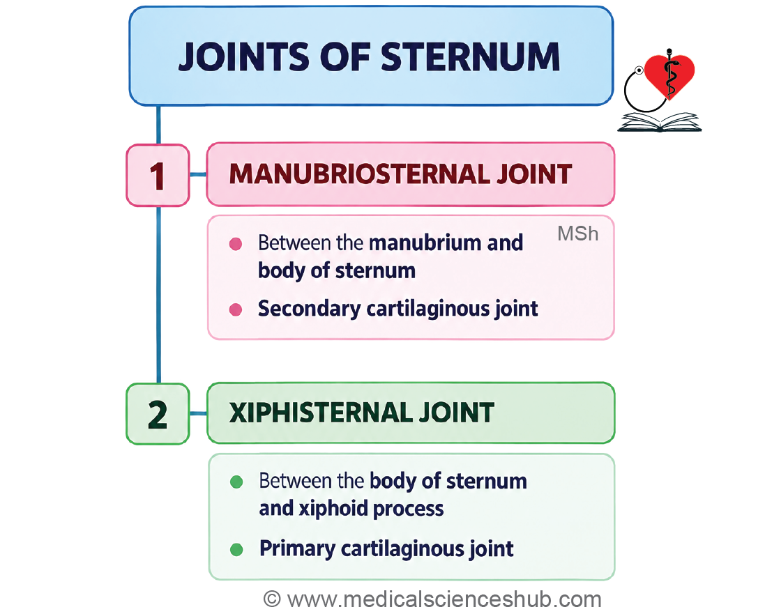
- The manubriosternal joint is formed between the inferior border of the manubrium and the superior border of the body of the sternum.
Type
- It is a secondary cartilaginous joint (symphysis).
Movement
- The joint permits a small degree of movement between the manubrium and the body of the sternum during respiration.
Special Feature
- The manubriosternal joint is not considered a typical secondary cartilaginous joint because it may undergo ossification with advancing age.
- In some individuals, the manubrium and body of the sternum fuse to form a bony union or synostosis.
Xiphisternal Joint
Articulation
- The xiphisternal joint is formed between the xiphoid process and the body of the sternum.
Type
- It is a primary cartilaginous joint (synchondrosis).
Ossification
- The xiphisternal joint may gradually ossify with age.
- It commonly develops into a bony union or synostosis during middle or later adult life.
Intervertebral Joints
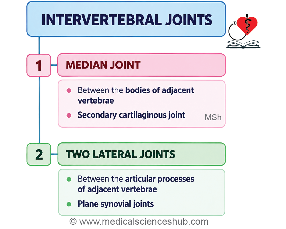
- Adjacent vertebrae are connected by three principal joints:
- One median joint is present between the vertebral bodies.
- Two facet joints, one on each side, are present between the articular processes.
Joint between vertebral bodies
- The joint between adjacent vertebral bodies is a secondary cartilaginous joint (symphysis). The opposing surfaces of the vertebral bodies are covered by thin layers of hyaline cartilage. An intervertebral disc is interposed between the bodies of adjacent vertebrae. The disc provides a strong connection while allowing a small amount of movement.
Joints between articular processes
- The joints between the superior and inferior articular processes of adjacent vertebrae are called facet or zygapophysial joints.
- They are plane synovial joints. Their articular surfaces are covered with hyaline cartilage. These joints primarily permit gliding movements between adjacent vertebrae.
Ligaments of vertebral column
- The intervertebral discs connect the bodies of adjacent vertebrae.
- Joint capsules surround and stabilize the facet joints.
- The anterior longitudinal ligament extends along the anterior surfaces of the vertebral bodies and intervertebral discs.
- The posterior longitudinal ligament runs along the posterior surfaces of the vertebral bodies within the vertebral canal.
- Superiorly, the posterior longitudinal ligament continues as the tectorial membrane.
- The intertransverse ligaments connect the transverse processes of adjacent vertebrae.
- The interspinous ligaments extend between adjacent spinous processes.
- The supraspinous ligament connects the tips of the spinous processes.
- In the cervical region, the supraspinous ligament continues as the ligamentum nuchae.
- The ligamenta flava connect the laminae of adjacent vertebrae.
Intervertebral disc
General Features
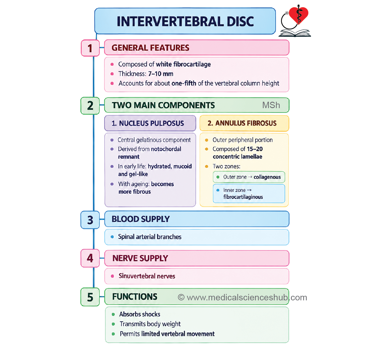
- An intervertebral disc is a fibrocartilaginous structure located between the bodies of adjacent vertebrae. Intervertebral discs extend from the C2–C3 level to the lumbosacral junction.
- Each disc forms part of a secondary cartilaginous joint between adjacent vertebral bodies. The shape of each disc generally corresponds to the shape of the adjacent vertebral bodies.
- The thickness of the discs varies according to the region of the vertebral column. The discs are relatively thick in the lumbar region and thinner in the upper thoracic region. In the cervical and lumbar regions, the discs are thicker anteriorly and contribute to the secondary curvatures of the vertebral column. In the thoracic region, the discs are more nearly uniform in thickness.
- Collectively, the intervertebral discs contribute approximately one-fifth to one-quarter of the total length of the vertebral column.
Structure
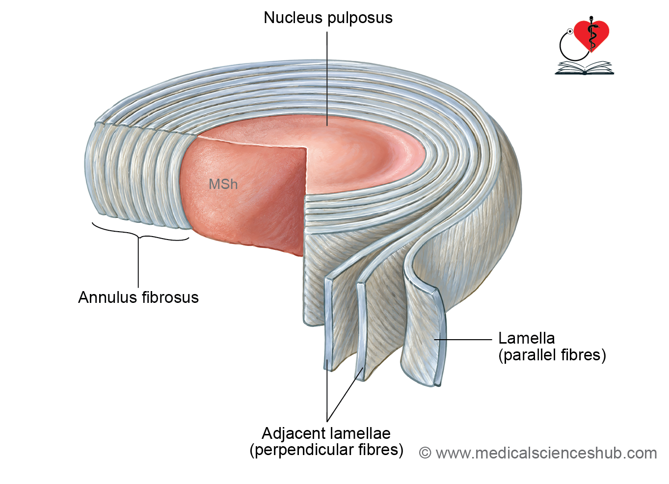
- Each intervertebral disc consists of two main components:
- The central nucleus pulposus.
- The peripheral annulus fibrosus.
Nucleus Pulposus
- The nucleus pulposus occupies the central region of the intervertebral disc. It has a soft, gelatinous consistency and contains a high proportion of water. It represents a remnant of the embryonic notochord.
- With increasing age, notochordal cells gradually disappear. The nucleus becomes less hydrated and undergoes progressive fibrous and degenerative changes with age.
Annulus Fibrosus
- The annulus fibrosus forms the outer part of the intervertebral disc. It consists of multiple concentric lamellae containing collagen and fibrocartilage. Collagen fibres within each lamella run in an oblique direction. The direction of the fibres alternates in successive lamellae, increasing the strength of the disc. The outer part is predominantly fibrous, while the inner part gradually blends with fibrocartilaginous tissue.
Blood Supply
- Blood vessels are mainly confined to the peripheral part of the annulus fibrosus. Most of the intervertebral disc is avascular. Nutrients reach the deeper parts of the disc mainly by diffusion through the vertebral end plates.
Innervation
- Sensory nerve fibres are mainly distributed to the outer part of the annulus fibrosus. Innervation is provided largely by recurrent meningeal, or sinuvertebral, nerves, with contributions from sympathetic fibres.
Functions
- Intervertebral discs contribute to the shape and curvatures of the vertebral column.
- They absorb and distribute forces acting on the vertebral column.
- They permit limited movement between adjacent vertebral bodies.
- They transmit body weight from one vertebra to the next.
- They help maintain flexibility while providing stability to the vertebral column.
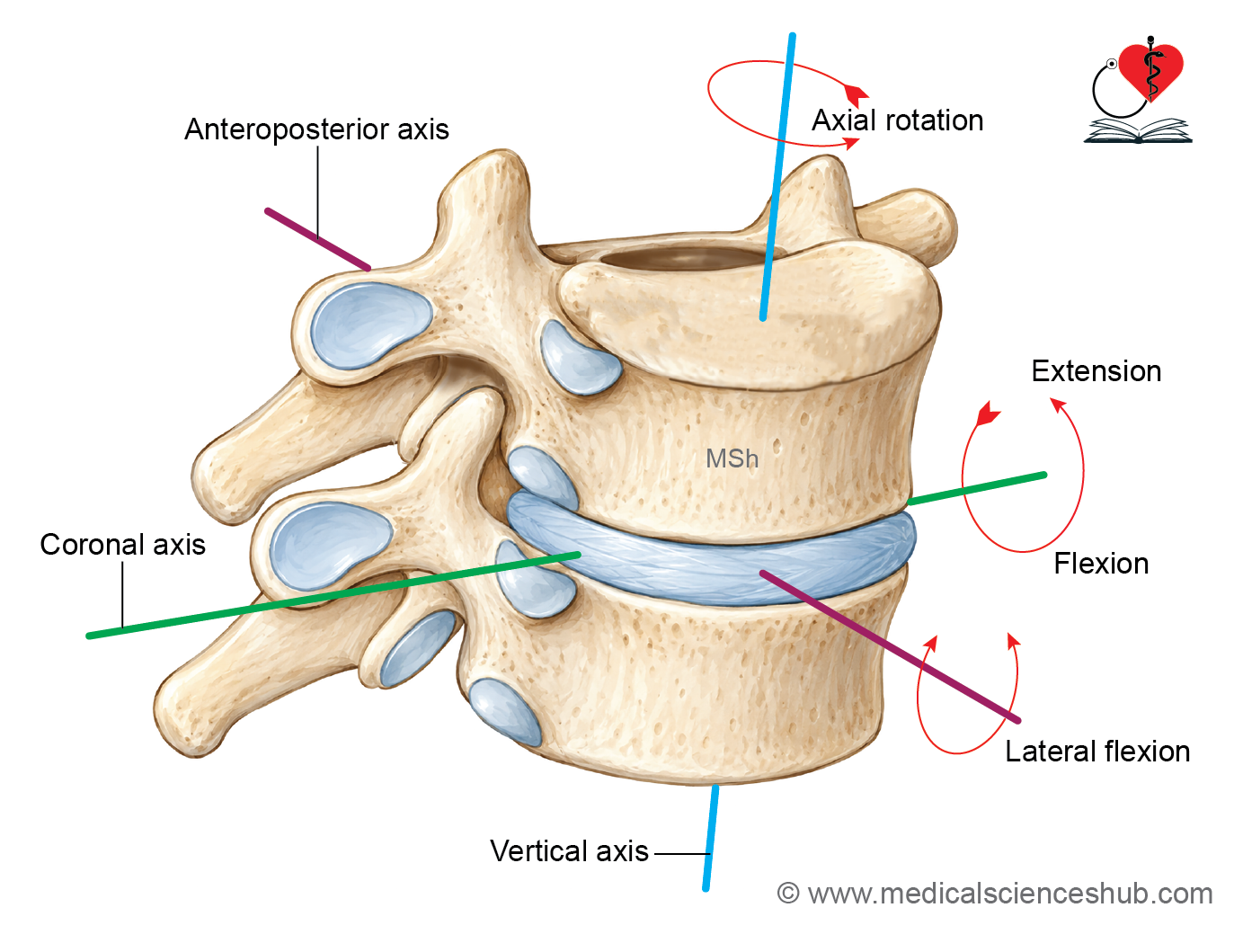
Movements of vertebral column
- Movement between two adjacent vertebrae is relatively limited.
- The combined movements occurring at multiple vertebral levels provide considerable mobility to the vertebral column.
- The main movements include flexion, extension, lateral flexion, and rotation.
- The range and direction of these movements are influenced mainly by the elasticity of the intervertebral discs and the orientation of the articular facets.
Regional Differences
- Flexion and extension are particularly well developed in the cervical and lumbar regions.
- Lateral flexion occurs in the cervical and lumbar regions and to a lesser extent in the thoracic region.
- Rotation is greatest in the cervical region, particularly in the upper cervical spine.
- Rotation is also permitted in the thoracic region because of the orientation of the articular facets.
- Rotation is markedly restricted in the lumbar region because of the orientation of its articular facets.
Intervertebral Disc Herniation
- Disc herniation, commonly called a slipped disc, occurs when the nucleus pulposus protrudes through a weakened or torn part of the annulus fibrosus. The herniated material may compress an adjacent spinal nerve root or other neural structures. Depending on the site and severity of compression, the patient may develop pain, numbness, muscle weakness, or, in severe cases, neurological deficits.
Degenerative Disc Disease
- Intervertebral discs undergo progressive structural changes with advancing age. The nucleus pulposus gradually loses water content, while the annulus fibrosus may become less flexible and develop fissures. These changes reduce the elasticity and shock-absorbing capacity of the disc. Progressive degeneration may also reduce the height of the intervertebral disc and alter the mechanics of the vertebral column.
- The term annulus is derived from Latin and means “ring,” referring to the ring-like arrangement of the annulus fibrosus around the nucleus pulposus.
📝 Test Your Knowledge – Practice MCQs
Attempt the chapter MCQ quiz and assess your understanding of key concepts.
Click to see: Medical Disclaimer and Copyright Notice