
Competencies
- AN21.4: Describe and demonstrate extent, attachments, direction of fibres, nerve supply and actions of intercostal muscles.
- AN21.9: Describe and demonstrate mechanics and types of respiration.
Introduction
- The thoracic wall is formed by the bony thoracic cage and the muscles occupying the spaces between the ribs.
- Externally, the thoracic wall is covered by skin, subcutaneous tissue, fascia, and muscles of the pectoral and back regions.
- The thoracic wall receives its main nerve and vascular supply through the intercostal nerves and vessels.
- The thoracic cage surrounds and protects vital organs, particularly the heart and lungs.
Surface Anatomy of Thorax
Bony Landmarks
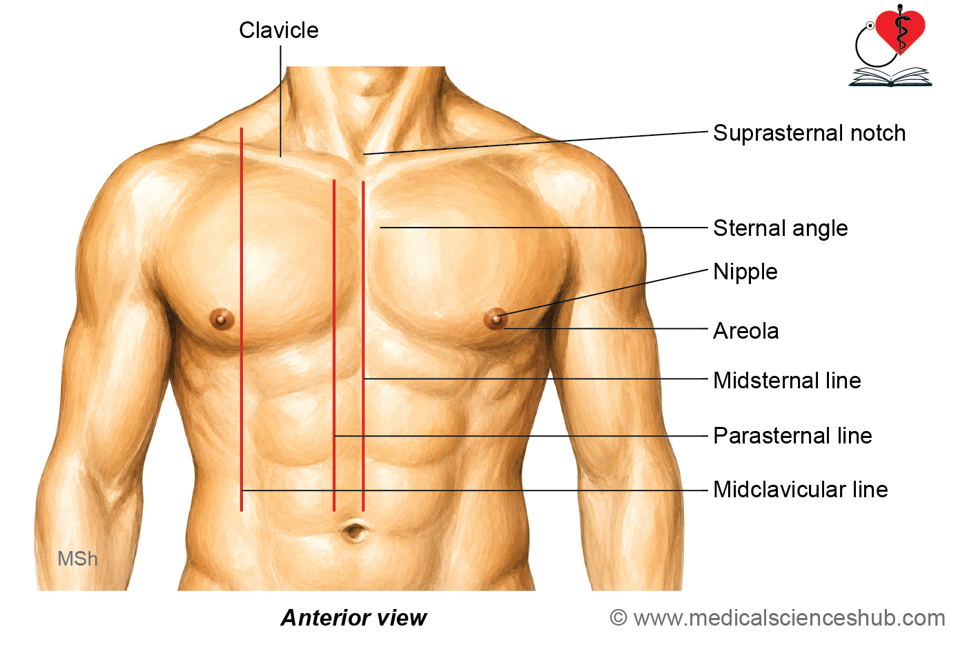
Clavicle
- The clavicle is located horizontally at the root of the neck. It lies immediately beneath the skin and can therefore be palpated along its entire length.
Suprasternal Notch
- The suprasternal notch is also known as the jugular or interclavicular notch. It is situated between the medial ends of the clavicles, just above the superior border of the manubrium sterni. The trachea can be palpated through this notch. The suprasternal notch corresponds approximately to the level of the lower border of the T2 vertebra.
Sternal Angle
- The sternal angle, or angle of Louis, is a palpable transverse ridge at the junction of the manubrium and body of the sternum.
- It is located approximately 5 cm below the suprasternal notch.
- Posteriorly, it corresponds to the level of the intervertebral disc between the T4 and T5 vertebrae.
- The second costal cartilage articulates with the sternum at this level.
- Clinically, the sternal angle serves as an important landmark for identifying the second rib and subsequently counting the ribs and intercostal spaces.
- The term angle of Louis is associated with Antoine Louis, a French surgeon and physiologist (1723–1792).
Costal Margin
- The costal margin, also called the costal arch or arcus costalis, forms the lower boundary of the anterior thoracic cage. It is formed mainly by the costal cartilages of the 7th to 10th ribs. The 11th and 12th ribs are floating ribs and do not contribute to the costal margin.
Infrasternal Angle
- The infrasternal angle is also called the subcostal angle or angulus infrasternalis. It is formed between the right and left costal margins. The xiphoid process projects downward into the apex of this angle.
Soft Tissue Landmarks
Nipple and Areola
- In an adult male, the nipple is usually located near the fourth intercostal space, approximately 10 cm lateral to the median plane. The exact position of the nipple may vary between individuals. Each nipple is surrounded by a circular pigmented area known as the areola.
Montgomery Tubercles
- The areola contains modified sebaceous glands that produce small surface elevations known as Montgomery tubercles. These tubercles usually become more prominent during pregnancy. They are named after William Fetherstone Montgomery, an Irish obstetrician (1797–1859).
Ictus Cordis
- The ictus cordis, also known as the apex beat or apical impulse, is the palpable impulse produced by the apex of the heart against the chest wall. In a normal adult, it is usually felt in the left fifth intercostal space, just medial to the midclavicular line. Its position is approximately 9–10 cm lateral to the median plane.
Anatomical lines
- Several imaginary vertical lines are used as surface landmarks to describe the position and extent of thoracic structures.
- These lines are particularly useful for describing the surface projection of internal organs and for clinical examination.
Lines on the Anterior Thoracic Wall
- Midsternal line: This vertical line passes through the centre of the sternum and lies in the median sagittal plane.
- Parasternal line: This vertical line runs close to the lateral border of the sternum.
- Midclavicular line: This vertical line passes through the midpoint of the clavicle. Inferiorly, it passes approximately through the tip of the ninth costal cartilage and the midinguinal point.
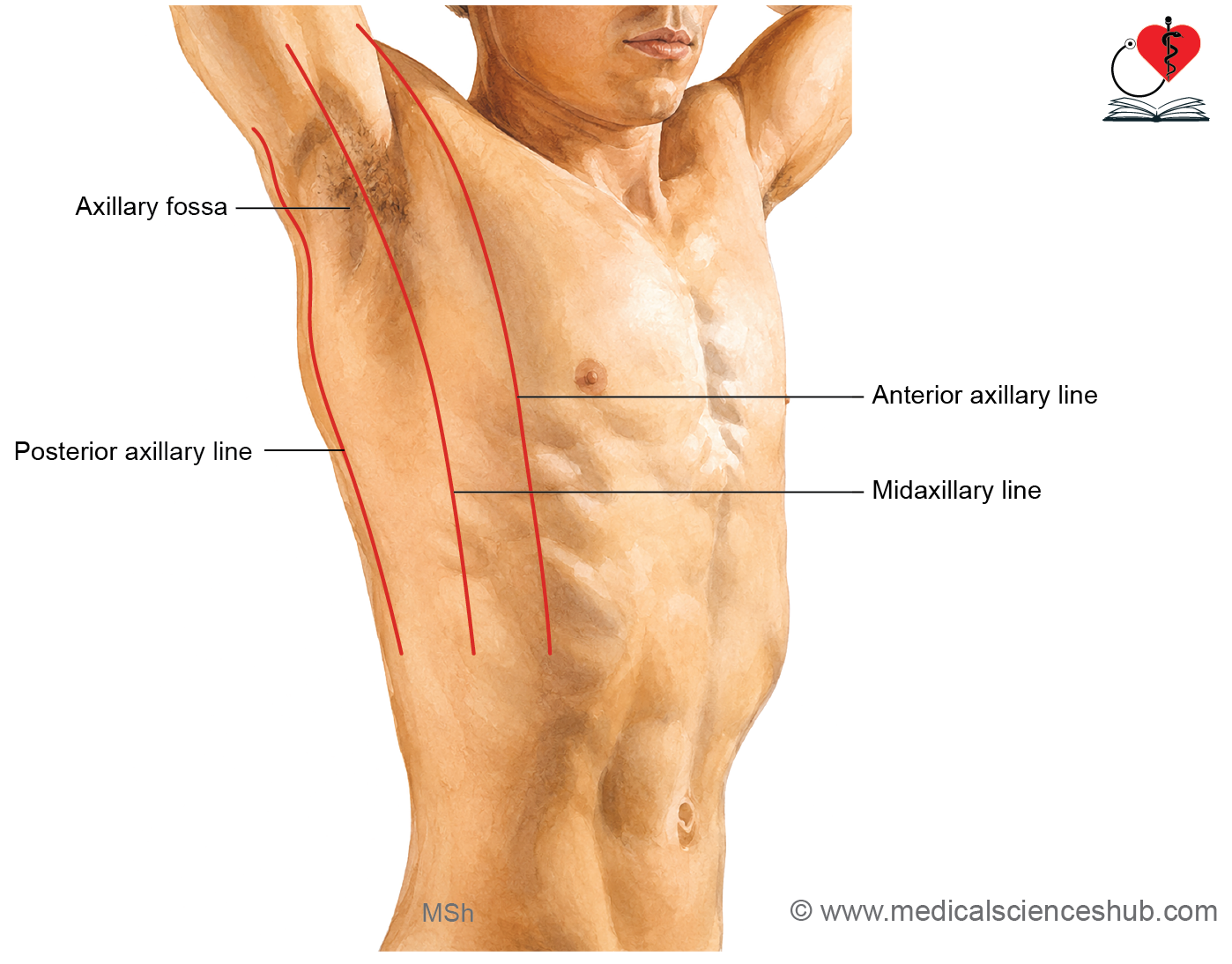
Lines on the Lateral Thoracic Wall
- Anterior axillary line: This vertical line follows the anterior axillary fold.
- Posterior axillary line: This vertical line follows the posterior axillary fold.
- Midaxillary line: This vertical line passes midway between the anterior and posterior axillary lines.
Lines on the Posterior Thoracic Wall
- Scapular line: This vertical line passes through the inferior angle of the scapula when the upper limb is in the anatomical position.
- Posterior median line: This line, also called the mid-dorsal line, runs vertically along the midline of the back in the median sagittal plane.
Structure of Thoracic Wall
From superficial to deep, the thoracic wall is covered by the following structures:
- Skin containing the nipples and areolae.
- Superficial fascia containing the mammary glands.
- Deep fascia, which is relatively indistinct.
- Muscles associated with the thoracic wall.
Skin
- The skin covering the posterior aspect of the thorax is thicker than that over the anterior and lateral aspects. This refers to the overall thickness of the skin and does not indicate the histological ‘thick skin’ type found on the palms and soles.
Superficial Fascia
- The superficial fascia of the thoracic region contains a moderate amount of fat, along with cutaneous nerves and blood vessels. In females, the superficial fascia contains the well-developed mammary gland. In males, the mammary gland is normally rudimentary.
Muscles Covering the Thoracic Wall
- The thoracic wall is covered by several muscles that are functionally related to the upper limb and back. These muscles are considered extrinsic muscles and do not form the thoracic wall proper.
Muscles Related to the Upper Limb
- Pectoralis major covers a large part of the anterior thoracic wall.
- Pectoralis minor lies deep to pectoralis major.
- Serratus anterior covers the lateral aspect of the thoracic wall.
These muscles connect the thoracic cage with the upper limb or pectoral girdle and therefore are not intrinsic muscles of the thoracic wall.
Muscles of the Back
- The posterior aspect of the thoracic wall is covered by several back muscles.
- These include trapezius, latissimus dorsi, levator scapulae, rhomboid major, and rhomboid minor.
- Serratus posterior superior and serratus posterior inferior also lie in relation to the posterior thoracic wall.
- The erector spinae group forms a prominent longitudinal muscle mass along the posterior aspect of the vertebral column.
INTERCOSTAL SPACES
- An intercostal space is the interval between two adjacent ribs and their costal cartilages.
- There are 11 pairs of intercostal spaces on each side of the thoracic wall.
- The upper nine intercostal spaces extend anteriorly as far as the sternum or costal margin, whereas the 10th and 11th spaces remain open anteriorly.
- Each intercostal space contains intercostal muscles and membranes, along with nerves, blood vessels, and lymphatics.
Typical Intercostal Spaces
- The third to sixth intercostal spaces are considered typical intercostal spaces.
- Their intercostal nerves and vessels are distributed mainly to the thoracic wall and do not continue into the upper limb or anterior abdominal wall. Other intercostal spaces show variations in the course or distribution of their neurovascular structures.
Contents of a Typical Intercostal Space
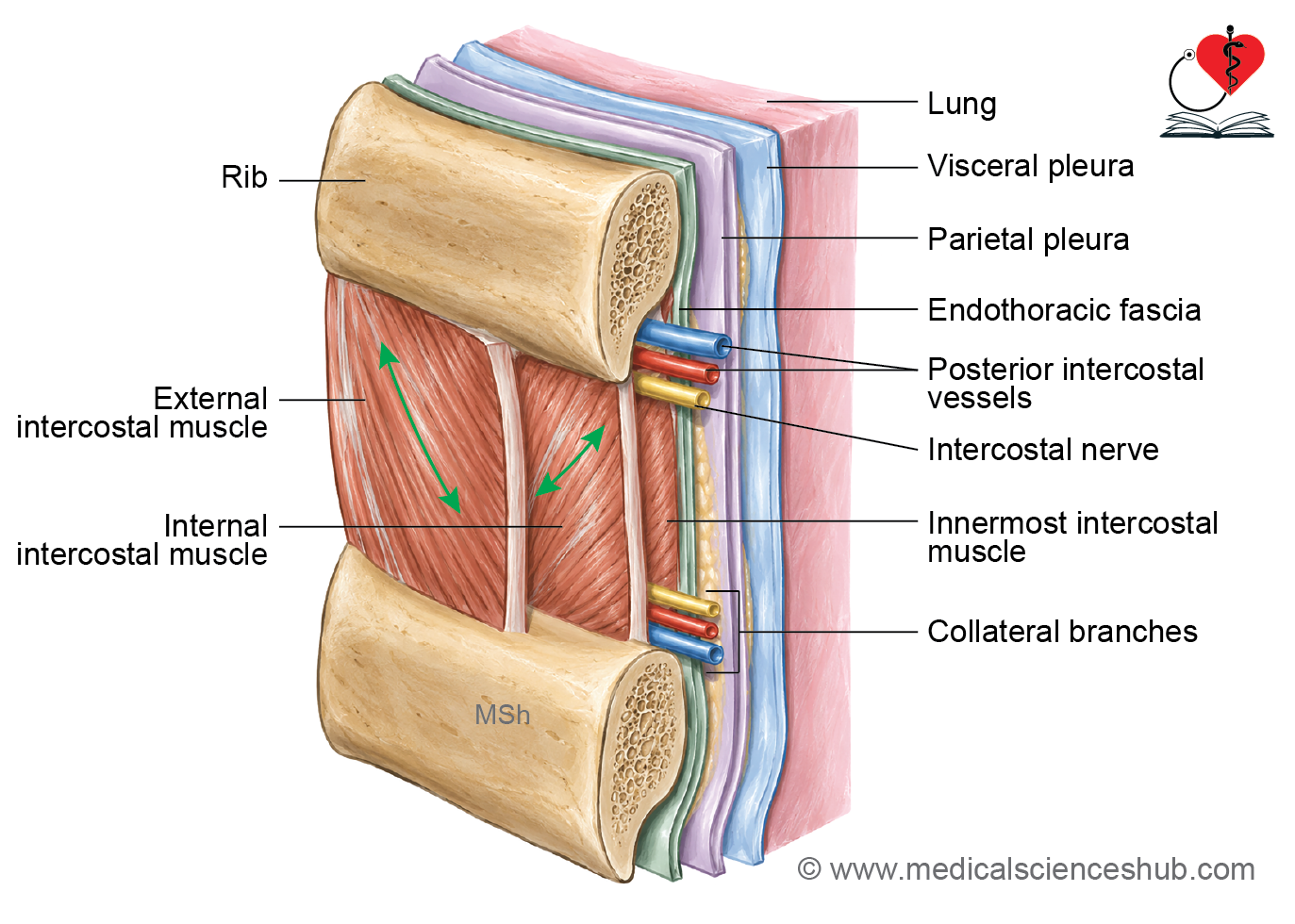
- A typical intercostal space contains three layers of intercostal muscles:
- External intercostal muscle.
- Internal intercostal muscle.
- Innermost intercostal muscle (intercostalis intimus).
- It contains an intercostal nerve derived from the anterior ramus of a thoracic spinal nerve.
- It contains posterior and anterior intercostal arteries. Intercostal veins accompany the arteries and nerves. Lymphatic vessels and lymph nodes are also present within the intercostal spaces.
Parts of an Intercostal Space
- Each intercostal space can be divided into posterior interosseous and anterior intercartilaginous parts.
- The interosseous part lies between the bony portions of two adjacent ribs.
- The intercartilaginous part lies between the costal cartilages of adjacent ribs.
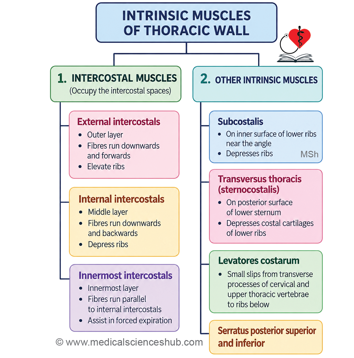
Intercostal Muscles
- The intercostal muscles occupy the intercostal spaces and are arranged in three layers from superficial to deep:
- External intercostal muscles form the outermost layer.
- Internal intercostal muscles form the middle layer.
- Innermost intercostal muscles form the deepest layer.
- These muscles help maintain the integrity of the intercostal spaces and assist in movements of the ribs during respiration.
External Intercostal Muscle
- The external intercostal muscle, also called intercostalis externus, forms the outermost muscular layer of an intercostal space.
- There are 11 pairs of external intercostal muscles.
Extent and Membrane
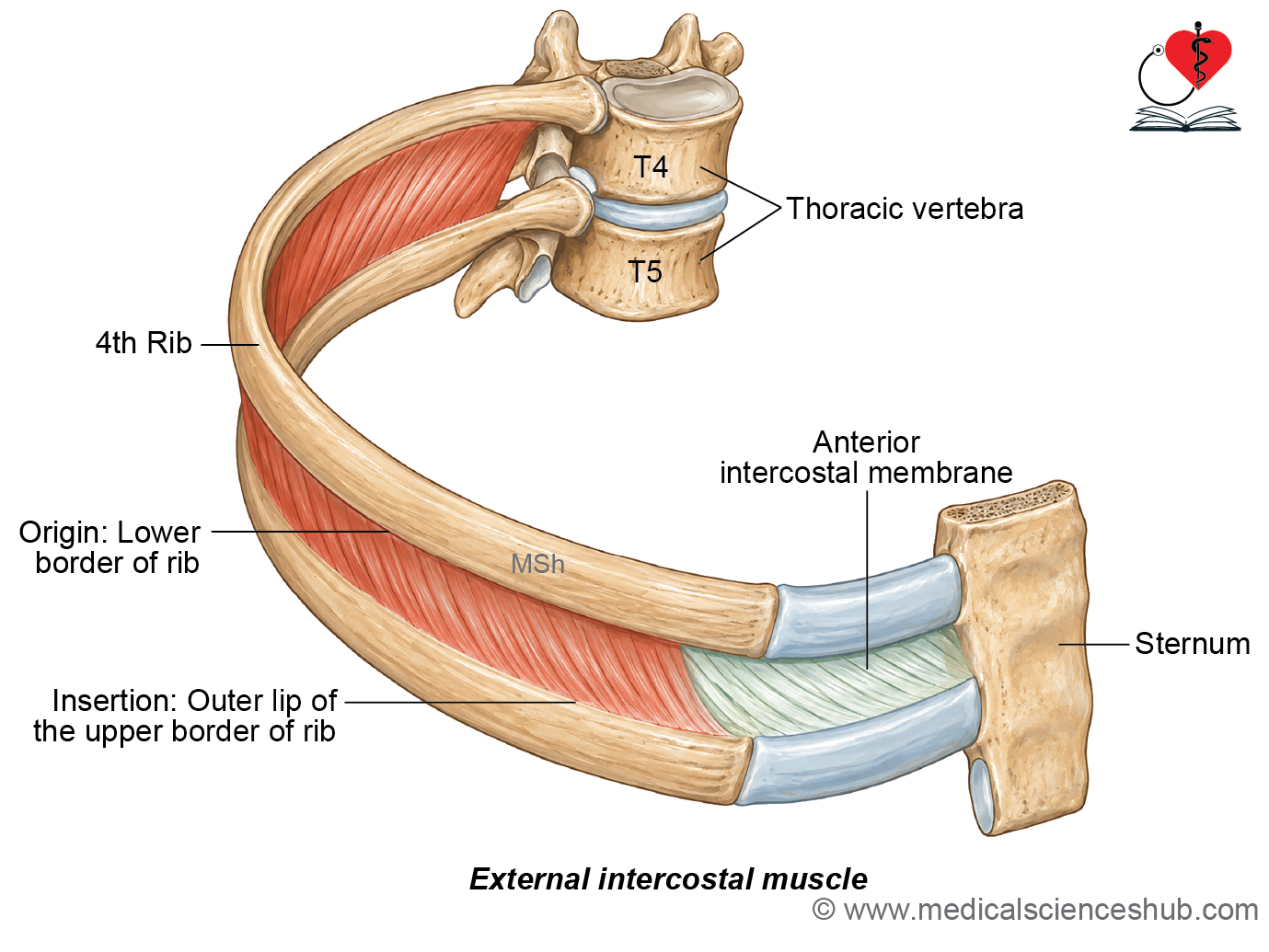
- Each external intercostal muscle extends from the tubercle of the rib posteriorly to the costochondral junction anteriorly. From the costochondral junction to the sternum, the muscle is replaced by the external intercostal membrane.
Attachments
- The muscle arises from the lower border of the rib above. Its fibers are inserted into the upper border of the rib below.
Direction of Fibers
- The fibers pass downward and forward from the rib above to the rib below. In the anterior part of the thoracic wall, they also have a medial inclination. The direction of these fibers helps distinguish the external intercostal muscle from the internal intercostal muscle.
Nerve Supply
- Each external intercostal muscle is supplied by the intercostal nerve of the corresponding intercostal space.
Action
- The external intercostal muscles primarily elevate the ribs and assist inspiration.
- They also help maintain the rigidity of the intercostal spaces during respiration.
Clinical Anatomy
Supracostalis Muscle: The supracostalis is an occasional muscular variation associated with the upper part of the thoracic wall. It is usually a thin muscle extending across several of the upper ribs. It lies deep to the pectoral muscles.
Internal Intercostal Muscle
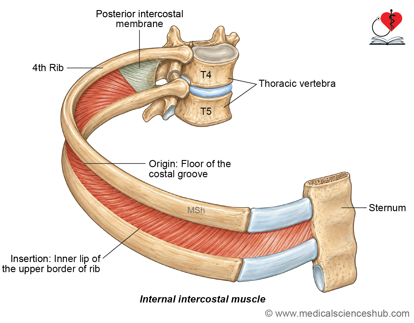
- The internal intercostal muscle, also called intercostalis internus, forms the middle muscular layer of an intercostal space.
- There are 11 pairs of internal intercostal muscles.
Extent and Membrane
- Each internal intercostal muscle extends from the lateral border of the sternum anteriorly to approximately the angle of the rib posteriorly.
- Posterior to the angle of the rib, the muscle is replaced by the internal intercostal membrane.
Attachments
- The muscle arises mainly from the costal groove and adjacent inner surface of the rib above. Its fibers are inserted into the upper border of the rib below.
Direction of Fibers
- The fibers generally pass downward and backward from the rib above to the rib below.
- Their direction is approximately perpendicular to that of the external intercostal fibers.
Nerve Supply
- Each internal intercostal muscle is supplied by the intercostal nerve of the corresponding intercostal space.
Actions
- The interosseous part of the internal intercostal muscle depresses the ribs and is particularly active during forced expiration.
- The interchondral part, located between adjacent costal cartilages, assists in elevating the ribs during inspiration.
- Internal intercostal muscles have little role in quiet expiration because normal quiet expiration occurs mainly through passive elastic recoil of the lungs and thoracic wall.
Innermost Intercostal Muscle
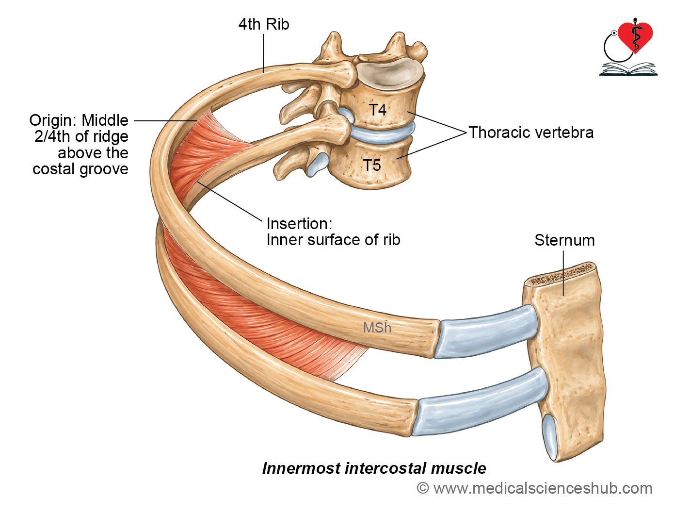
- The innermost intercostal muscle, or intercostalis intimus, forms the deepest layer of the intercostal musculature.
- There are usually 11 pairs, although the muscle may be poorly developed or absent in the upper two or three intercostal spaces.
- It is best developed in the middle portion of an intercostal space.
Extent and Attachments
- The innermost intercostal muscle occupies mainly the middle part of the intercostal space.
- It arises from the inner surface of the rib above, near the region of the costal groove.
- Its fibers descend to attach to the inner surface of the rib below.
Direction of Fibers
- The fibers run in the same general direction as those of the internal intercostal muscle.
- They pass downward and backward from the rib above to the rib below.
Nerve Supply
- The muscle is supplied by the corresponding intercostal nerve.
Actions
- The innermost intercostal muscle acts together with the internal intercostal muscle.
- It assists in depressing the ribs during forced expiration.
- It also helps maintain the stability of the intercostal spaces during respiratory movements.
Neurovascular Plane
- The intercostal neurovascular bundle lies between the internal and innermost intercostal muscles.
- This interval is known as the neurovascular plane of the intercostal space.
- The main intercostal vein, artery, and nerve usually lie in the costal groove along the inferior border of the rib above.
Relation to the Pleura
- The innermost intercostal muscle is separated from the parietal pleura by the endothoracic fascia.
- The endothoracic fascia forms a thin connective tissue layer between the inner surface of the thoracic wall and the parietal pleura.
Anatomical Classification
- Traditionally, the deepest muscular layer of the thoracic wall was described as having three components: innermost intercostal, subcostalis, and transversus thoracis.
- In current anatomical descriptions, the innermost intercostal, subcostalis, and transversus thoracis are regarded as separate muscles.
Subcostalis
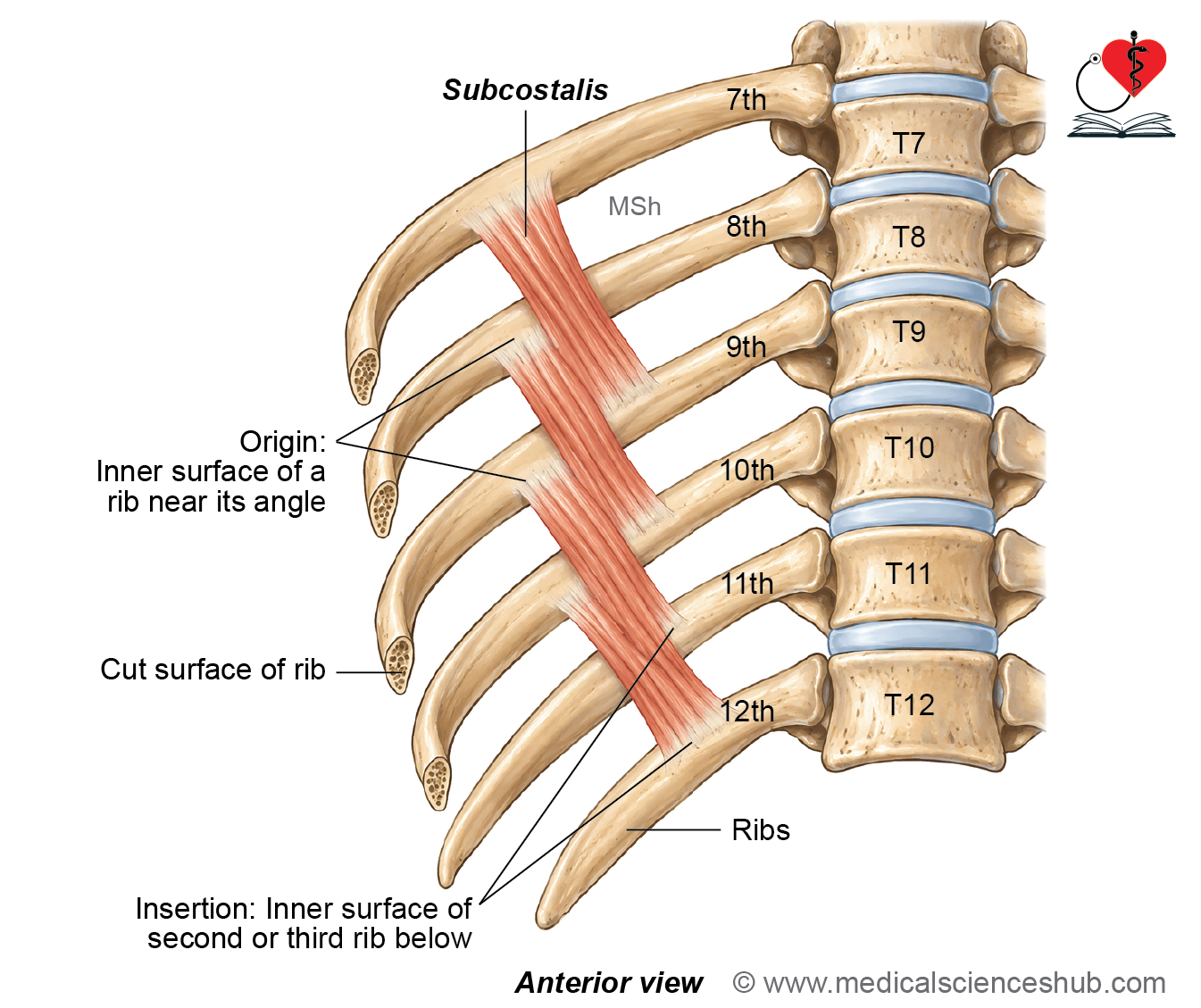
- The subcostalis muscles lie on the internal surface of the posterior thoracic wall. They occupy approximately the same plane as the innermost intercostal muscles. Their number is variable. They are better developed in the lower part of the thorax.
Attachments
- Each subcostalis muscle arises from the inner surface of a rib near its angle. Its fibers pass downward and medially, crossing one or two intercostal spaces. The muscle is inserted into the inner surface of the second or third rib below its origin.
Direction of Fibers
The fibers follow approximately the same direction as those of the internal and innermost intercostal muscles.
Nerve Supply
- The subcostalis muscles are supplied by the adjacent intercostal nerves.
Action
- The subcostalis muscles assist in depressing the ribs, particularly during forced expiration.
Sternocostalis (Transverse Thoracis)
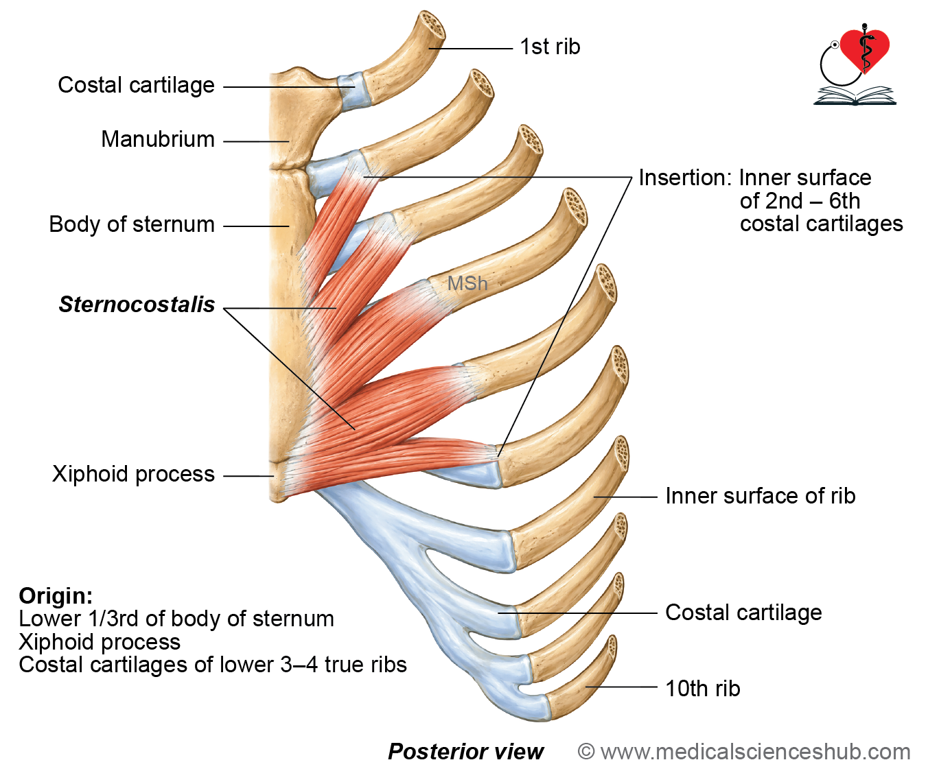
- The transversus thoracis is a thin muscle situated on the inner surface of the anterior thoracic wall. It lies immediately posterior to the sternum and adjacent costal cartilages. It is also known as sternocostalis or triangularis sterni. The muscle usually consists of four or five radiating slips on each side.
Origin
- The transversus thoracis arises from:
- The posterior surface of the lower part of the body of the sternum.
- The posterior surface of the xiphoid process.
- The posterior surfaces of the adjacent lower costal cartilages.
Insertion
- Its muscular slips are inserted into the inner surfaces of the second to sixth costal cartilages.
Direction of Fibers
- The fibers pass upward and laterally from their sternal origin toward the costal cartilages.
Nerve Supply
- The muscle is supplied by the adjacent intercostal nerves.
Action
- The transversus thoracis weakly pulls the second to sixth costal cartilages downward. It therefore assists in depression of the ribs during forced expiration.
Clinical Anatomy
Sternalis (rectus sternalis)
- The sternalis, also known as rectus sternalis, is a variant muscle of the anterior thoracic wall. It has also been described as japonicus, parasternalis, and sternalis brutorum.
It is present in approximately 8% of the population, although its frequency varies among different populations. The muscle usually lies superficial to the pectoralis major and runs approximately parallel to the lateral border of the sternum.
Attachments
• The attachments of the sternalis muscle are variable. Superiorly, it may arise from the upper part of the sternum or adjacent structures. Inferiorly, it may attach to the pectoral fascia, costal cartilages, lower true ribs, or rectus sheath.
Function
• The sternalis muscle has no clearly established physiological function.
Clinical Importance
The sternalis may be encountered during surgical procedures involving the anterior thoracic wall. When adequately developed, it may be used as a muscle flap in reconstructive and plastic surgery. Its presence is also important in radiological imaging because it may occasionally be mistaken for an abnormal soft-tissue mass.
Table
Mechanics of Respiration
- Respiration consists of two alternating phases: inspiration and expiration.
- Both phases are associated with changes in the dimensions and volume of the thoracic cavity.
- Inspiration is normally an active process in which contraction of respiratory muscles increases thoracic volume and draws air into the lungs. Quiet expiration is mainly a passive process produced by relaxation of the inspiratory muscles and elastic recoil of the lungs and thoracic wall. During quiet breathing, expiration generally lasts longer than inspiration.
Rate of Respiration
- The respiratory rate varies with age and is usually higher in younger individuals. In infants, the respiratory rate is approximately 30–40 breaths per minute. In children, it is approximately 20–30 breaths per minute. In adults at rest, the normal respiratory rate is approximately 12–20 breaths per minute.
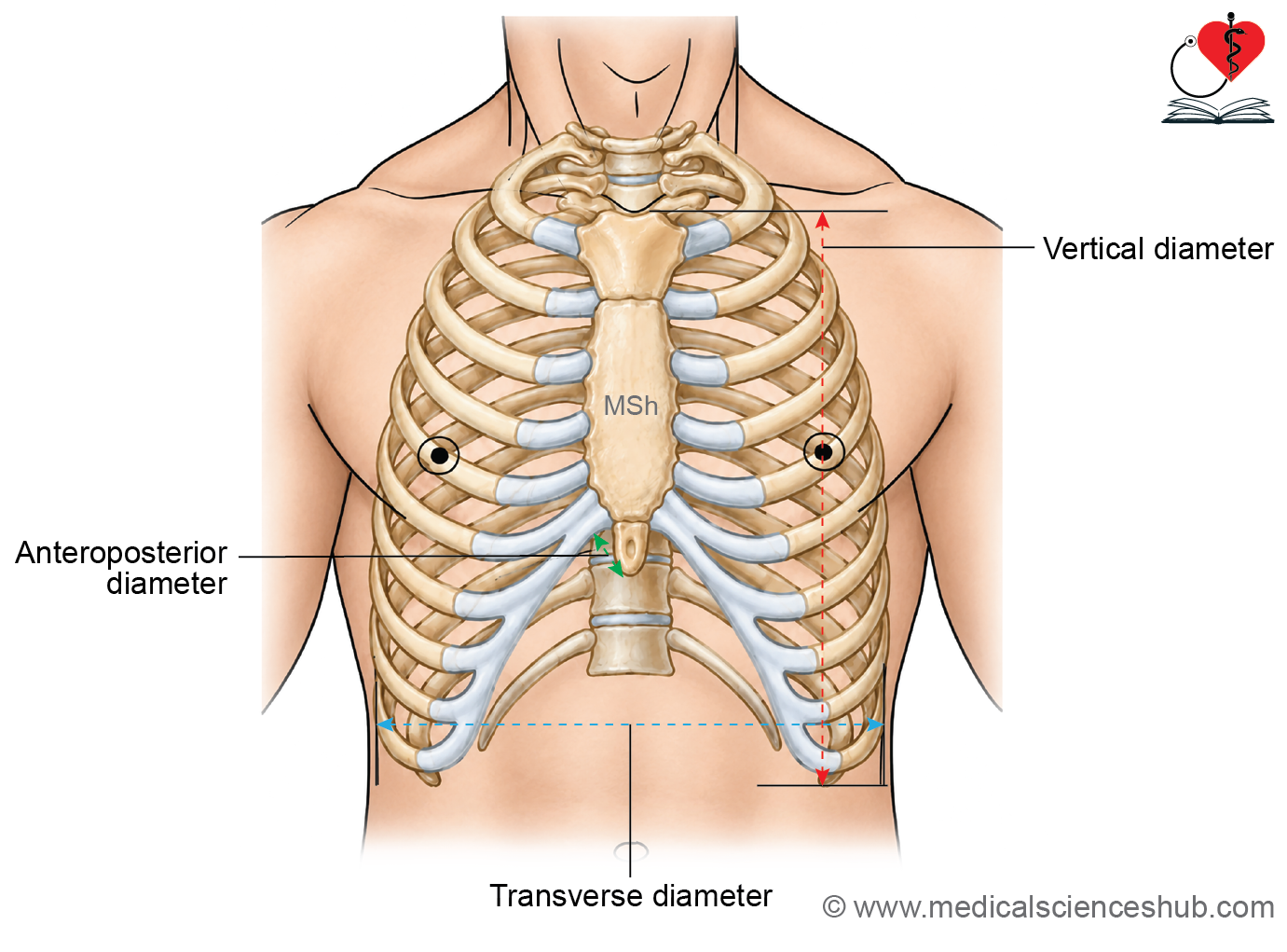
Diameters of Thoracic Cavity
- Respiratory movements alter three principal dimensions of the thoracic cavity:
- Anteroposterior diameter.
- Transverse diameter.
- Vertical diameter.
- During inspiration, these dimensions increase and enlarge the thoracic cavity. During expiration, the dimensions decrease as the thoracic cavity returns toward its resting volume.
Inspiration
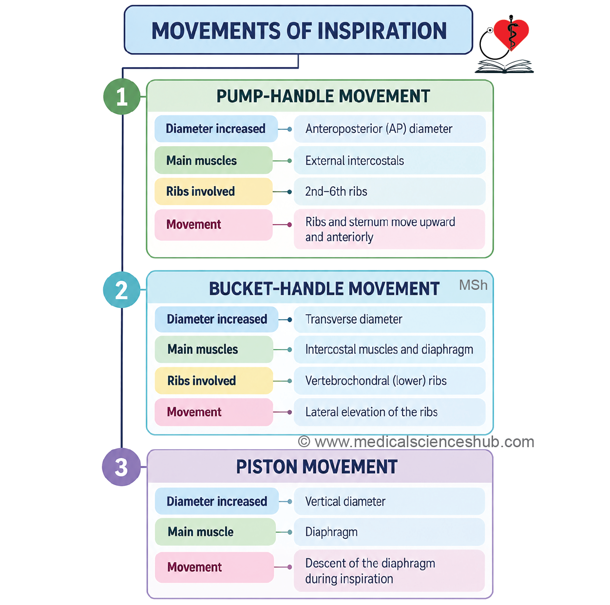
- During inspiration, expansion of the thoracic cavity lowers intrathoracic pressure and allows air to enter the lungs. Enlargement of the thoracic cavity occurs through coordinated movements of the ribs, sternum, and diaphragm. Three characteristic movements contribute to this expansion: pump-handle, bucket-handle, and piston movements.
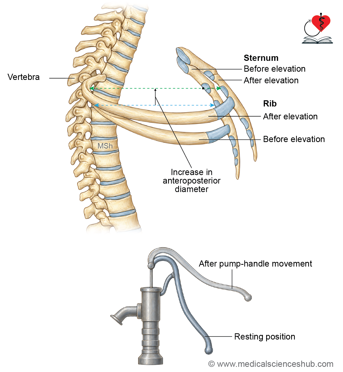
Anteroposterior diameter
- The anteroposterior diameter of the thorax increases mainly through the pump-handle movement of the upper ribs. During this movement, the anterior ends of the ribs and the sternum move upward and forward. This movement increases the distance between the sternum and the vertebral column.
- The external intercostal muscles assist in elevating the ribs during inspiration. Movements occur mainly at the costovertebral and costotransverse joints, with associated movement of the costal cartilages and sternum.
Pump-Handle Movement
- A typical rib slopes downward from its posterior end toward its anterior end. During inspiration, elevation of the rib causes its anterior end to move upward and forward.
- The sternum moves forward along with the anterior ends of the ribs. This movement resembles the raising of the handle of a water pump and is therefore called the pump-handle movement.
The resulting forward displacement of the sternum increases the anteroposterior diameter of the thoracic cavity. The movement is particularly prominent in the upper ribs.
Transverse diameter
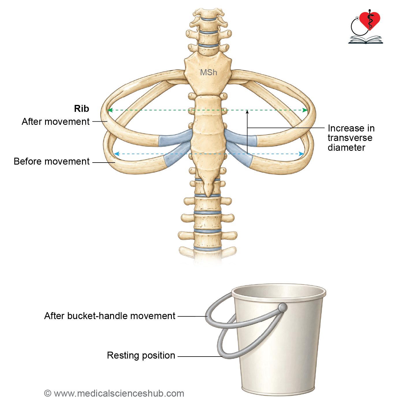
- The transverse diameter of the thoracic cavity increases mainly through the bucket-handle movement of the ribs. This movement is more prominent in the lower ribs.
- During inspiration, elevation of the ribs causes their lateral portions to move upward and outward. The outward displacement of the ribs increases the side-to-side dimension of the thoracic cavity. The movement resembles the elevation of a bucket handle.
- The intercostal muscles contribute to this movement by elevating and stabilizing the ribs. Movements occur principally at the costovertebral and costotransverse joints.
Bucket-Handle Movement
The curved shaft of a rib lies at a lower level than its anterior and posterior ends. When the rib is elevated, its lateral portion moves outward as well as upward. This outward movement enlarges the transverse diameter of the thoracic cavity.
Vertical diameter
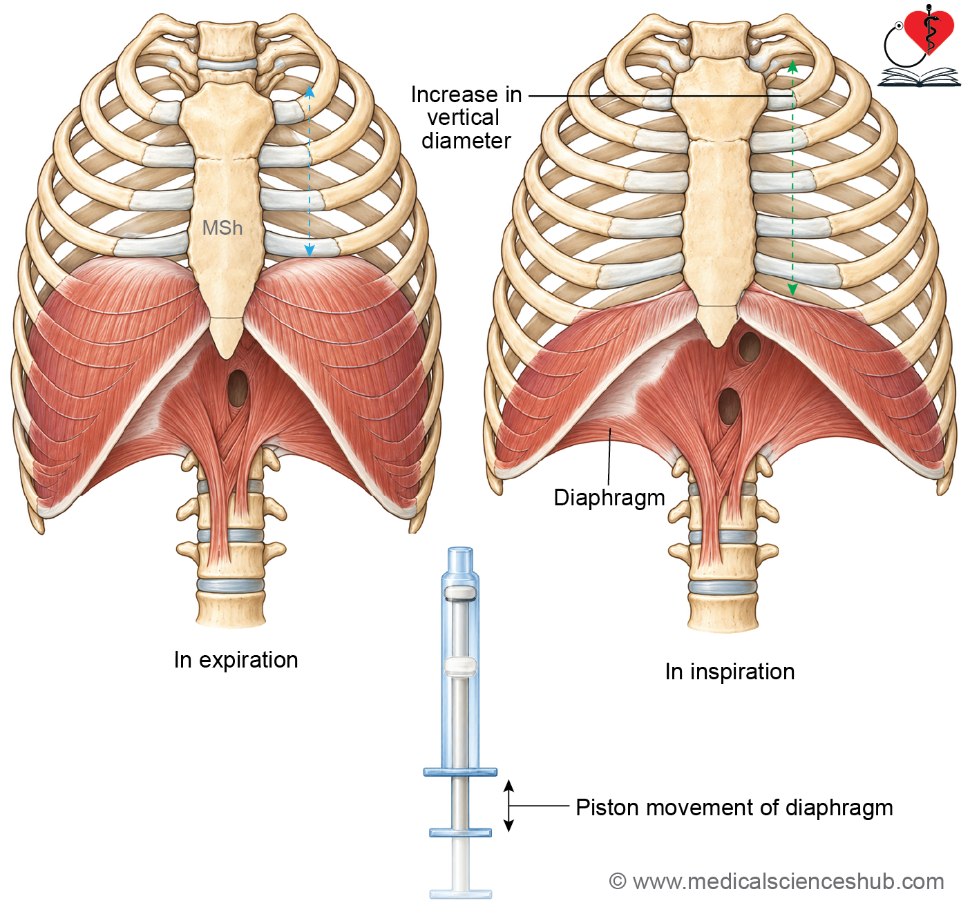
- The vertical diameter of the thoracic cavity increases mainly through the piston movement of the diaphragm.
- The diaphragm forms the muscular floor of the thoracic cavity. During inspiration, the diaphragm contracts and its domes descend. The central tendon moves downward as the diaphragm becomes flatter.
- This descent increases the vertical dimension of the thoracic cavity. The abdominal viscera are displaced downward and slightly forward as the diaphragm descends. The diaphragm is the principal muscle responsible for quiet inspiration.
Expiration
- Quiet expiration is predominantly a passive process. It begins when the diaphragm and other inspiratory muscles relax.
- Relaxation of the diaphragm allows its domes to rise toward their resting position.
- The ribs and sternum also return toward their resting positions. Elastic recoil of the lungs and thoracic wall reduces the volume of the thoracic cavity. The resulting increase in intrathoracic pressure drives air out of the lungs.
Forced Expiration
- Forced expiration is an active process and requires contraction of expiratory muscles.
- The internal intercostal muscles, particularly their interosseous parts, assist in depressing the ribs. The muscles of the anterior abdominal wall are major contributors to forced expiration by increasing intra-abdominal pressure and pushing the diaphragm upward.
- Transversus thoracis and serratus posterior inferior may provide additional assistance in depressing the ribs. Quadratus lumborum can assist by stabilizing or depressing the 12th rib during forceful respiratory movements.
Movements of the Diaphragm
The diaphragm moves only a short distance during quiet respiration. Its excursion becomes considerably greater during deep or forced breathing. The extent of diaphragmatic movement varies with posture, depth of respiration, and individual factors.
Clinical Anatomy
Rider’s bone is a sesamoid bone that may develop within the tendon of the adductor longus. In some individuals, it represents calcification of the tendon. It has been described in horse riders, where repeated friction and mechanical stress on the tendon may contribute to its formation.
Types of Respiration (Breathing)
- Breathing can be described according to its rate, depth, and the amount of muscular effort involved.
- Based on these features, respiration may be described as quiet, deep, or forced breathing.
1. Quiet Breathing
- Quiet breathing occurs during rest without conscious respiratory effort. Its rate and depth vary with age, physical condition, and metabolic requirements.
Inspiration
- Quiet inspiration is an active process. The diaphragm is the principal muscle of quiet inspiration. The external intercostal muscles assist by elevating and stabilizing the ribs.
Expiration
- Quiet expiration is mainly a passive process. It occurs through relaxation of the inspiratory muscles and elastic recoil of the lungs and thoracic wall. Under normal resting conditions, no major expiratory muscle contraction is required.
2. Deep Breathing
- Deep breathing involves a greater volume of air entering and leaving the lungs with each respiratory cycle. It may occur voluntarily during breathing exercises, including yoga and other respiratory practices. During slow deep breathing, the depth of respiration increases while the respiratory rate may decrease.
Inspiration
- The diaphragm and external intercostal muscles continue to play major roles. With greater inspiratory effort, accessory muscles may also assist in expanding the thoracic cavity. These may include the scalene muscles, sternocleidomastoid, levatores costarum, and serratus posterior superior.
Expiration
- Expiration following a moderately deep inspiration may occur largely through elastic recoil. During more vigorous deep breathing, expiratory muscles may also become active.
3. Forced Breathing
- Forced breathing occurs when ventilation must increase substantially, such as during strenuous physical activity. It may also occur in respiratory disorders associated with increased difficulty in moving air into or out of the lungs. Both inspiration and expiration become active processes during forceful breathing.
Forced Inspiration
- The diaphragm and external intercostal muscles contract more strongly.
- Accessory inspiratory muscles are recruited when additional thoracic expansion is required. These may include the sternocleidomastoid, scalene muscles, pectoral muscles when the upper limb is fixed, and other muscles capable of elevating the ribs.
Forced Expiration
Forced expiration requires active muscular contraction. The internal intercostal muscles assist by depressing the ribs. The anterior abdominal muscles increase intra-abdominal pressure and push the diaphragm upward. Additional muscles may assist depending on the intensity of expiration and the position of the body.
Clinical Anatomy
Rider’s bone is a sesamoid bone that may develop within the tendon of the adductor longus. In some individuals, it represents calcification of the tendon. It has been described in horse riders, where repeated friction and mechanical stress on the tendon may contribute to its formation.
📝 Test Your Knowledge – Practice MCQs
Attempt the chapter MCQ quiz and assess your understanding of key concepts.
Click to see: Medical Disclaimer and Copyright Notice