
Competencies
- AN8.5: Identify and name various bones in articulated hand, specify the parts of metacarpals and phalanges and enumerate the peculiarities of pisiform.
- AN8.6: Features of individual bones (upper limb): Describe scaphoid fracture and explain the anatomical basis of avascular necrosis. This chapter includes carpals, metacarpals, phalanges, peculiarities of pisiform, and avascular necrosis of scaphoid.
Introduction
Bones of Hand
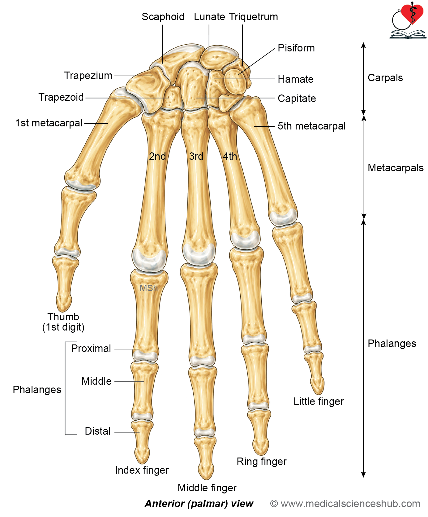
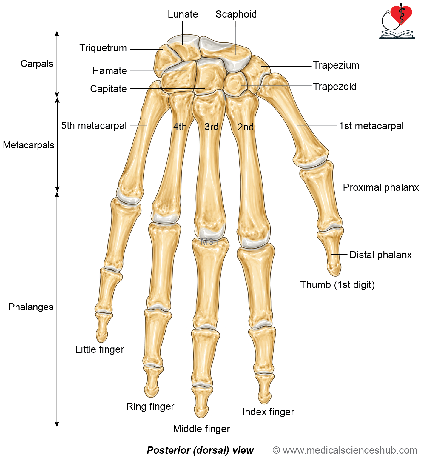
- Each hand contains a total of 27 bones.
- The carpus is formed by 8 carpal bones, arranged in two rows.
- The metacarpus consists of 5 metacarpal bones, which form the framework of the palm.
- The digits contain 14 phalanges, including proximal, middle, and distal phalanges. The thumb has only proximal and distal phalanges.
- Therefore, each hand is composed of 8 carpal bones, 5 metacarpals, and 14 phalanges, making a total of 27 bones.
Carpal Bones
- The carpal bones are 8 short bones that form the skeleton of the wrist.
- They connect the hand with the forearm and are arranged in two rows.
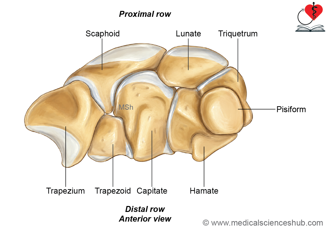
- The proximal row, from lateral to medial, consists of the scaphoid, lunate, triquetrum, and pisiform.
- The distal row, from lateral to medial, consists of the trapezium, trapezoid, capitate, and hamate.
- The bones of the proximal row, except the pisiform, articulate proximally with the distal end of the radius and the articular disc to form the wrist joint.
- Distally, the scaphoid, lunate, and triquetrum articulate with the bones of the distal carpal row.
- The pisiform does not participate in the wrist joint. It is a sesamoid bone within the tendon of flexor carpi ulnaris and articulates only with the triquetrum.
- The distal row of carpal bones articulates distally with the bases of the metacarpal bones, forming the carpometacarpal joints.
Scaphoid Bone
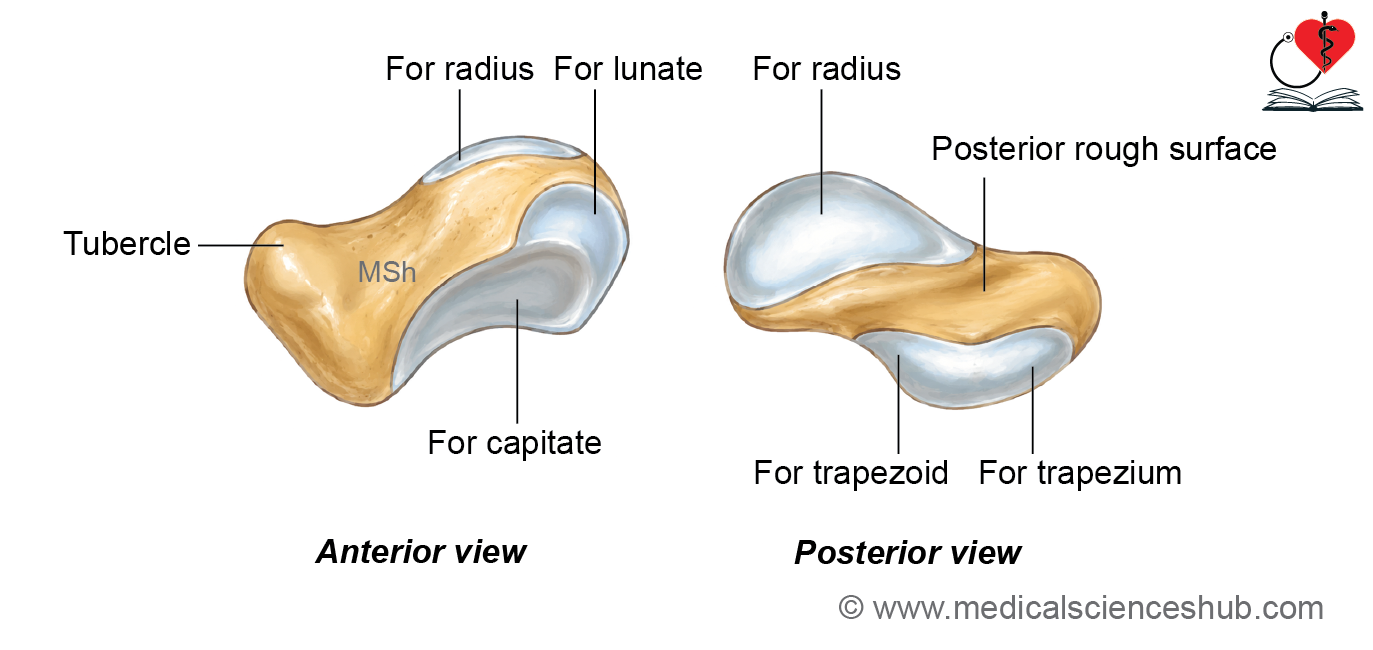
- The scaphoid is the largest and most lateral bone of the proximal row of carpal bones.
- It is boat-shaped, and its long axis is directed downward, forward, and laterally.
Surfaces
- The scaphoid has six surfaces: anterior, posterior, superior, inferior, medial, and lateral.
- The anterior (palmar) surface is gently concave and non-articular. It bears the scaphoid tubercle, which provides attachment to the flexor retinaculum and gives origin to the abductor pollicis brevis muscle.
- The posterior (dorsal) surface is non-articular and contains a narrow groove.
- The superior surface is convex, smooth, and articulates with the lateral part of the distal articular surface of the radius, contributing to the wrist joint.
- The inferior surface articulates with the trapezium and trapezoid.
- The medial surface has two articular facets: a small superior semilunar facet for the lunate and a larger deep concave facet for the capitate.
- The lateral surface is narrow, non-articular, and continuous with the scaphoid tubercle. It provides attachment to the radial collateral ligament of the wrist joint.
CLINICAL INTEGRATION
Scaphoid fracture and avascular necrosis
- Scaphoid fracture is the most common fracture of the carpal bones.
- A scaphoid fracture commonly occurs after a fall on an outstretched hand.
- Patients typically present with tenderness and swelling in the anatomical snuffbox.
- The initial investigation is a plain radiograph of the wrist, although early fractures may not always be visible.
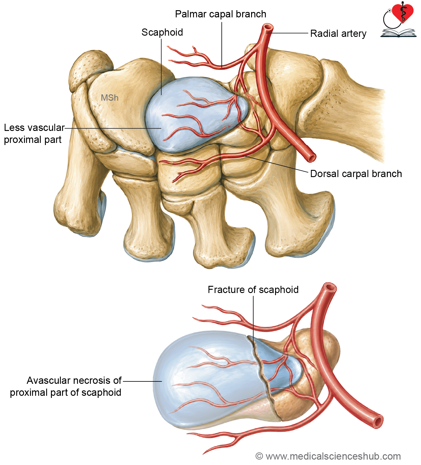
- The scaphoid receives approximately 20 to 30 percent of its blood supply from the palmar branch and 70 to 80 percent from the dorsal branches of the radial artery.
- Most of the proximal part of the scaphoid is supplied through vessels entering the distal part. Therefore, a fracture through the waist or proximal part of the bone may interrupt this retrograde blood supply, leading to avascular necrosis of the proximal fragment.
- Non-union is another important complication of a scaphoid fracture because of its relatively poor blood supply.
Lunate Bone
- The lunate is a semilunar (moon-shaped) bone located in the proximal row of the carpal bones.
Surfaces
- It has six surfaces: superior, inferior, lateral, medial, anterior, and posterior.
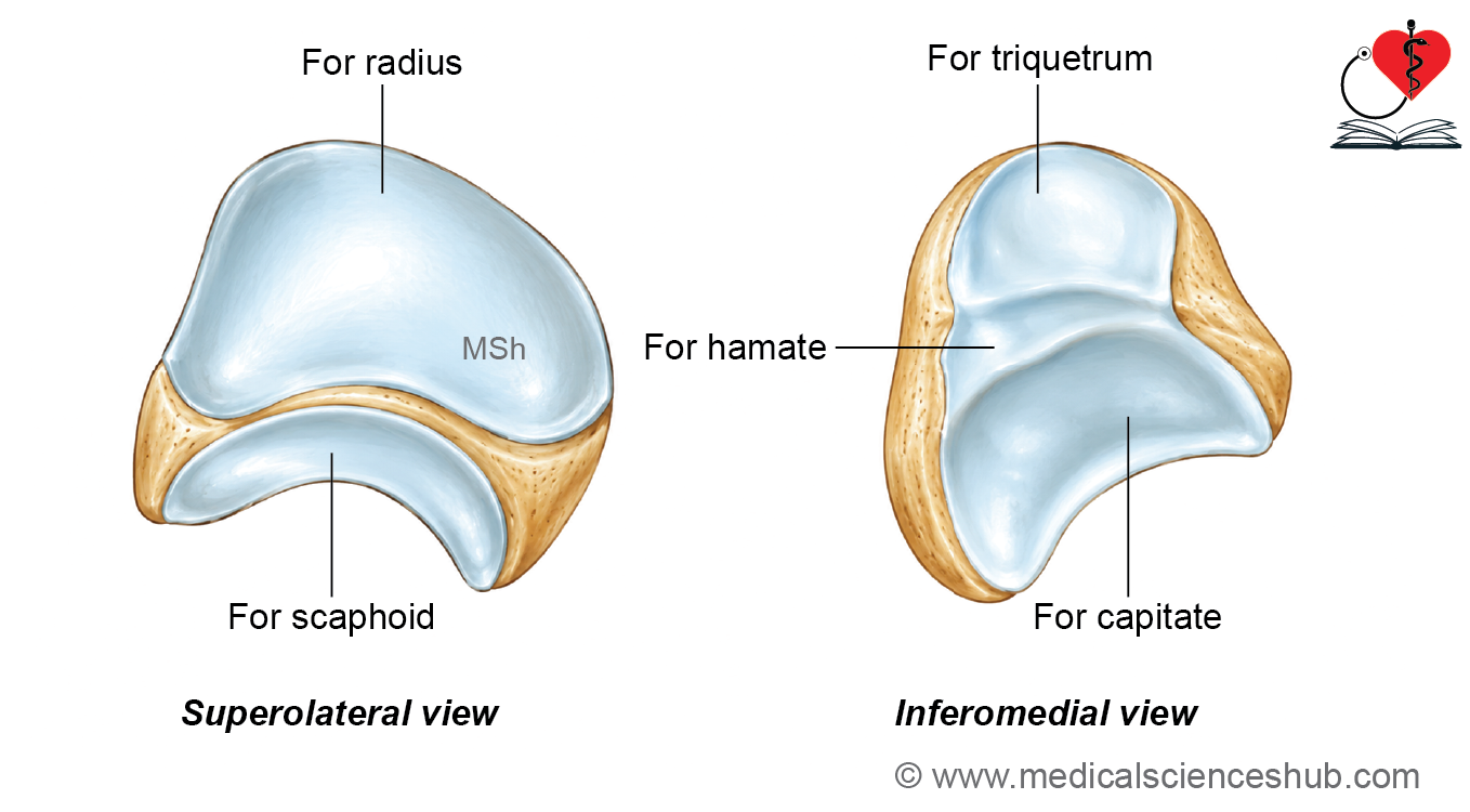
- The superior (proximal) surface is convex and articulates with the medial part of the distal articular surface of the radius, contributing to the wrist joint.
- The inferior (distal) surface is concave and has two articular facets. The lateral deep concave facet articulates with the capitate, whereas the medial narrow facet articulates with the hamate.
- The lateral surface bears a semilunar articular facet that articulates with the scaphoid.
- The medial surface has a quadrilateral articular facet that articulates with the triquetrum.
- The anterior (palmar) surface is non-articular, larger, and triangular.
- The posterior (dorsal) surface is non-articular, smaller, and quadrilateral.
Triquetrum
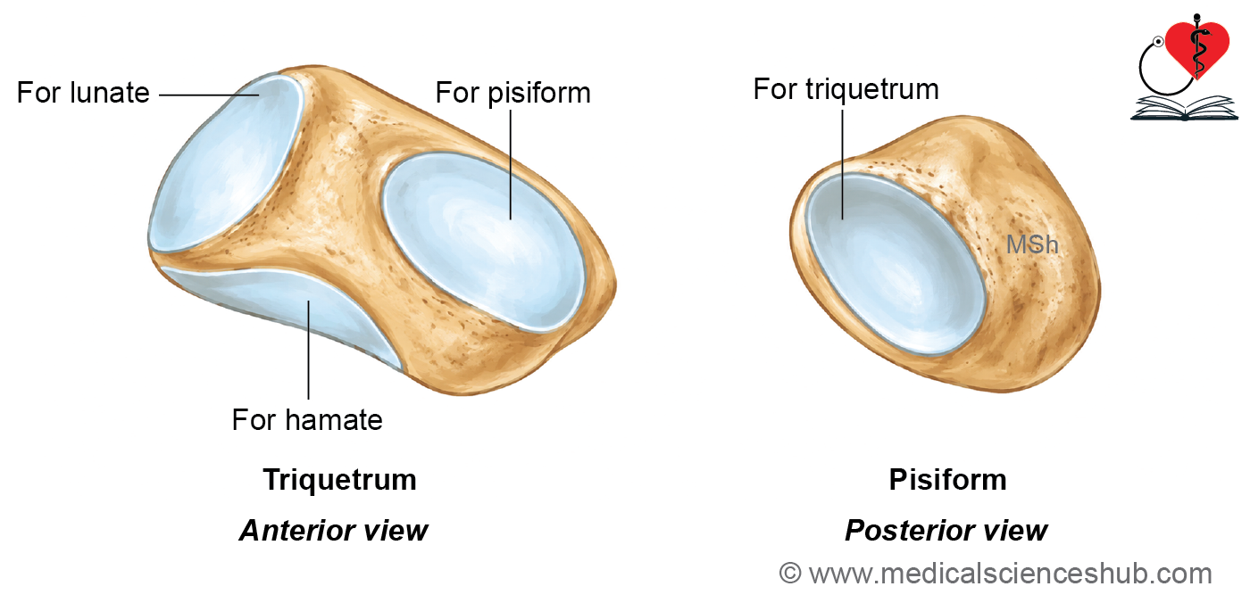
The triquetrum is a wedge-shaped bone located in the proximal row of the carpal bones..
Surfaces
- It has six surfaces: superior, inferior, anterior, posterior, lateral, and medial.
- The superior (proximal) surface bears an articular facet that articulates with the triangular articular disc of the distal radioulnar joint.
- The inferior (distal) surface has a concavo-convex articular facet for the hamate.
- The anterior (palmar) surface contains a circular articular facet for the pisiform, which serves as an important identifying feature.
- The posterior (dorsal) surface is non-articular and is continuous with the superior surface.
- The lateral surface, which forms the base of the wedge, has a square-shaped articular facet for the lunate.
- The medial surface forms the apex of the wedge and provides attachment to the ulnar collateral ligament of the wrist joint.
Pisiform
- The pisiform is the smallest and most medial bone of the proximal row of the carpal bones. It is a sesamoid bone that develops within the tendon of flexor carpi ulnaris.
Surfaces
- The posterior surface bears a circular superior articular facet for articulation with the triquetrum, while its inferior part is non-articular. This is an important identifying feature.
- The anterior surface is ridge-like and receives the insertion of flexor carpi ulnaris. It also provides attachment to the pisohamate ligament.
- The medial surface is convex and larger than the lateral surface. It gives origin to the abductor digiti minimi and provides attachment to the extensor retinaculum.
- The lateral surface is flat and has an indistinct vertical ridge that provides attachment to the flexor retinaculum.
- The pisiform serves as the insertion site of flexor carpi ulnaris and contributes to the attachment of the flexor retinaculum, enhancing the mechanical advantage of the muscle.
Peculiarities of Pisiform Bone
- The pisiform is a small, pea-shaped sesamoid bone that develops within the tendon of flexor carpi ulnaris.
- Proximally, it receives the insertion of the flexor carpi ulnaris tendon. Distally, it provides attachment to the pisohamate ligament.
- The pisiform articulates only with the anterior surface of the triquetrum.
- Unlike the other carpal bones, it does not possess articular facets on its medial, lateral, or inferior surfaces.
- The pisiform is the last carpal bone to ossify.
- It develops from a single secondary ossification center, consistent with its sesamoid nature.
- As a sesamoid bone, the pisiform typically lacks a nutrient foramen.
Trapezium
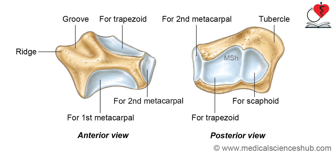
The trapezium is the most lateral bone of the distal row of carpal bones.
Surfaces
- It has six surfaces: anterior, posterior, superior, inferior, lateral, and medial.
- The anterior (palmar) surface has a lateral crest, a central groove, and a sharp medial margin. The crest and sharp margin provide attachment to the superficial and deep layers of the flexor retinaculum, converting the groove into an osseofibrous canal for the tendon of flexor carpi radialis. The crest also gives origin to the abductor pollicis brevis, opponens pollicis, and flexor pollicis brevis muscles.
- The posterior (dorsal) surface is long, narrow, and non-articular.
- The superior surface has a concave articular facet for the scaphoid.
- The inferior surface has a saddle-shaped (concavoconvex) articular facet for the first metacarpal, forming the first carpometacarpal joint that permits the wide range of thumb movements.
- The lateral surface is rough and non-articular, providing attachment to the radial collateral ligament of the wrist joint.
- The medial surface has an elongated articular facet for the trapezoid and a distal rectangular facet for the base of the second metacarpal.
Trapezoid
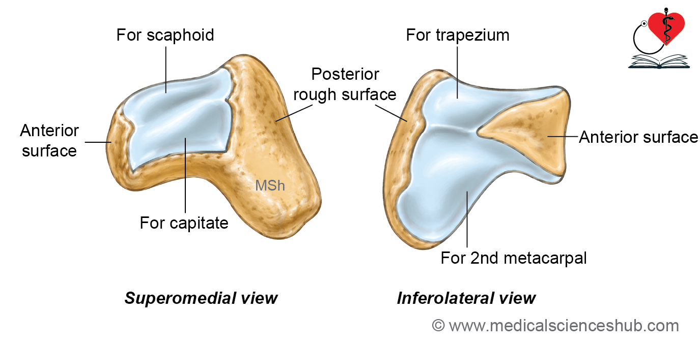
- The trapezoid is a shoe-shaped carpal bone located in the distal row between the trapezium and the capitate.
Surfaces
- It has six surfaces: anterior, posterior, superior, inferior, lateral, and medial.
- The anterior (palmar) surface is rough and non-articular.
- The posterior (dorsal) surface is also rough, non-articular, and larger than the anterior surface. It gives origin to some fibers of the oblique head of the adductor pollicis muscle.
- The superior surface has a rectangular articular facet for the scaphoid.
- The inferior surface has a concave articular facet for the base of the second metacarpal.
- The lateral surface bears an oblique convex articular facet for the trapezium.
- The medial surface has a square-shaped articular facet for the capitate.
Capitate
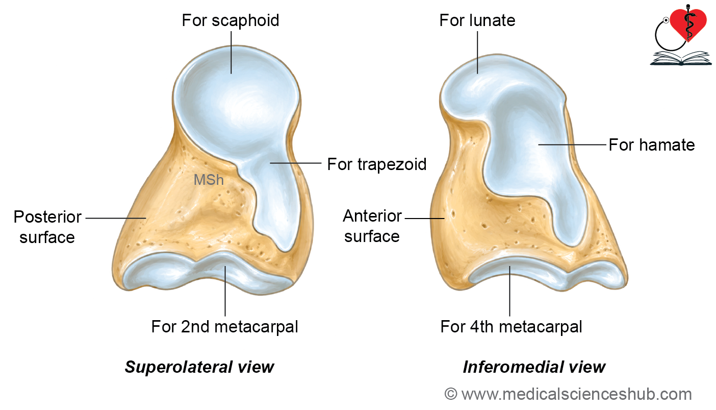
- The capitate is the largest of the carpal bones and is located in the center of the distal row.
Surfaces
- It has six surfaces: superior, inferior, lateral, medial, anterior, and posterior.
- The superior surface, also called the head, is rounded and serves as an important identifying feature. It articulates with the scaphoid, lunate, and trapezoid.
- The inferior surface is triangular and has two articular facets: a large central facet for the base of the third metacarpal and a smaller posteroinferior strip-like facet for the fourth metacarpal.
- The lateral surface, or neck, is constricted and bears an articular facet for the trapezoid.
- The medial surface is broad and flat, with an articular facet near its posterior border for the hamate.
- The anterior (palmar) surface is rough and non-articular. It provides attachment for a few fibers of the oblique head of the adductor pollicis muscle.
- The posterior (dorsal) surface is also rough and non-articular and is longer than the anterior surface.
Hamate
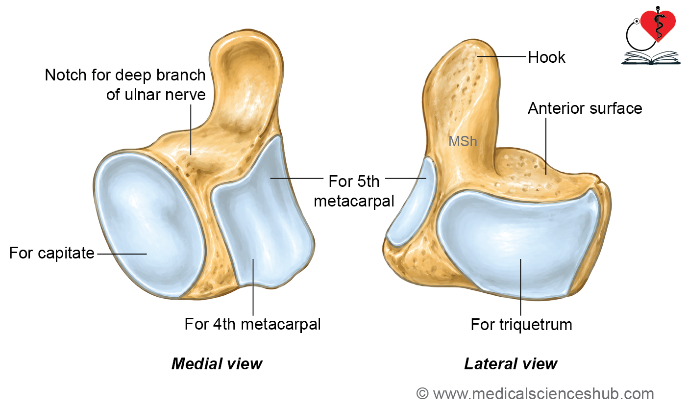
The hamate is a wedge-shaped carpal bone with a prominent hook-like projection called the hamulus.
- It is the most medial bone of the distal row of carpal bones.
Surfaces
- The hamate has six surfaces: anterior, posterior, superior, inferior, medial, and lateral.
- The anterior (palmar) surface bears the hamulus, which is concave laterally and convex medially. The tip of the hamulus provides attachment to the flexor retinaculum. The medial side of the hamulus gives origin to the opponens digiti minimi and flexor digiti minimi brevis muscles.
- The posterior (dorsal) surface is rough and non-articular.
- The superior surface, which forms the apex of the wedge, has an articular facet for the lunate.
- The inferior surface is divided by a faint ridge into a smaller lateral facet for the fourth metacarpal and a larger medial facet for the fifth metacarpal.
- The medial surface has a concavoconvex articular facet for the triquetrum.
- The lateral surface bears an oval articular facet for the capitate.
Ossification
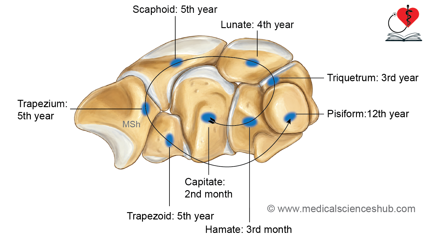
- The carpal bones generally ossify from secondary ossification centers, except the capitate and hamate, which ossify from primary ossification centers.
- The capitate is the first carpal bone to ossify, with its primary center appearing during the second month of intrauterine life.
- The hamate develops from a primary ossification center that appears during the third month of intrauterine life.
- The triquetrum begins ossification around the third year after birth.
- The lunate ossifies around the fourth year.
- The scaphoid, trapezium, and trapezoid usually begin ossification during the fifth year.
- The pisiform is the last carpal bone to ossify, with its ossification center appearing between 9 and 12 years of age, most commonly around the 12th year.
CLINICAL INTEGRATION
Age determination
- The appearance of carpal ossification centers is useful for age determination during childhood.
- Normally, 2 carpal bones have begun ossification by 2 years of age.
- By the third year, 3 carpal bones have ossified.
- By the fourth year, 4 carpal bones have ossified.
- Between the fifth and eleventh years, 7 carpal bones have ossified.
- By the twelfth year, all 8 carpal bones have ossified.
Scaphoid fracture and avascular necrosis
- A scaphoid fracture commonly occurs after a fall on an outstretched hand.
- Patients typically present with tenderness and swelling in the anatomical snuffbox.
- The initial investigation is a plain radiograph of the wrist, although early fractures may not always be visible.
- The scaphoid receives approximately 20 to 30 percent of its blood supply from the palmar branch and 70 to 80 percent from the dorsal branches of the radial artery.
- Most of the proximal part of the scaphoid is supplied through vessels entering the distal part. Therefore, a fracture through the waist or proximal part of the bone may interrupt this retrograde blood supply, leading to avascular necrosis of the proximal fragment.
- Non-union is another important complication of a scaphoid fracture because of its relatively poor blood supply.
Metacarpals
- There are five metacarpal bones, which form the skeletal framework of the palm.
- The metacarpals are classified as miniature long bones.
- They are numbered from lateral to medial as the first to fifth metacarpals, beginning with the thumb and ending with the little finger.
General Features of Metacarpals
- Each metacarpal consists of a base, a shaft, and a head.
- The shaft is curved, being concave anteriorly (towards the palm) and convex posteriorly (towards the dorsum).
- The dorsal surface of the shaft has a flat, triangular area.
- The head is located at the distal end and is larger than the base. It articulates with the proximal phalanx to form the metacarpophalangeal joint.
- The base is situated at the proximal end and articulates with the carpal bones and adjacent metacarpals.
First Metacarpal Bone
- The first metacarpal is the shortest and stoutest of the five metacarpal bones.
- Unlike the other metacarpals, its dorsal surface faces laterally and does not have a flat triangular area.
- The palmar surface has a longitudinal ridge that divides it into a larger anterolateral area and a smaller anteromedial area.
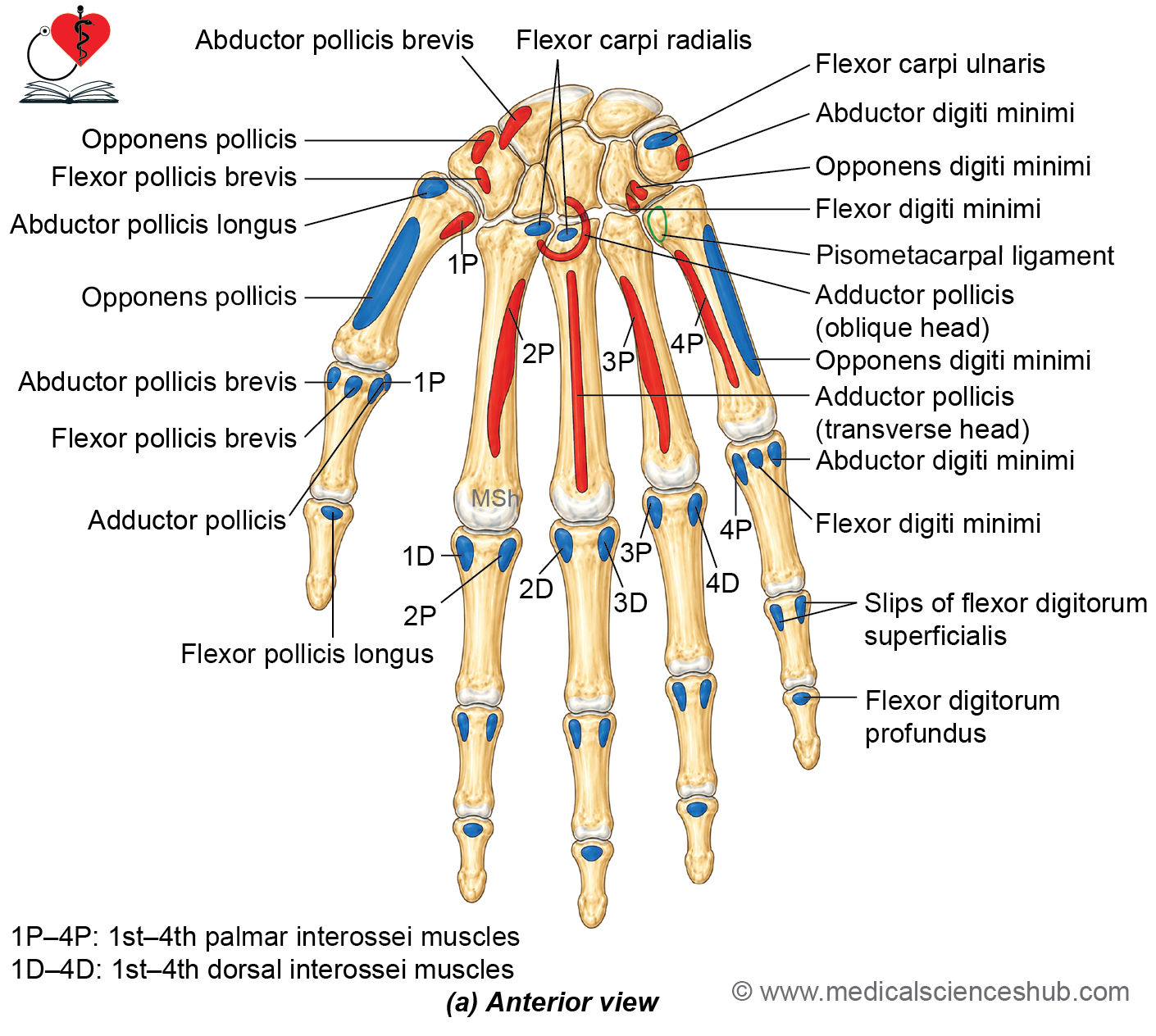
- The larger anterolateral area provides insertion for the opponens pollicis, whereas the smaller anteromedial area gives origin to the lateral head of the first dorsal interosseous muscle.
- The base has a saddle-shaped (concavoconvex) articular facet for the trapezium, forming the first carpometacarpal joint.
- The lateral aspect of the base receives the insertion of abductor pollicis longus, while the medial aspect gives origin to the first palmar interosseous muscle.
- The head is convex and broader from side to side than the heads of the other metacarpals.
- The first metacarpal is rotated approximately 90 degrees relative to the other metacarpals. Consequently, its dorsal surface faces laterally, its lateral surface faces anteriorly, and thumb extension occurs at approximately 90 degrees to the plane of the palm.
Second Metacarpal Bone
- The second metacarpal is the longest of the five metacarpal bones.
- Its base is grooved and primarily articulates with the trapezoid.
- The anterolateral part of the base also has a quadrangular articular facet for the trapezium.
- The medial side of the base bears an articular facet for the third metacarpal.
Attachments
- The anterior aspect of the base receives the insertion of flexor carpi radialis and gives origin to some fibers of the oblique head of adductor pollicis.
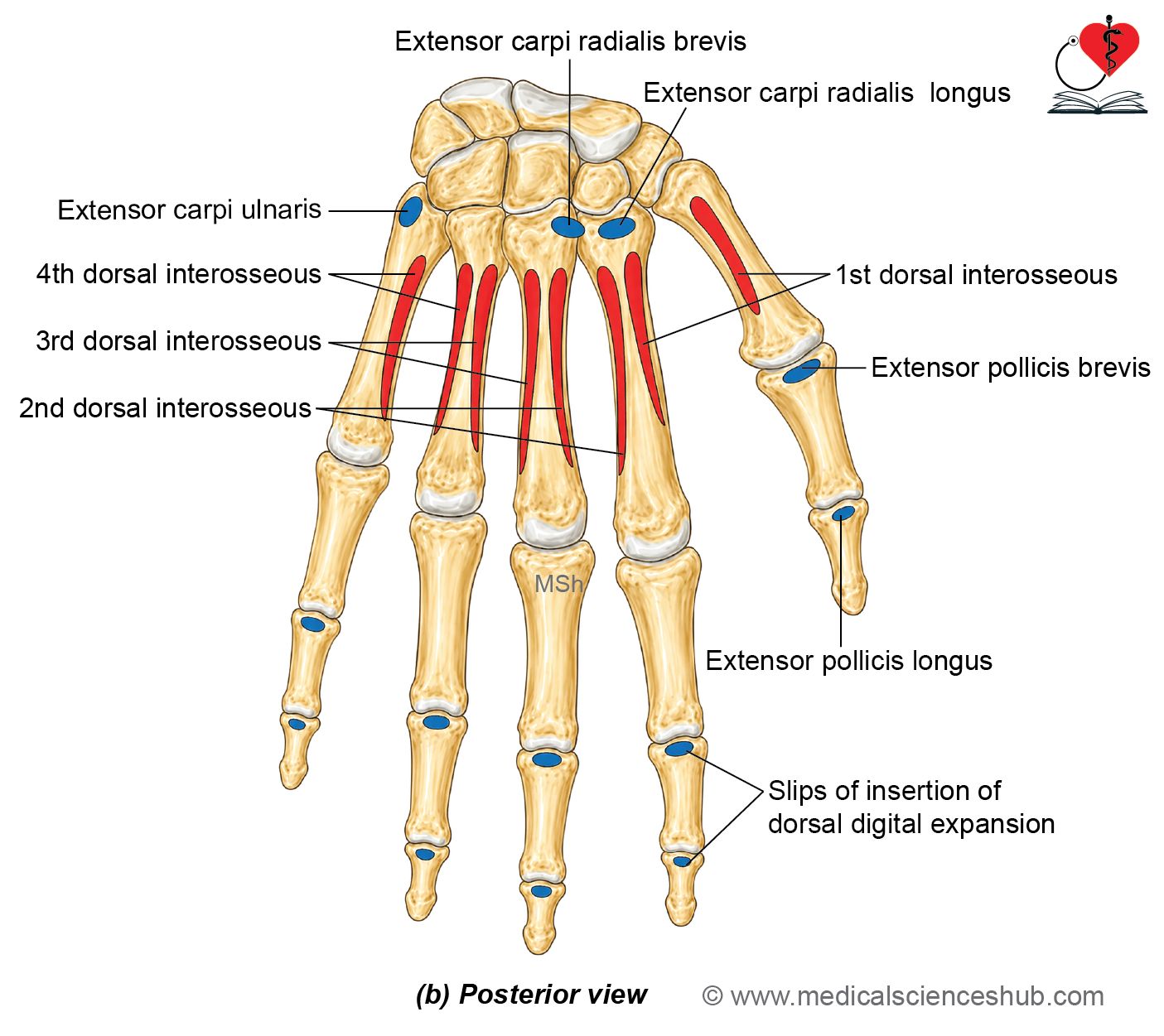
- The posterior aspect of the base receives the insertion of extensor carpi radialis longus.
- The lateral side of the shaft gives origin to the medial head of the first dorsal interosseous muscle.
- The medial surface of the shaft is divided by a vertical ridge into anterior and posterior areas.
- The anterior area gives origin to the second palmar interosseous muscle.
- The posterior area gives origin to the lateral head of the second dorsal interosseous muscle.
Third Metacarpal Bone
- The third metacarpal is distinguished by a prominent styloid process at the base.
- The base primarily articulates with the capitate.
- A strip-like articular facet on the lateral side of the base articulates with the second metacarpal.
- Two oval articular facets on the medial side of the base articulate with the fourth metacarpal.
Attachments
- The anterior aspect of the base receives the insertion of flexor carpi radialis and gives origin to some fibers of the oblique head of adductor pollicis.
- The extensor carpi radialis brevis inserts on the posterior aspect of the styloid process.
- The lateral aspect of the shaft gives origin to the medial head of the second dorsal interosseous muscle.
- The medial aspect of the shaft gives origin to the lateral head of the third dorsal interosseous muscle.
- A ridge on the anterior surface of the shaft provides origin to the transverse head of the adductor pollicis muscle.
Fourth Metacarpal Bone
- The fourth metacarpal has a quadrilateral base.
- Two circular articular facets on the lateral side of the base articulate with the third metacarpal.
- The superior surface of the base articulates with the hamate.
- A strip-like articular facet on the medial side of the base articulates with the fifth metacarpal.
Attachments
- A ridge on the lateral surface of the shaft divides it into anterior and posterior areas.
- The anterior area gives origin to the third palmar interosseous muscle.
- The posterior area gives origin to the medial head of the third dorsal interosseous muscle.
- The medial surface of the shaft gives origin to the lateral head of the fourth dorsal interosseous muscle.
Fifth Metacarpal Bone
- The fifth metacarpal has a non-articular tubercle on the medial aspect of its base.
- The tubercle receives the insertion of extensor carpi ulnaris.
- The superior surface of the base articulates with the hamate.
- A strip-like facet on the lateral side of the base articulates with the fourth metacarpal.
Attachments
- The medial surface of the shaft gives origin to the opponens digiti minimi muscle.
- The anterior part of the lateral surface gives origin to the fourth palmar interosseous muscle.
- The posterior part of the lateral surface gives origin to the medial head of the fourth dorsal interosseous muscle.
Ossification of Metacarpals
- Each metacarpal is classified as a miniature long bone because it develops from one primary and one secondary ossification center, unlike typical long bones, which usually have two secondary ossification centers.
- The primary ossification center appears in the shaft during the ninth week of intrauterine life.
- The secondary ossification center appears by approximately 2 years of age.
- In the first metacarpal, the secondary center is located at the base.
- In the second to fifth metacarpals, the secondary centers are located in the heads of the bones.
- These secondary centers usually fuse with the shaft between 16 and 18 years of age.
- Therefore, the epiphysis is present at the head of all metacarpals except the first, in which the epiphysis is located at the base.
Phalanges
Each hand contains 14 phalanges. The thumb has 2 phalanges (proximal and distal), whereas each of the other four fingers has 3 phalanges (proximal, middle, and distal).
General Features of Phalanx
Every phalanx consists of a base, a shaft, and a head.
- The base is the expanded proximal end and bears a concave oval articular facet for articulation with the adjacent metacarpal or proximal phalanx.
- The shaft gradually tapers distally. Its anterior (palmar) surface is flat from side to side and concave along its long axis, whereas the posterior (dorsal) surface is convex.
- The head forms the distal end of the phalanx. The heads of the proximal and middle phalanges have pulley-shaped articular surfaces that articulate with the bases of the succeeding phalanges.
- The distal phalanx ends in a non-articular tapering terminal part. On its anterior aspect, the distal phalanx bears a rough horseshoe-shaped tubercle, which provides support for the fingertip pulp and attachment for the fibrous septa of the digital pad.
Attachments
- The proximal phalanx receives attachment of the fibrous flexor sheath along the sides of its shaft.
- The lumbricals and interossei muscles insert on the sides of the base of the proximal phalanx.
- In the middle phalanx, the two slips of flexor digitorum superficialis insert on either side of the shaft. The fibrous flexor sheath is also attached to the shaft.
- The extensor digitorum tendon inserts on the dorsal surface of the base of the middle phalanx.
- In the distal phalanx, the flexor digitorum profundus inserts on the anterior (palmar) surface of the base. The two slips of the dorsal digital expansion unite and attach to the posterior (dorsal) surface of the base.
- In the thumb, the base of the proximal phalanx receives insertion of abductor pollicis brevis and flexor pollicis brevis laterally, while adductor pollicis and the first palmar interosseous insert medially. The extensor pollicis brevis inserts on the dorsal aspect of the base.
- The base of the distal phalanx of the thumb receives the insertion of flexor pollicis longus on its palmar surface and extensor pollicis longus on its dorsal surface.
- In the little finger, the medial side of the base of the proximal phalanx receives insertion of abductor digiti minimi and flexor digiti minimi brevis.
Ossification
- Each phalanx develops from one primary and one secondary ossification center.
- The primary ossification center forms the shaft. It appears during the eighth week of intrauterine life in the distal phalanges, the tenth week in the proximal phalanges, and the twelfth week in the middle phalanges.
- The secondary ossification center develops at the base of each phalanx.
- The secondary center usually appears by 2 years of age and fuses with the shaft by approximately 16 years of age.
CLINICAL INTEGRATION
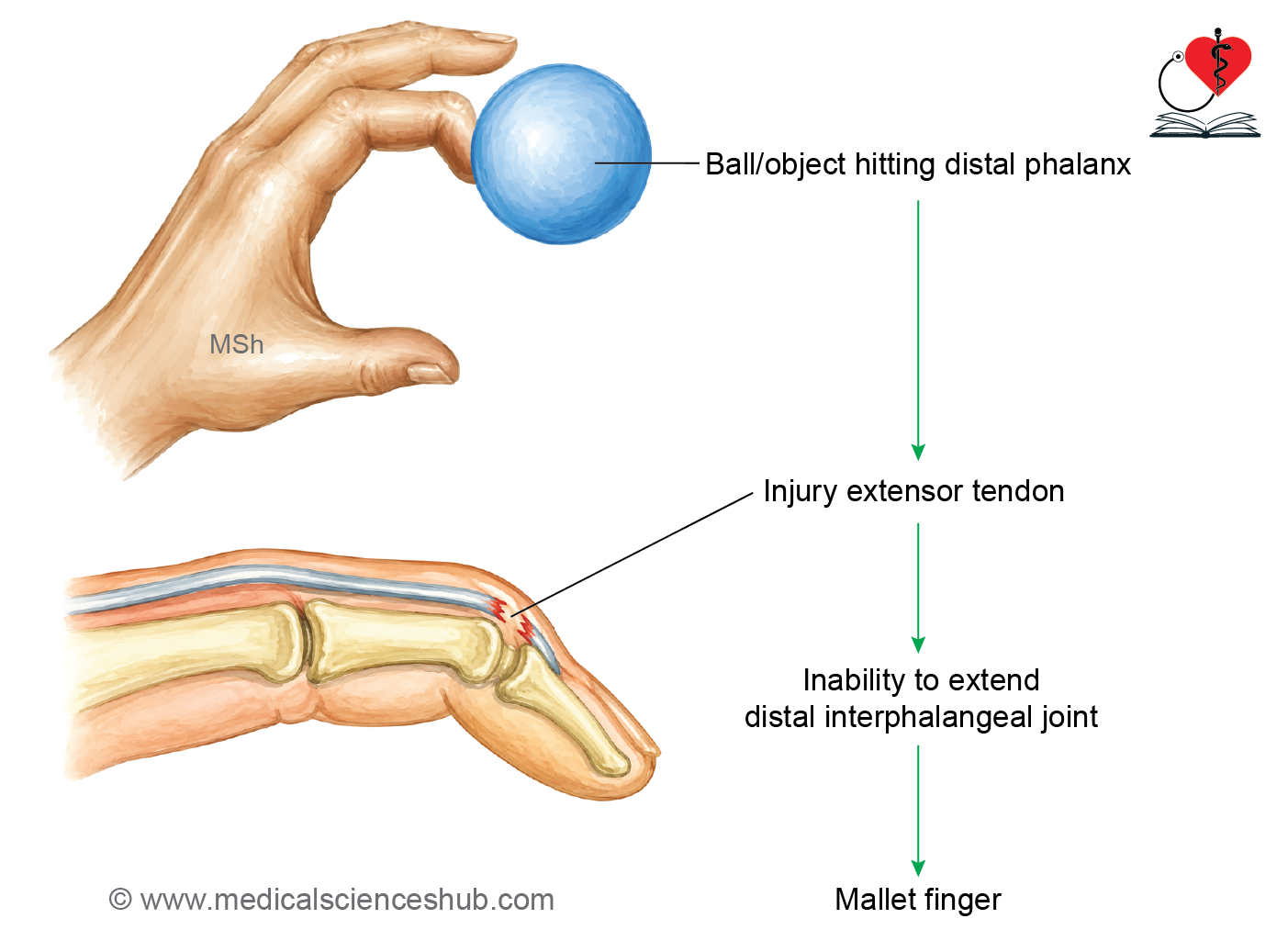
- Mallet finger (hammer finger) results from injury or rupture of the extensor tendon at the distal phalanx, causing inability to actively extend the distal interphalangeal joint.
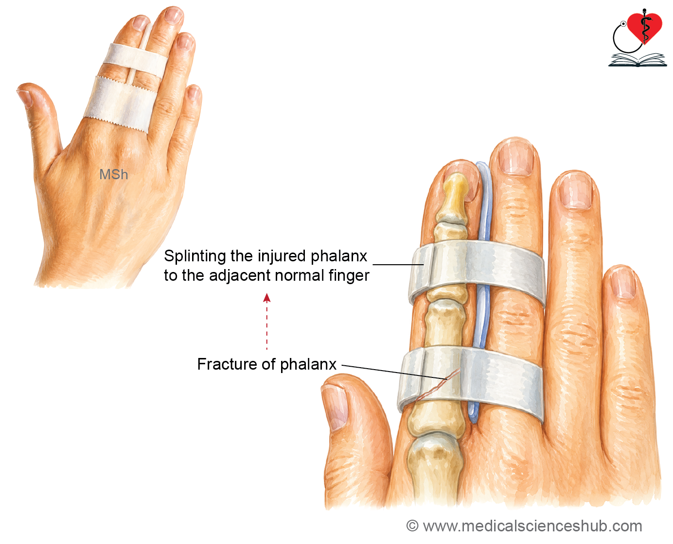
- The distal phalanx is the most commonly fractured bone of the hand, usually following a crush injury.
- A buddy splint is commonly used to treat stable phalangeal fractures by securing the injured finger to the adjacent uninjured finger, which acts as a natural splint while allowing controlled movement.
Important Questions
- List the peculiarities of pisiform bone.
- Write a short note on avascular necrosis of scaphoid.
- What is mallet finger.
📝 Test Your Knowledge – Practice MCQs
Attempt the chapter MCQ quiz and assess your understanding of key concepts.
Click to see: Medical Disclaimer and Copyright Notice