
Competency
- AN14.1: Bone: Lower limb: Identify the given bone, its side, and its important features, and keep it in anatomical position.
Introduction
Each lower limb contains 31 bones. These bones are arranged into the pelvic girdle, thigh, knee, leg, and foot.
- Pelvic girdle: Hip bone: 1
- Thigh: Femur: 1
- Knee: Patella: 1
- Leg
- Tibia: 1
- Fibula: 1
- Foot
- Tarsal bones: 7
- Metatarsal bones: 5
- Phalanges: 14
Hip Bone
- The hip bone is also known as the os coxae or innominate bone (os innominatum). It is a large, flat, and irregularly shaped bone.
- The hip bone forms the pelvic girdle. It connects the lower limb to the axial skeleton.
- In adults, each hip bone is formed by the fusion of three bones: the ilium, ischium, and pubis.
Parts of Hip Bone
- The hip bone is made up of three parts:
- Ilium: The broad, upper part of the hip bone.
- Ischium: The lower and back part of the hip bone.
- Pubis: The lower and front part of the hip bone.
- The hip bone has a broad upper part, a narrow middle part, and a broad lower part.
- The middle constricted region contains a large, cup-shaped socket called the acetabulum, which articulates with the head of the femur.
- The lower expanded part contains a large opening known as the obturator foramen.
- The pubis is located in front of and towards the midline of the obturator foramen.
- The ischium is situated behind and lateral to the obturator foramen.
- The lower end of the ischium is thickened to form the ischial tuberosity, which supports body weight while sitting.
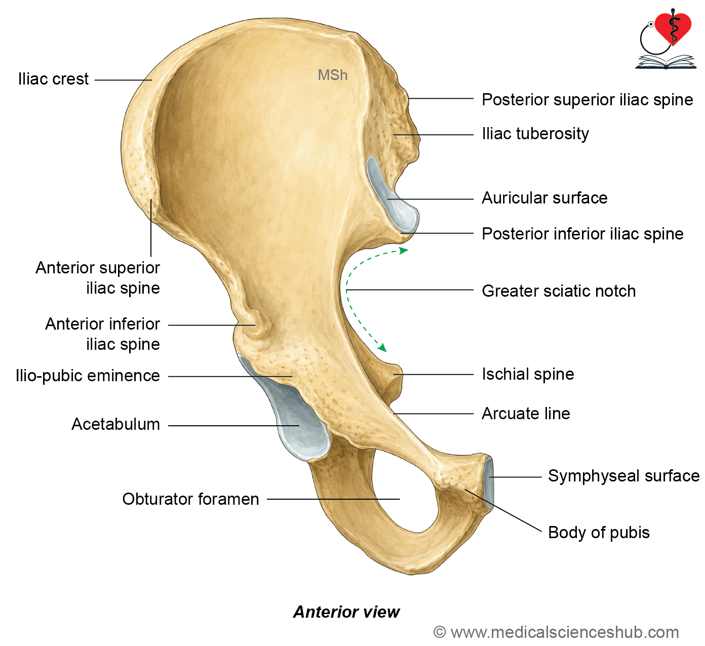
Anatomical Position and Side Determination
- Hold the hip bone so that the acetabulum faces laterally and downward.
- Keep the ilium directed upward.
- The obturator foramen should lie below the acetabulum.
- The pubis should be in front and towards the midline, while the ischium should be behind and laterally.
- Finally, tilt the bone until the anterior superior iliac spine (ASIS) and the pubic tubercle lie in the same coronal plane.
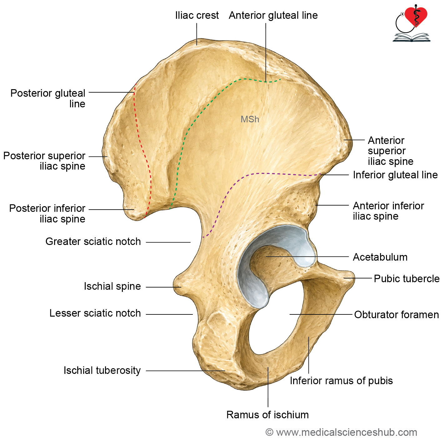
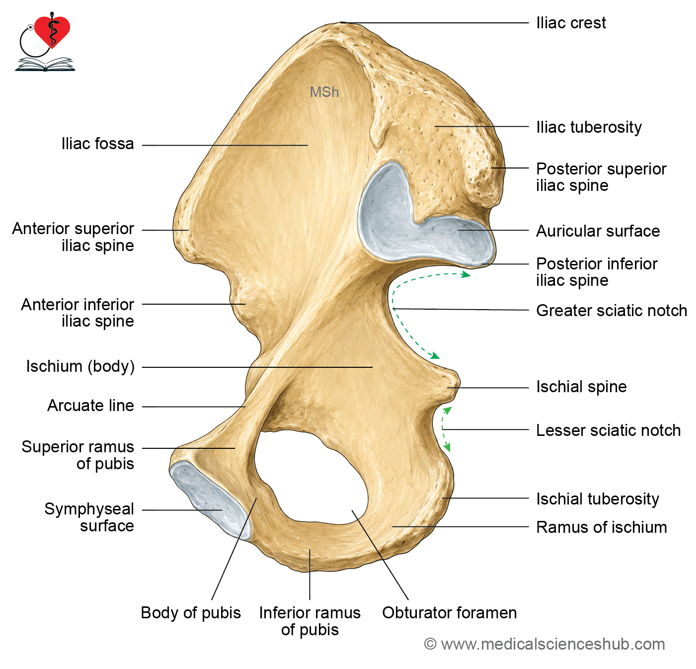
Ilium
- The ilium is the broad, flat, upper part of the hip bone.
- It extends above the acetabulum.
Parts of Ilium
- The ilium has two ends: the iliac crest (upper end) and the lower end.
- It has three borders: anterior, posterior, and medial.
- It also has three surfaces: the gluteal (external), iliac fossa (internal), and sacropelvic surfaces.
Features of Ilium
Ends of Ilium
The ilium has two ends: the iliac crest (upper end) and a lower end.
Iliac crest
This is the thick, curved upper border of the ilium.
Curvatures
- Seen from the side, it curves upward (convex).
- From front to back, it curves inward for the first two-thirds (concave) and then curves outward for the last third (convex).
Ends of Iliac Crest
- Anterior superior iliac spine (ASIS): · This is the front end of the iliac crest. It serves as an attachment point for:
- The outer end of the inguinal ligament.
- The sartorius muscle, which originates here and extends slightly onto the notch just below this spine.
- Posterior superior iliac spine (PSIS): This is the back end of the iliac crest. It is smaller and less noticeable than the ASIS. It provides attachment for the sacrotuberous ligament.
Segments of Iliac Crest
- The iliac crest is divided into two segments:
- Ventral segment: Forms the anterior two-thirds of the crest.
- Dorsal segment: Forms the posterior one-third of the crest.
- Ventral segment of the iliac crest: The ventral segment has three parts:
- Outer lip
- Intermediate area
- Inner lip
- Iliac tubercle: The iliac tubercle is a prominent bony projection on the outer lip of the ventral segment. It is located about 5 cm behind the anterior superior iliac spine (ASIS). It serves as an important anatomical landmark and provides attachment for muscles and fascia.
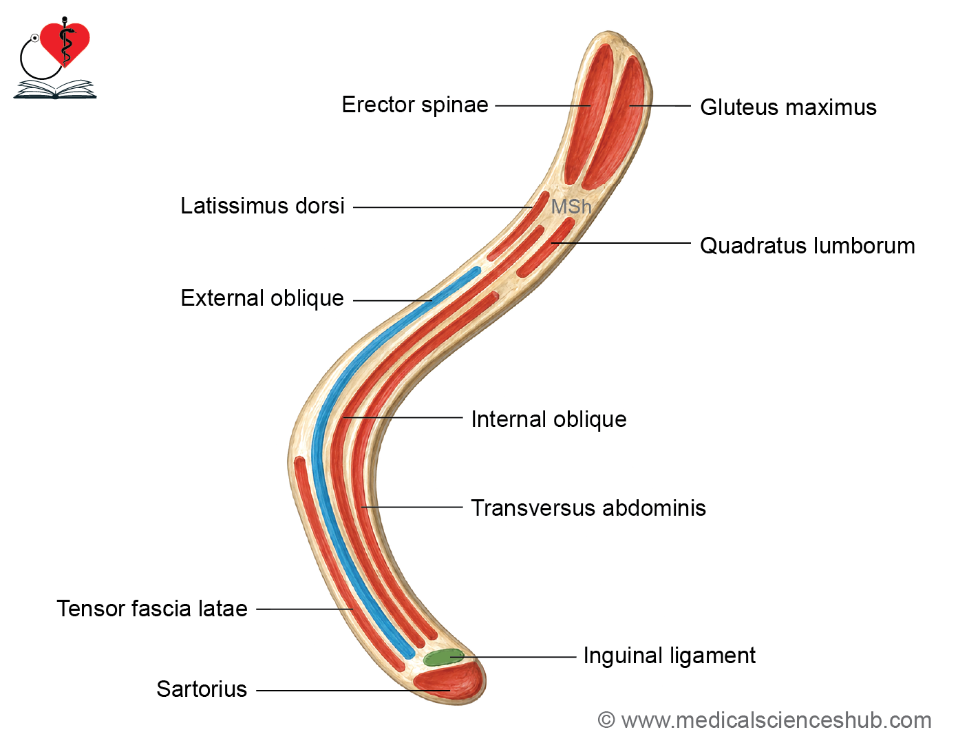
Attachments of Iliac Crest
Outer Lip
- Tensor fascia latae originates just in front of the tubercle.
- External oblique muscle attaches along the front two-thirds.
- Latissimus dorsi originates just behind the highest point of the crest.
- Iliotibial tract attaches at the tubercle.
Middle Area
- Internal oblique muscle originates here.
Inner Lip
- Transverse abdominis originates from the front two-thirds.
- Quadratus lumborum originates from the back one-third.
Dorsal Segment
- A ridge divides it into an outer and an inner sloping area.
- Outer sloping area: Gluteus maximus originates here.
- Inner sloping area: Erector spinae originates here.
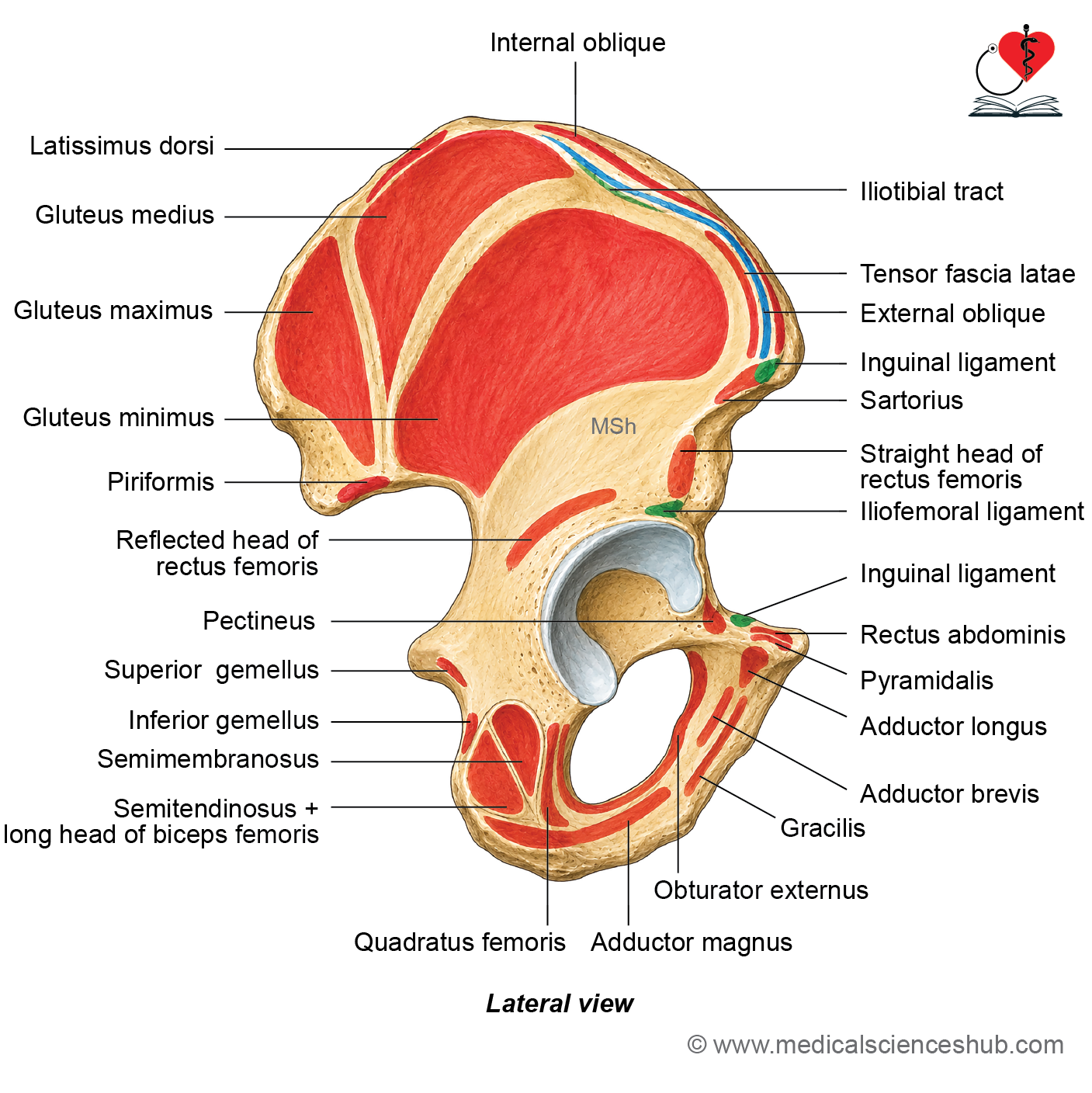
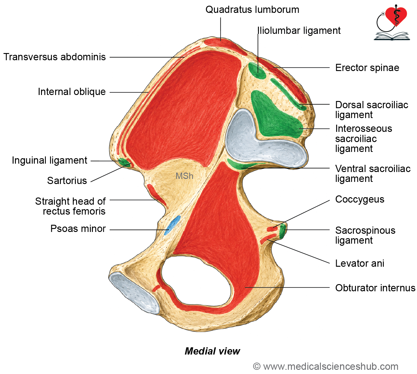
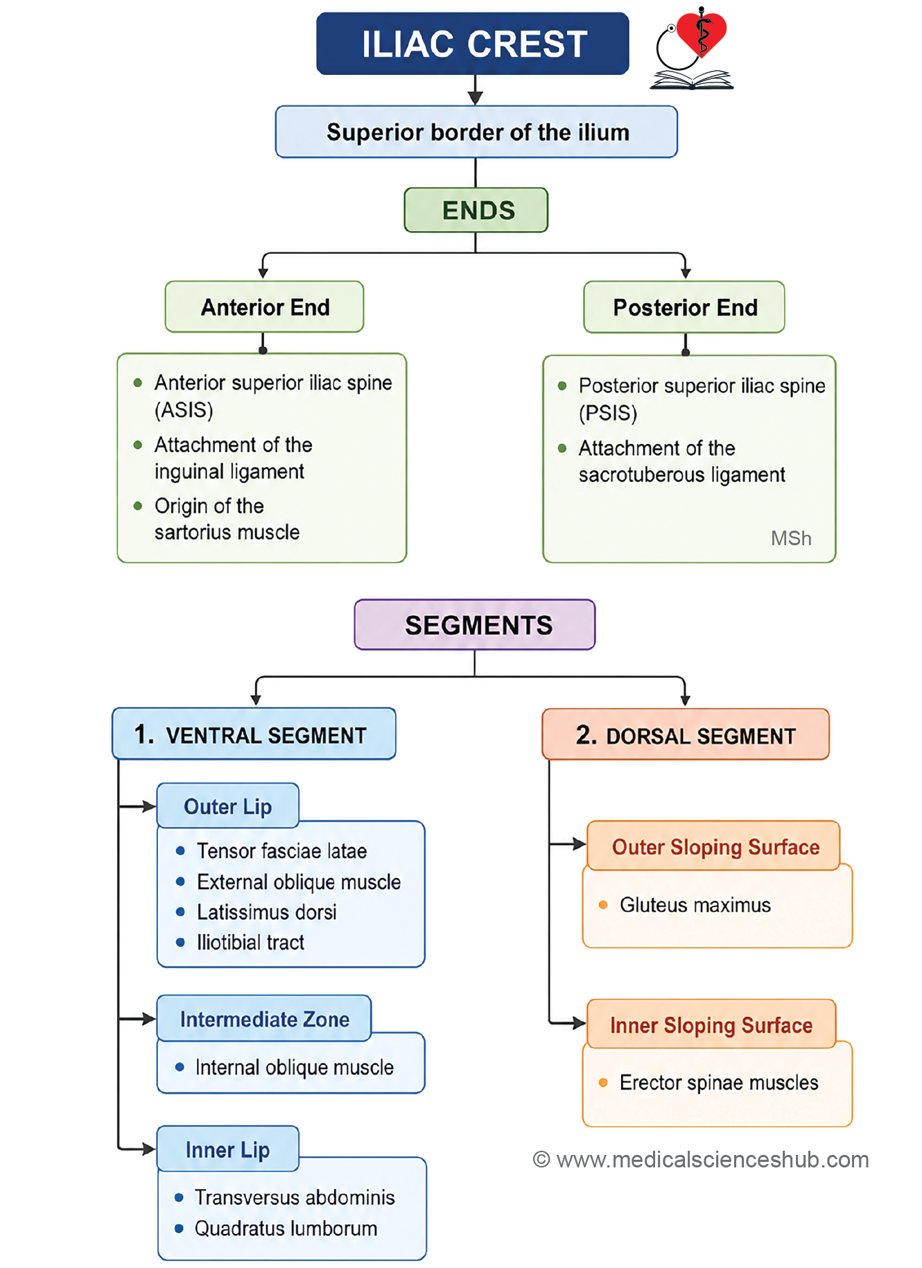
CLINICAL INTEGRATION
- The iliac crest can be felt under the skin at the lower boundary of the trunk. At the back, its highest point lies level with the gap between the L3 and L4 vertebrae.
- Hematopoiesis: Even in adults, the iliac crest continues to produce blood cells.
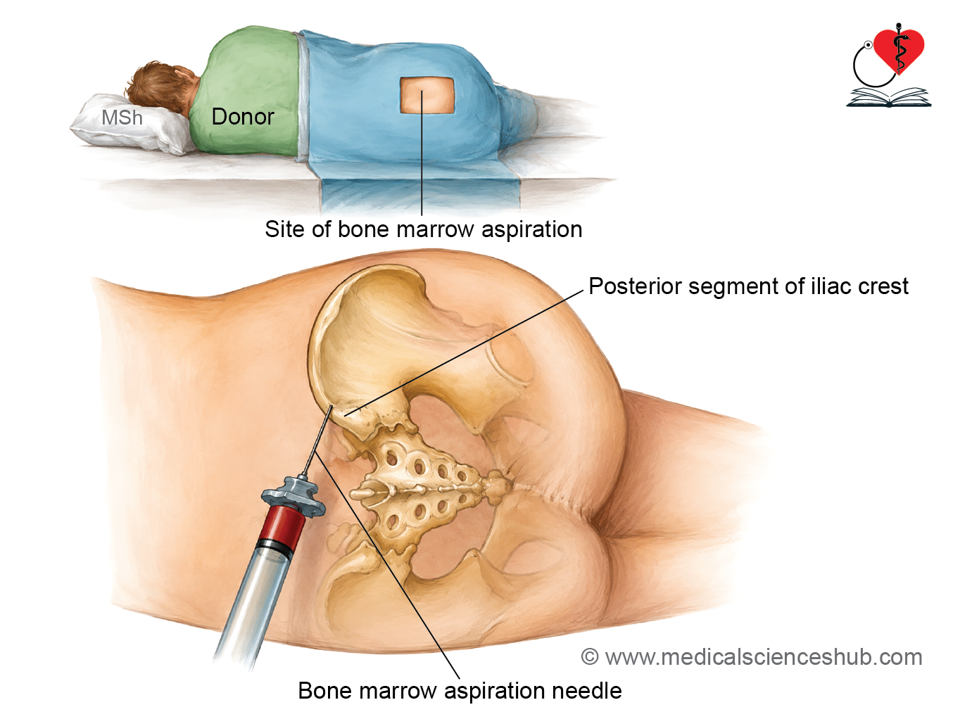
- Bone marrow aspiration: This procedure removes a small sample of marrow cells for microscopic study, helping diagnose conditions like anemia and leukemia. The back part of the iliac crest is the usual site used for this.
- Bone marrow donation: For bone marrow transplants, cells are collected from a donor’s posterior iliac crest. These donated cells help treat patients with aplastic anemia, those recovering after chemotherapy, and similar conditions.
Lower End of Ilium
- The lower end of the ilium is smaller than the upper end. It joins with the ischium and pubis at the acetabulum (the hip socket). The ilium contributes the upper two-fifths of the acetabulum.
Borders of Ilium
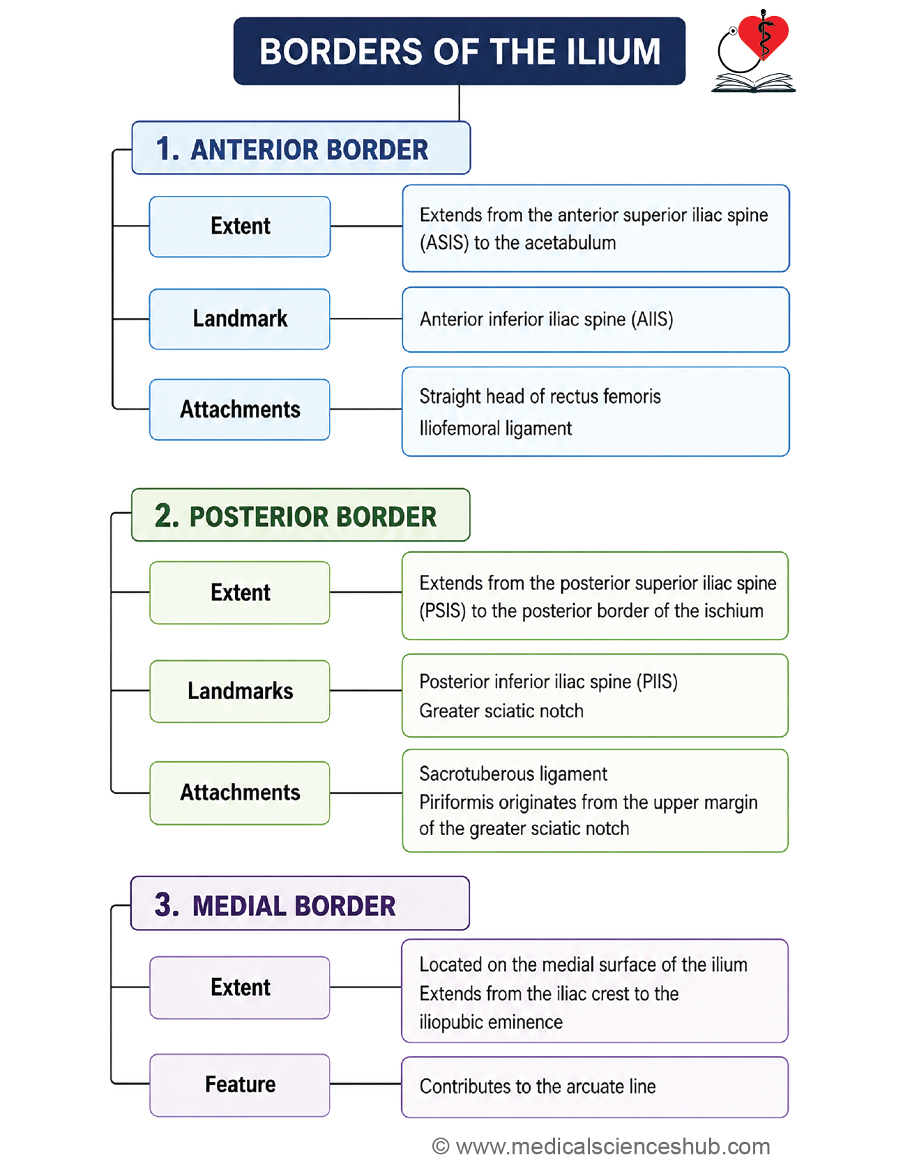
Anterior Border of Ilium
- Extent: The anterior border runs from the anterior superior iliac spine down to the acetabulum.
- Features: The upper part of the anterior border has a notch. Anterior Inferior Iliac Spine (AIIS) is a small forward-pointing projection found on the lower part of the anterior border.
- Attachments
- The straight head of the rectus femoris muscle originates from the upper part of the AIIS.
- The iliofemoral ligament attaches to the lower part of the AIIS.
Posterior Border of Ilium
- Extent: The posterior border runs from the posterior superior iliac spine down to the posterior border of the ischium.
- Features:
- Posterior Inferior Iliac Spine (PIIS): A bony bump located a few centimeters below the posterior superior iliac spine.
- Greater Sciatic Notch: A deep notch below the PIIS. Just below this notch lies the ischial spine.
- Attachments
- The sacrotuberous ligament attaches to the posterior border, between the PSIS and PIIS.
- A few fibers of the piriformis muscle originate from the upper edge of the greater sciatic notch.
- Greater Sciatic Foramen
- The sacrospinous and sacrotuberous ligaments close off the greater sciatic notch, turning it into the greater sciatic foramen.
Medial Border of Ilium
- Extent: The medial border lies on the inner (pelvic) surface of the ilium, running from the iliac crest down to the iliopubic eminence.
- It separates the iliac fossa from the sacropelvic surface of the ilium.
- Arcuate Line: The lower part of the medial border forms a smooth, rounded ridge called the arcuate line.
Surfaces of Ilium
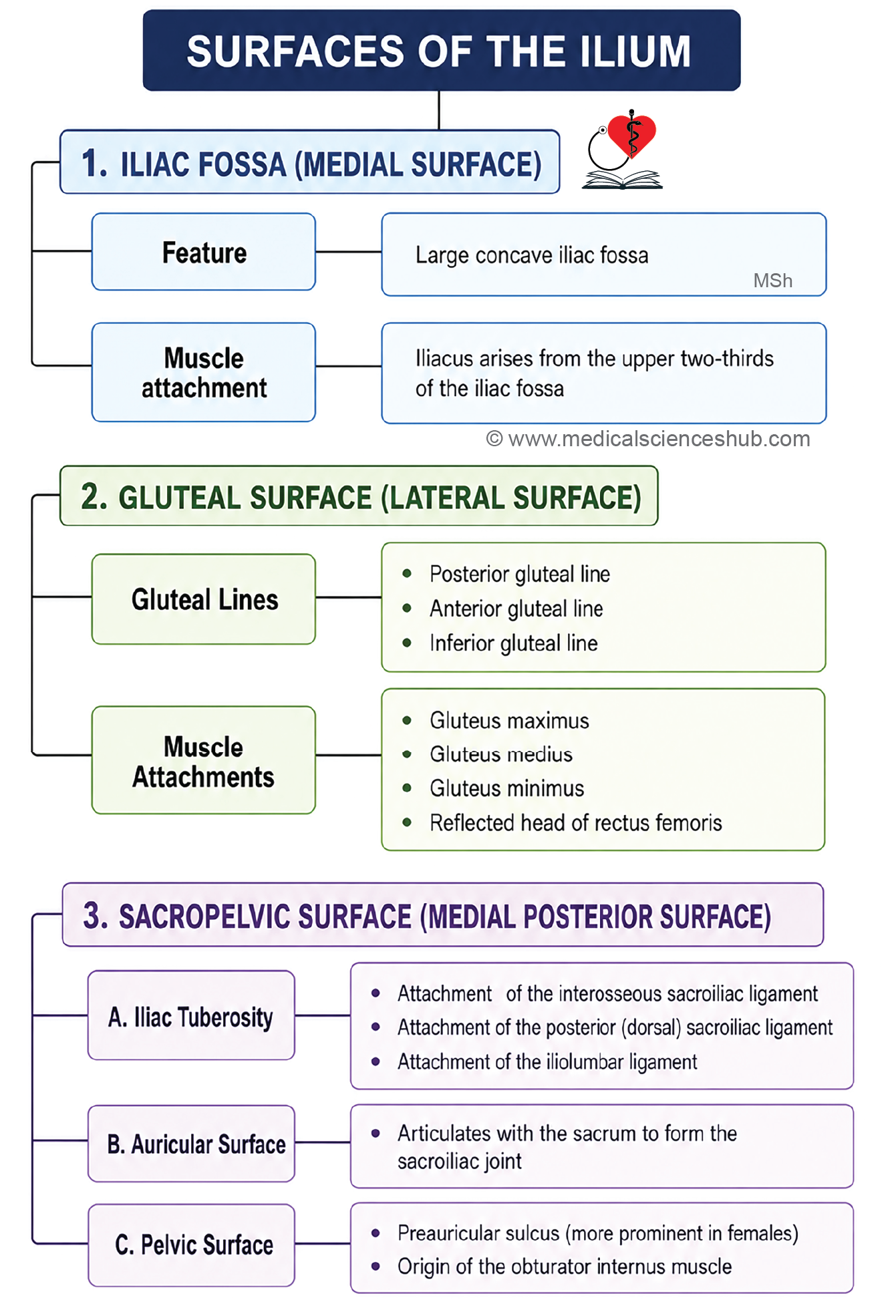
The ilium has three surfaces: gluteal surface, iliac surface, and sacropelvic surface.
Iliac surface (iliac fossa) of ilium
- A shallow, smooth, concave hollow on the inner surface of the ilium.
- Boundaries:
- Above: iliac crest.
- Front: anterior border of the ilium.
- Back: medial border of the ilium.
- It forms the outer wall of the false pelvis.
- Attachment: The iliacus muscle originates from the upper two-thirds of this fossa.
Gluteal surface of ilium
- This is the outer surface of the ilium, divided into four regions by three gluteal lines.
- Gluteal Lines
- Posterior gluteal line: The shortest line, running from a point 5 cm in front of the PSIS to a point in front of the PIIS.
- Anterior (middle) gluteal line: Runs from the greater sciatic notch to the iliac crest, just in front of the iliac tubercle.
- Inferior gluteal line: Runs from the upper part of the AIIS to the lower part of the greater sciatic notch.
- Attachments
- Gluteus maximus (upper fibers) originates behind the posterior gluteal line.
- Gluteus medius originates between the anterior and posterior gluteal lines.
- Gluteus minimus originates between the anterior and inferior gluteal lines.
- The reflected head of rectus femoris originates from a shallow groove between the inferior gluteal line and the acetabulum.
Sacropelvic Surface of Ilium
- Located on the back-inner side of the ilium, behind the medial border.
- It has a rough, uneven texture and is made up of three parts:
- Iliac tuberosity
- Auricular surface
- Pelvic surface
Iliac tuberosity
- It is rough patch just below the dorsal part of the iliac crest.
- Attachments
- Interosseous sacroiliac ligament.
- Dorsal sacroiliac ligament, at the back.
- Iliolumbar ligament, above.
Auricular surface
- An ear-shaped joint surface (L- or C-shaped) that feels rough despite being an articular surface.
- It lies below and in front of the iliac tuberosity.
- Covered by fibrocartilage.
- Articulation: This surface joins with the matching auricular surface of the sacrum to form the sacroiliac joint.
Pelvic Surface of Ilium
- The pelvic surface continues on into the pelvic surface of the ischium.
- It is smooth and lies below and in front of the auricular surface.
- Preauricular Sulcus: A shallow groove located just in front of the auricular surface. This groove is deeper and more clearly marked in females than in males.
- Attachments
- The edges of the preauricular sulcus attach to the anterior sacroiliac ligament.
- The obturator internus muscle originates from the lower part of the pelvic surface.
Pubis
- The ilium, or flank, forms the upper flat, wide plate of the hip bone and sits above the acetabulum. The pubis, also called the pubic bone or os pubis, makes up the front-lower part of the hip bone.
Parts of Pubis
The pubis is made up of three parts:
- Body
- Superior ramus
- Inferior ramus
Body of Pubis
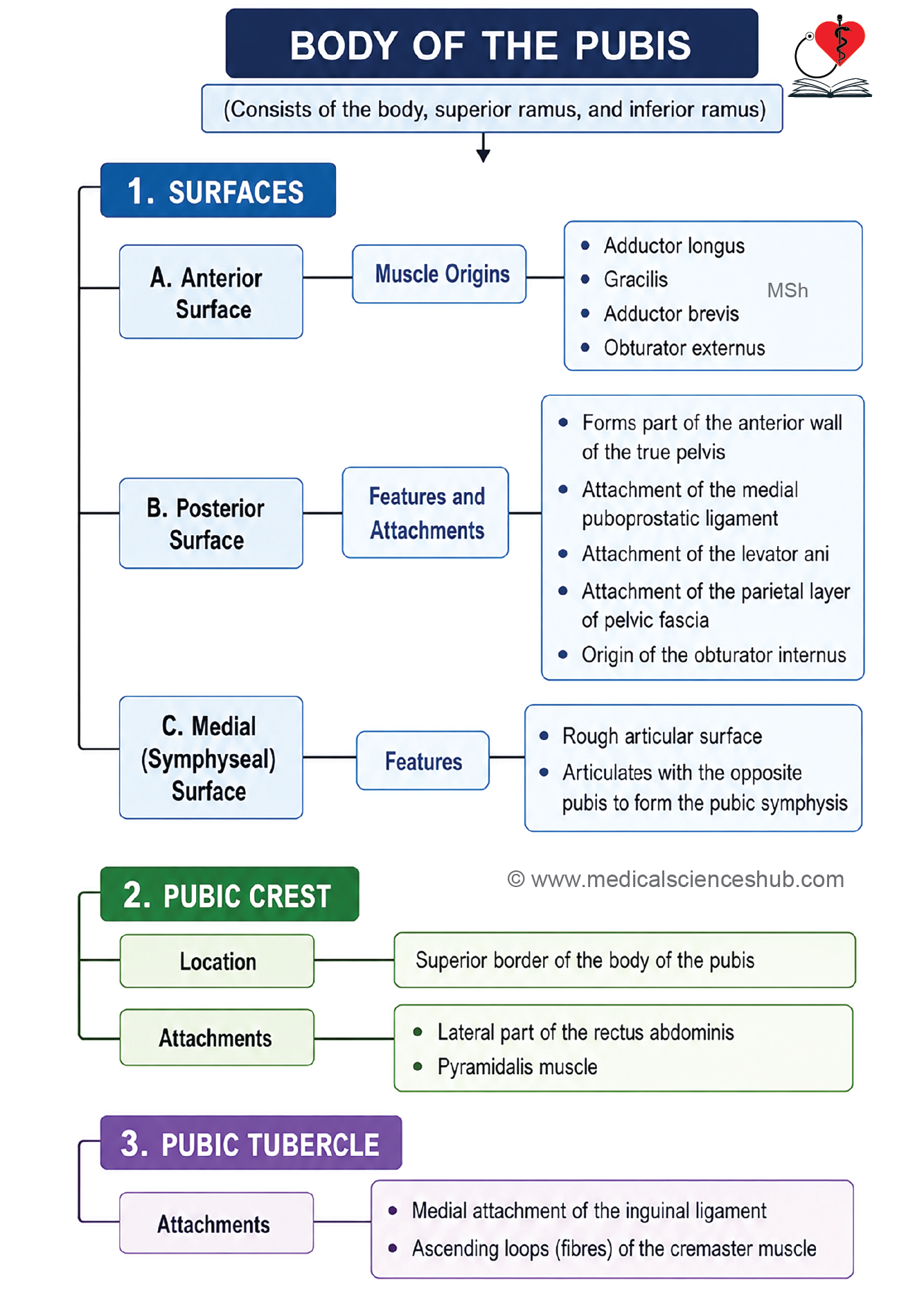
- The body of the pubis is flattened from front to back and has a four-sided (quadrilateral) shape.
- It consists of:
- Three surfaces: anterior, posterior, and medial.
- The pubic crest.
- The pubic tubercle.
Anterior Surface of Pubis
- This surface faces downward, forward, and outward.
Attachments
- Adductor longus originates from the corner between the pubic crest and the pubic symphysis.
- Gracilis originates from the lower part of the pubic body, with its origin extending onto the inferior ramus.
- Adductor brevis originates just to the side of gracilis.
- Obturator externus originates near the edge of the obturator foramen.
<H5>Posterior surface of pubis
- This surface is smooth and forms the front wall of the true pelvis.
- A pad of fat, called the retropubic pad of fat, separates it from the urinary bladder.
Attachments
Moving from the inner to the outer side, the posterior surface gives attachment to:
- The medial puboprostatic ligament.
- The levator ani muscle (origin).
- The parietal layer of pelvic fascia.
- The obturator internus muscle (origin).
Symphyseal (Medial) Surface of Pubis
- This surface is rough and joins with the pubic bone on the other side to form the symphysis pubis. It is covered by hyaline cartilage.
Pubic crest
- The thick upper border of the pubic body forms a ridge called the pubic crest.
- Attachments: The lateral head of the rectus abdominis muscle and the pyramidalis muscle originate from the outer part of the pubic crest.
Public tubercle
- The outer end of the pubic crest forms a bony projection called the pubic tubercle.
Attachments
- The inner end of the inguinal ligament attaches here.
- The ascending loops of the cremaster muscle attach here.
- In males, the spermatic cord crosses over this point.
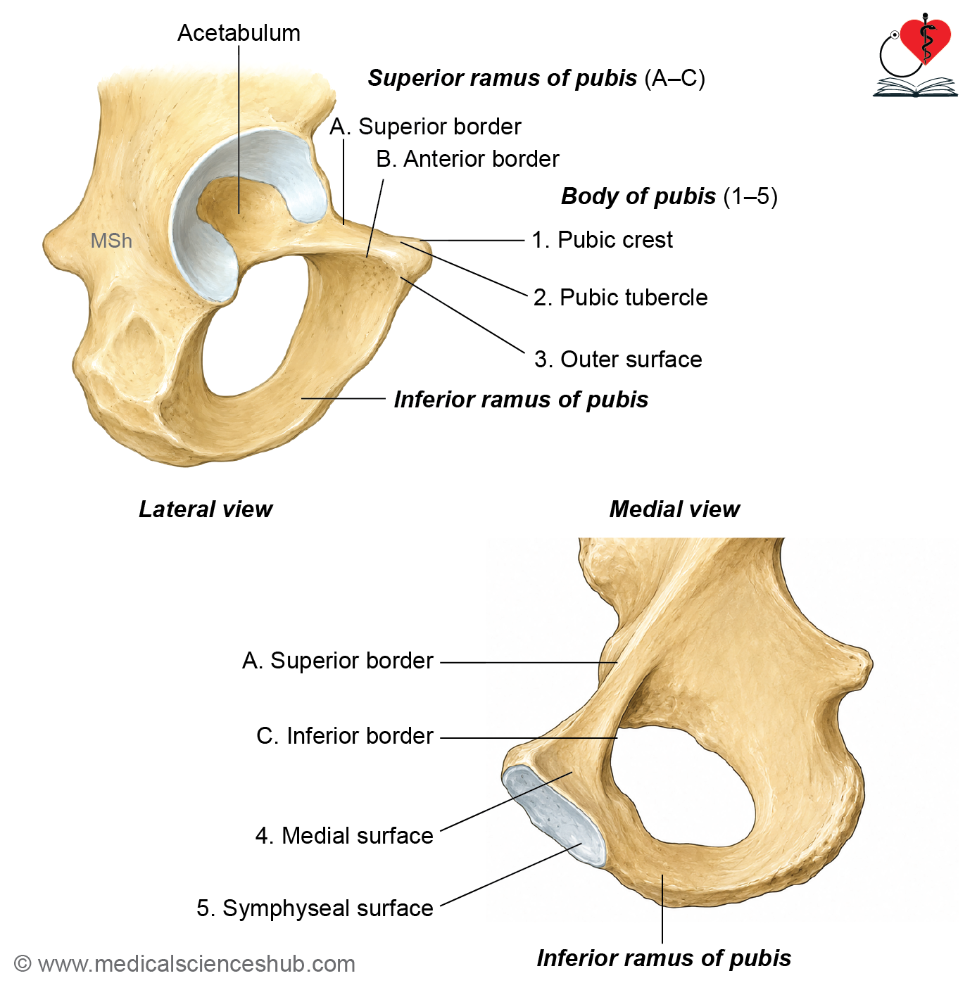
Superior Ramus of Pubis
- The superior ramus lies above the obturator foramen and extends from the pubic body to the acetabulum.
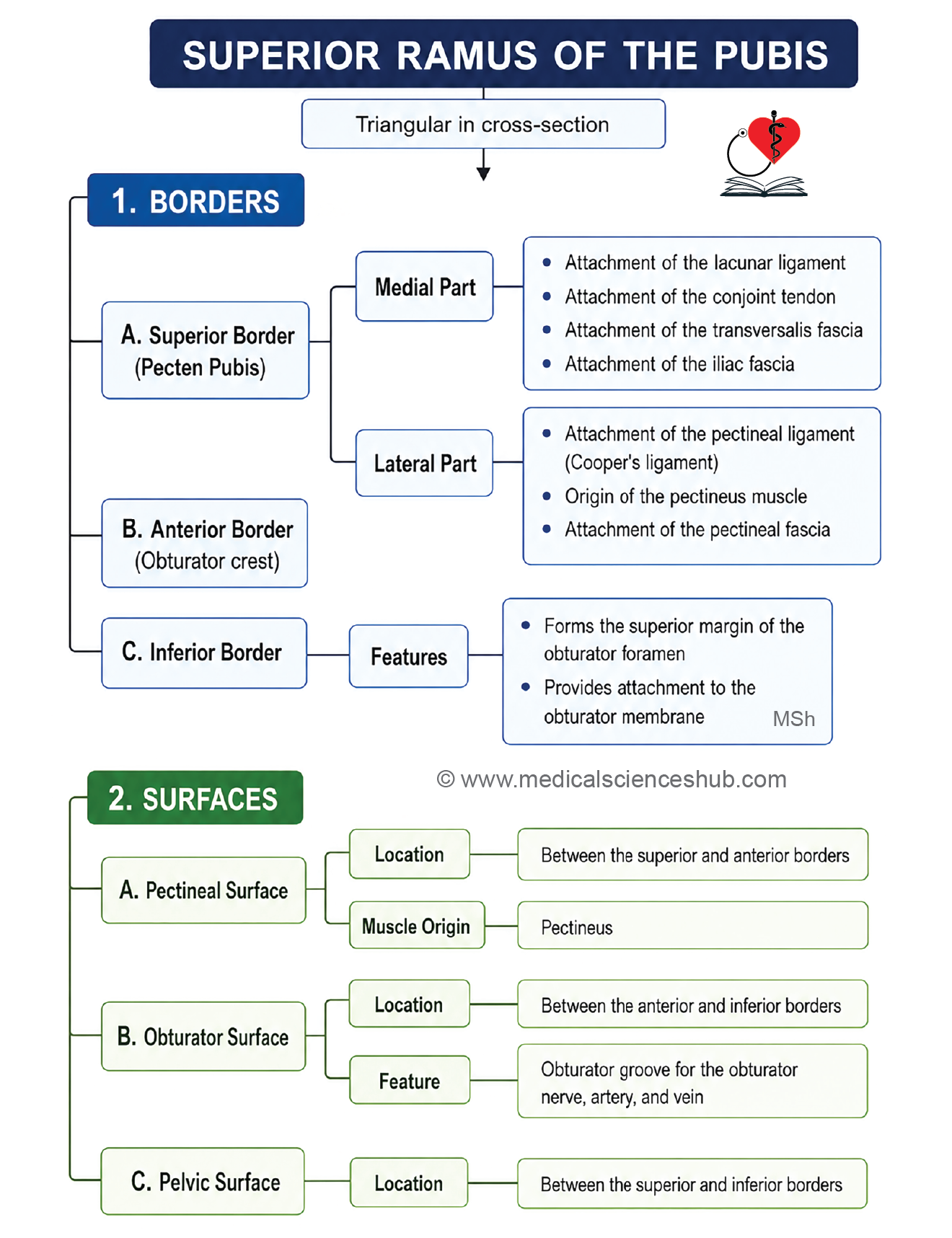
- It has a triangular cross-section, with three borders and three surfaces.
- Borders: anterior (obturator crest), superior (pectineal line), and inferior.
- Surfaces: pectineal, pelvic, and obturator.
Superior Border (Pectineal Line) of Superior Pubic Ramus
- Also called the pecten pubis, this border is sharp and well-defined.
- It runs from the pubic tubercle to the iliopubic eminence, then continues as the arcuate line.
Attachments
- The inner part of the pectineal line attaches to:
- Lacunar ligament (pectineal part of the inguinal ligament).
- Conjoint tendon of the anterior abdominal wall muscles.Fascia transversalis.
- Fascia iliaca.
- The outer part of the pectineal line attaches to:
- Pectineal ligament.
- Pectineus muscle.
- Pectineal fascia.
Anterior Border (Obturator Crest) of Superior Pubic Ramus
- It runs from the pubic tubercle to the acetabular notch.
Inferior border
- A sharp border that forms the upper edge of the obturator foramen.
- Attachment: The obturator membrane attaches here.
Pectineal Surface of Superior Pubic Ramus
- Located between the anterior border (obturator crest) and superior border (pectineal line).
- Triangular in shape, it runs from the pubic tubercle to the iliopubic eminence.
- Attachment: The pectineus muscle originates from this surface.
Obturator Surface of Superior Pubic Ramus
- Located between the anterior border (obturator crest) and inferior border.
- Contains a groove, the obturator groove, through which pass:
- The obturator nerve.
- The obturator artery.
- The obturator vein.
Pelvic Surface of Superior Pubic Ramus
- Located between the pectineal line and inferior border, and is smooth.
Relations
- In males, this surface is crossed by the obliterated umbilical artery and the vas deferens.
- In females, it is crossed by the round ligament of the uterus and the obliterated umbilical artery.
Inferior Ramus of Pubis
- The inferior ramus extends from the pubic body downward and joins the ramus of the ischium, together forming what is called the conjoint ramus.
- Conjoint ramus = inferior ramus of pubis + ramus of ischium.
Ischium
• The ischium forms the thick, back-lower part of the hip bone. It has two parts: a body and a ramus.
Body of Ischium
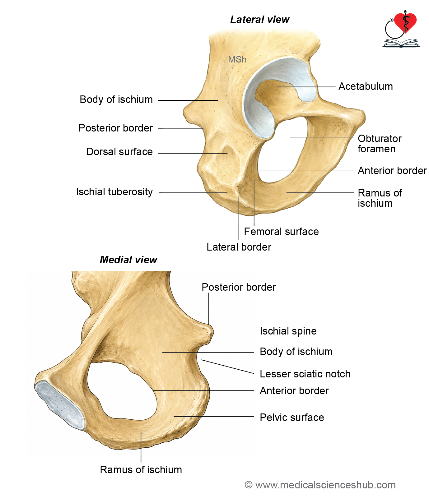
The body of the ischium is a thick mass of bone made up of:
- Two ends: upper and lower.
- Three borders: anterior, posterior, and lateral.
- Three surfaces: femoral, dorsal, and pelvic.
Ends of body of ischium
Upper end of ischium
- It Joins with the ilium and pubis at the acetabulum, forming the back-lower two-fifths of the acetabulum.
Lower end (ischial tuberosity)
- The lower end of the ischium forms the ischial tuberosity.
Borders of body of ischium
Anterior Border
- Forms the back edge of the obturator foramen.
- Attachment: The obturator membrane attaches here.
Posterior Border
- Continues upward into the posterior border of the ilium.
- Moving from top to bottom, it forms the lower part of the greater sciatic notch, the ischial spine, and the lesser sciatic notch.
- Ischial spine: It a triangular bony projection.
Attachments
- The sacrospinous ligament attaches to the edges of the ischial spine.
- The levator ani muscle originates from the inner surface of the ischial spine.
- Structures Crossing the Spine
- The back surface of the ischial spine is crossed by three structures, remembered by the mnemonic PIN:
- Pudendal nerve.
- Internal pudendal vessels.
- Nerve to obturator internus.
- The back surface of the ischial spine is crossed by three structures, remembered by the mnemonic PIN:
- Lesser Sciatic Notch: The lesser sciatic notch is a shallow, smooth notch located between the ischial spine and the ischial tuberosity.
- Attachments
- The superior gemellus muscle originates from the upper edge of the notch.
- The inferior gemellus muscle originates from the lower edge of the notch.
- The tendon of the obturator internus muscle passes through the lesser sciatic notch. The notch is lined with hyaline cartilage.
- Attachments
- Lateral Border of the Ischial Body: It forms the outer edge of the ischial tuberosity. The upper part of this border is not well-defined.
Surfaces of body of ischium
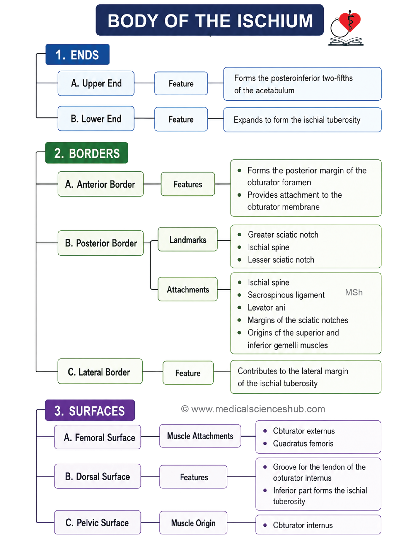
- The body of the ischium has three surfaces: femoral, dorsal, and pelvic.
- Femoral Surface: The femoral surface lies between the anterior and lateral borders.
- Attachments
- The obturator externus muscle originates along the edge of the obturator foramen.
- The quadratus femoris muscle originates from the lateral border of the upper part of the ischial tuberosity.
- Attachments
- Dorsal Surface: The dorsal surface continues upward into the gluteal surface of the ilium. A transverse groove divides this surface into upper and lower parts, and this groove allows passage for the tendon of the obturator internus muscle along with the two gemelli muscles. The lower, rough part of this surface forms the ischial tuberosity.
- Pelvic Surface: The pelvic surface lies between the anterior and posterior borders.
- Attachment: The obturator internus muscle originates from this surface.
Ramus of Ischium
- The ramus of the ischium joins with the ramus of the pubis to form the conjoint ischiopubic ramus.
Ischial tuberosity
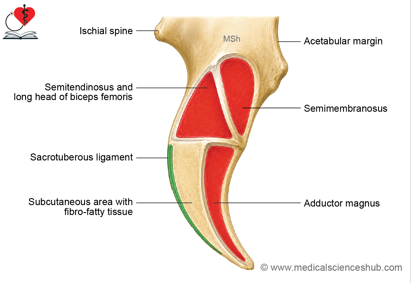
The ischial tuberosity, also called the tuber ischiadicum or “sit bone,” is a large mass of bone belonging to the ischium.
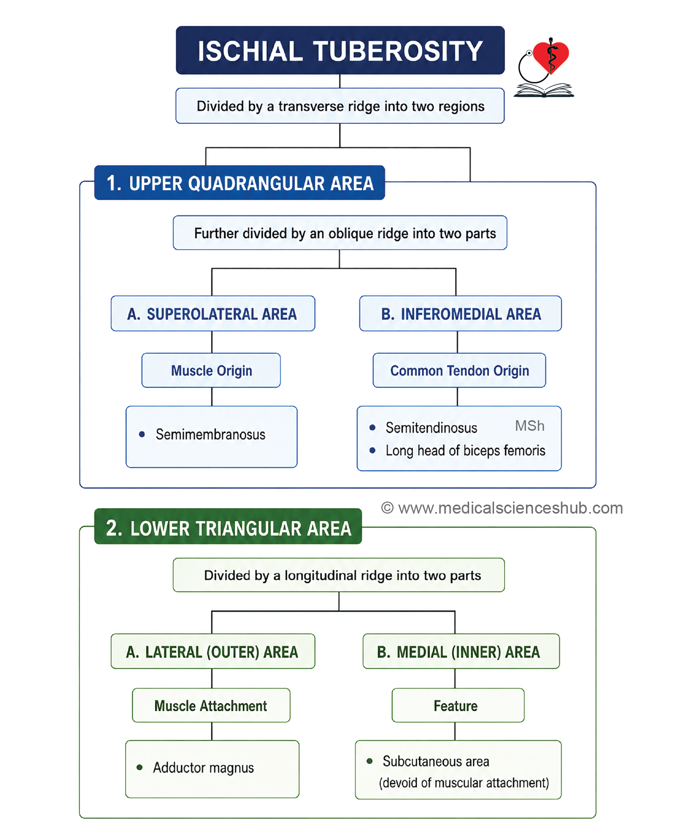
A transverse ridge divides it into an upper quadrangular area and a lower triangular area.
Upper Area
An oblique ridge further divides the upper area into two parts:
- A superolateral part, from which the semimembranosus muscle originates.
- An inferomedial part, from which the semitendinosus muscle and the long head of biceps femoris originate.
Lower Area
A longitudinal ridge divides the lower area into two parts:
- An outer part, from which the ischial head of adductor magnus originates.
- An inner part, which is covered by fibrofatty tissue and bears the body’s weight while sitting.
Margins of the Ischial Tuberosity
The medial margin gives attachment to the sacrotuberous ligament, while the lateral margin gives attachment to the ischiofemoral ligament.
Conjoint Ischiopubic Ramus
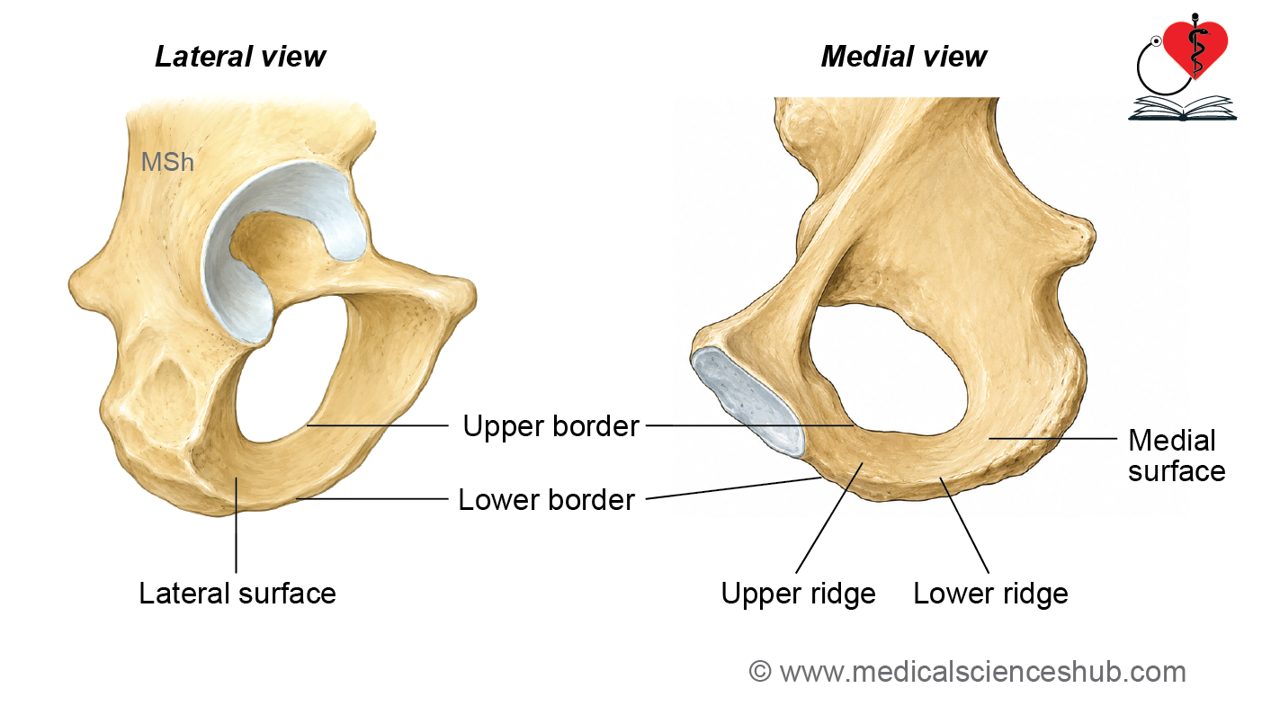
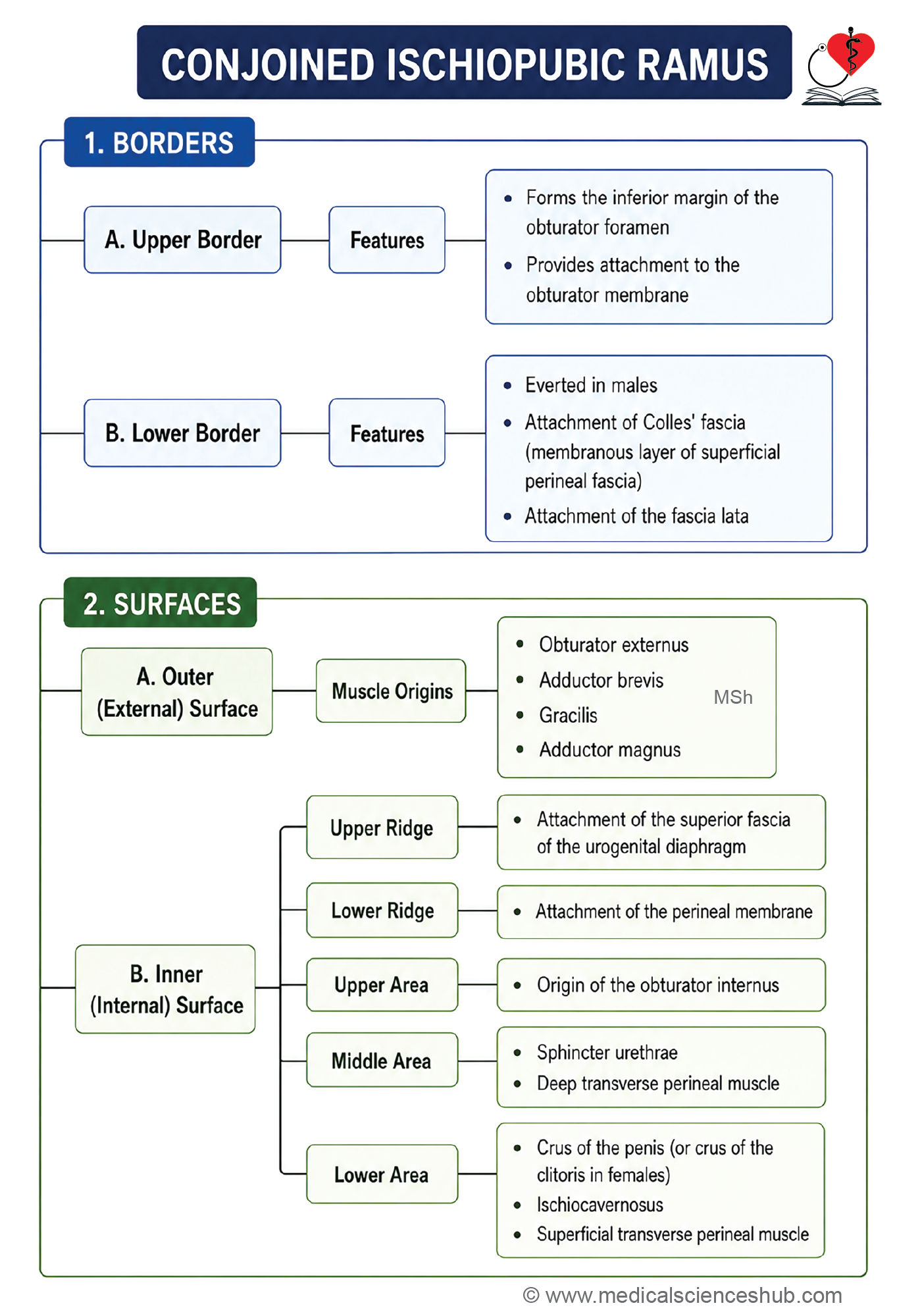
The inferior ramus of the pubis joins with the ramus of the ischium to form the conjoint ischiopubic ramus. It has two borders (upper and lower) and two surfaces (inner and outer).
Upper Border
- Forms the lower edge of the obturator foramen.
- Attachment: The obturator membrane attaches here.
Lower Border
- Forms the boundary of the subpubic angle and part of the pubic arch.
- In males, this border is more turned outward than in females, due to the attachment of the crus of the penis.
- Attachments: The fascia lata and the membranous layer of the superficial perineal fascia (Colles’ fascia) attach here.
Outer Surface
- This surface is rough.
- Attachments:
- The obturator externus muscle originates near the edge of the obturator foramen.
- The adductor brevis muscle originates here.
- The gracilis muscle originates here.
- The adductor magnus muscle originates here.
Inner Surface
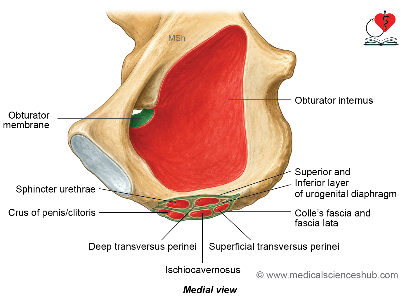
- This surface is convex and rough, and is divided by two ridges into three areas: upper, middle, and lower.
- Attachments:
- The upper ridge attaches to the superior fascia of the urogenital diaphragm.
- The lower ridge attaches to the perineal membrane (inferior fascia of the urogenital diaphragm).
- The upper area gives origin to the obturator internus muscle.
- The middle area gives origin to the sphincter urethrae and deep transverse perineal muscles.
- The lower area gives attachment to the crus of the penis or clitoris, and gives origin to the ischiocavernosus and superficial transverse perineal muscles.
Obturator foramen
- The obturator foramen is a large opening in the hip bone, located below the acetabulum, between the pubis and ischium.
- Sex Difference: The obturator foramen is large and oval-shaped in males, while it is triangular in females.
- Attachment: The obturator membrane attaches along the edges of the obturator foramen.
- Obturator Canal: At the obturator groove, a small gap exists between the obturator foramen and the obturator membrane, forming the obturator canal. This canal transmits the obturator nerve and obturator vessels.
Acetabulum
- The acetabulum is a cup-shaped, hemispherical cavity — the word “acetabulum” comes from Latin, meaning “little vinegar cup.” The acetabulum faces outward, downward, and forward.
- Acetabular Notch: The rim of the acetabulum is incomplete at its lower part, and this gap is called the acetabular notch.
- Attachments
- The transverse acetabular ligament bridges the gap at the acetabular notch.
- The edges of the acetabular fossa, near the notch, give attachment to the two limbs of the ligamentum teres femoris.
- Attachments
- Bony Contributions: The acetabulum is formed by three bones:
- The pubis contributes the front one-fifth.
- The ischium contributes the back two-fifths.
- The ilium contributes the upper two-fifths.
- Articulation: The acetabulum receives the head of the femur, together forming the ball-and-socket type synovial hip joint.
- Lunate Surface: This is a horseshoe-shaped joint surface covered by hyaline cartilage.
- Acetabular Fossa: This is the non-articular central part of the lunate surface. It is filled with a pad of fat covered by synovial membrane.
- Contents: Branches of the obturator vessels and nerve pass through the acetabular notch.
- Acetabular Labrum: This is a ring of cartilage attached to the rim of the acetabulum.
Ossification of Hip Bone
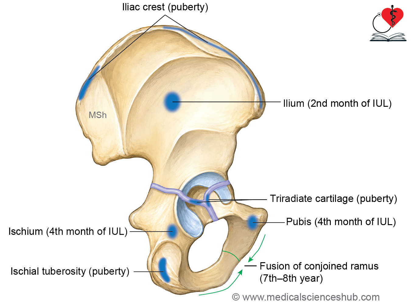
- The hip bone develops from three primary centers of ossification and five secondary centers.
- Primary Centers
- One center for the ilium appears by the 2nd month of intrauterine life.
- One center for the ischium appears by the 4th month of intrauterine life.
- One center for the pubis appears by the 4th month of intrauterine life.
- The fusion of these three primary centers forms a Y-shaped cartilage, called the triradiate cartilage, in the floor of the acetabulum. Separately, the ischium and pubis fuse together between the 7th and 8th year of age to form the conjoined ramus.
- Secondary Centers
- There are five secondary centers of ossification:
- One for the ischial tuberosity.
- Two for the iliac crest.
- Two for the Y-shaped triradiate cartilage.
- All secondary centers appear around puberty, and all the ossification centers fuse completely by the age of 25 years.
CLINICAL ANATOMY
- Forensic Fact: The pubis ossifies later than the ilium and ischium, so it is often missing from prenatal skeletal remains.
- Fusion at the Acetabulum: The three bones fuse together at the acetabulum between 16 and 18 years of age.
- Risser Sign: The Risser sign is a way of judging skeletal maturity by looking at how much the iliac crest and iliac bone have ossified on an X-ray. This sign helps doctors decide the best timing for surgery in cases of spinal deformity.
- Avulsion Fractures of the Pelvic Apophysis: In athletic teenagers, strong muscle pulling can tear away a piece of bone at its growth center, causing an avulsion fracture. For example, forceful contraction of the rectus femoris muscle can cause an avulsion fracture of the anterior inferior iliac spine.
Table 2.1: Differences between the male and female hip bones
| Feature | Male Hip Bone | Female Hip Bone |
|---|---|---|
| Overall build | Heavier, with well-marked muscle attachment sites | Lighter, with less distinct muscle attachment sites |
| Greater sciatic notch | Narrow | Wide |
| Obturator foramen | Large and oval-shaped | Small and triangular-shaped |
| Ischial spine | Turns inward | Does not turn inward |
| Preauricular sulcus | Faint or barely visible | Deep and clearly visible |
| Ischiopubic ramus | Thick and turned outward | Thin and not turned outward |
| Acetabulum | Larger, more than 5 cm across | Smaller, less than 5 cm across |
| Iliac fossa | Deeper | Shallower |
📝 Test Your Knowledge – Practice MCQs
Attempt the chapter MCQ quiz and assess your understanding of key concepts.
Click to see: Medical Disclaimer and Copyright Notice