
Competency
- AN21.3: Describe and demonstrate the boundaries of thoracic inlet, cavity, and outlet.
Introduction
- The thorax, derived from the Greek word thorakos meaning “chest,” is the region of the body located between the neck and the abdomen.
- The thorax is composed of two main components:
- Thoracic cage, which forms the supporting framework of the thorax.
- Thoracic cavity, which contains the major organs and structures within the thorax.
- The thoracic cage protects vital organs, particularly the heart and lungs, from external injury.
- The thoracic cavity is separated from the abdominal cavity by the thoracoabdominal diaphragm, a muscular partition that also plays an important role in respiration.
Surface Anatomy of Thorax
Bony Landmarks
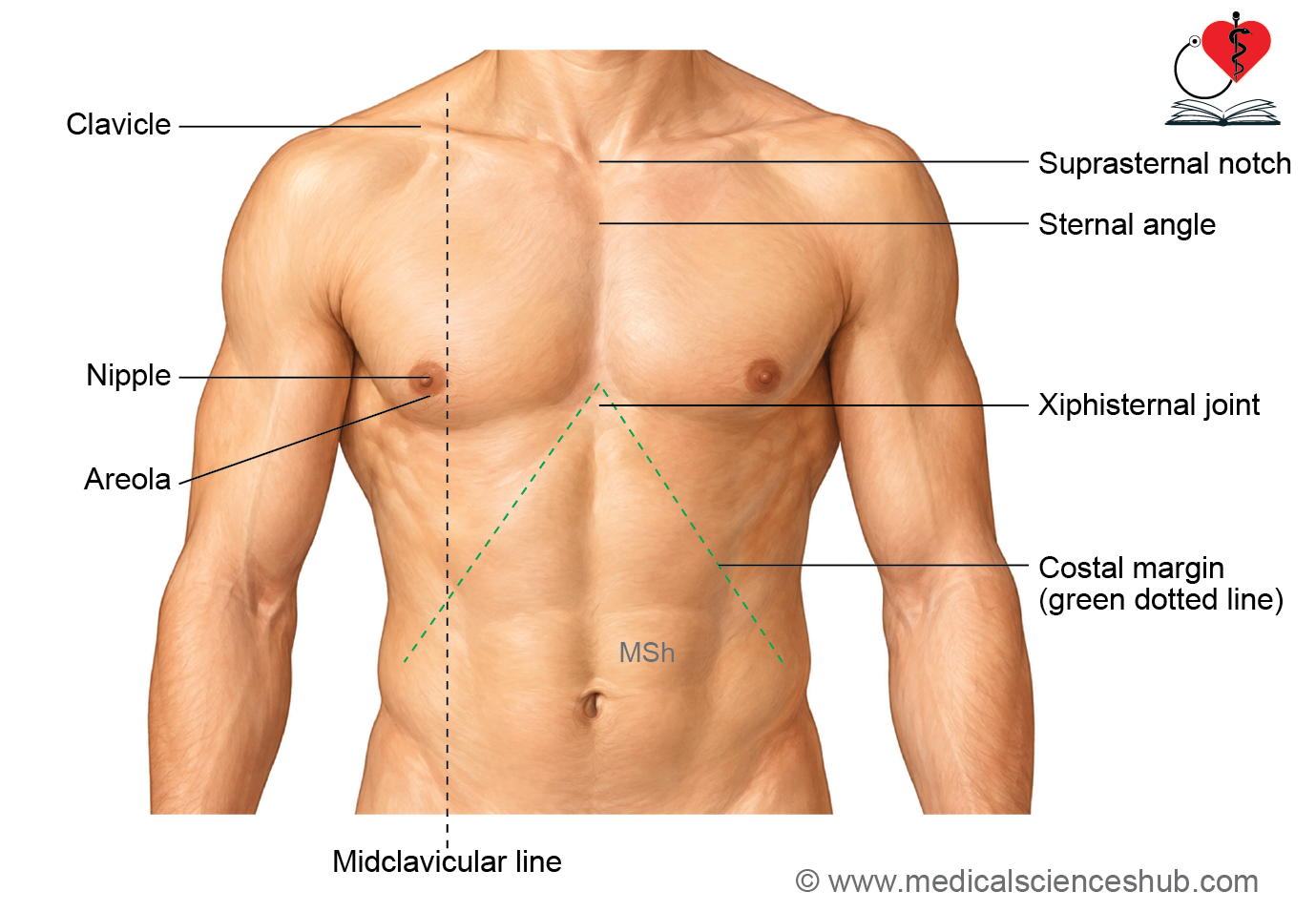
- Sternum: The sternum lies subcutaneously in the midline and consists of three parts: the manubrium, body, and xiphoid process.
- Suprasternal notch: Also called the jugular or interclavicular notch, it is located between the clavicles at the superior border of the manubrium. The trachea can be palpated through this notch. Its vertebral level corresponds approximately to the lower border of T2.
- Sternal angle: The sternal angle (angle of Louis) is an important palpable landmark at the junction between the manubrium and body of the sternum.
- Xiphisternal joint: The xiphisternal joint is situated between the costal margins. The infrasternal (subcostal) angle surrounds this region, with the xiphoid process projecting into the angle.
- Second costal cartilage: The second costal cartilage articulates with the sternum at the level of the sternal angle.
- Costal margin: The costal margin is formed by the cartilages of the 7th to 10th ribs and the free anterior ends of the 11th and 12th ribs.
- Thoracic vertebral spines: The spine of C7 (vertebra prominens) can be palpated at the root of the neck. The thoracic vertebral spines can then be followed inferiorly.
- The T3 spine is approximately level with the root of the spine of the scapula, while the T7 spine corresponds approximately to the inferior angle of the scapula.
Soft Tissue Landmarks
- Nipple: In males, the nipple is usually located in the 4th intercostal space, approximately 10 cm from the midsternal line. Its position in females is variable.
- Apex beat: The apex beat (apical impulse or ictus cordis) is the palpable outward movement produced by the cardiac apex. It is normally felt in the left 5th intercostal space, just medial to the midclavicular line.
Thoracic Cage
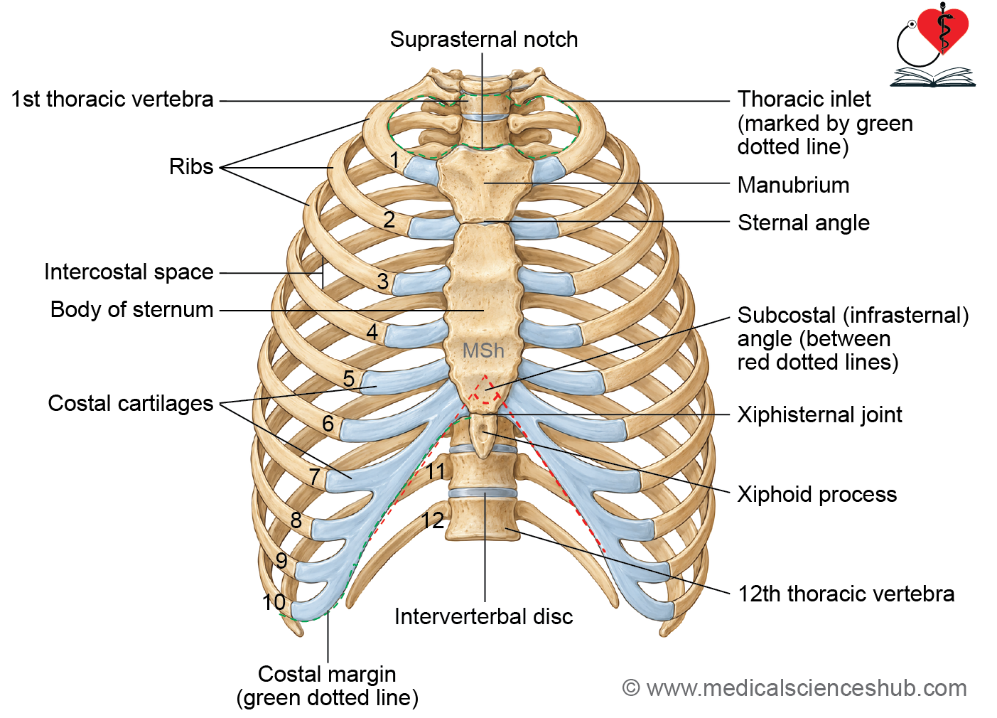
- The thoracic cage is the osseocartilaginous skeletal framework that supports the thoracic wall.
- It is an elastic and flexible cage that protects the heart and lungs and contributes to respiratory movements.
Formation of Thoracic Cage
- Anteriorly: It is formed by the sternum.
- Posteriorly: It is formed by the 12 thoracic vertebrae and intervertebral discs.
- Laterally: It consists of 12 pairs of ribs with their costal cartilages.
- Posteriorly, the ribs articulate with the thoracic vertebrae.
Classification of Ribs
- True ribs (1st–7th): These ribs attach directly to the sternum through their own costal cartilages.
- False ribs (8th–10th): These ribs attach indirectly to the sternum through the costal cartilage of the rib above.
- Floating ribs (11th–12th): These ribs have free anterior ends and do not articulate with the sternum or other costal cartilages.
Shape
- The normal thoracic cavity is shaped like a truncated cone, being narrower superiorly and wider inferiorly.
- In transverse section, its shape varies with age and sex:
- Adults: It is typically reniform or kidney-shaped.
- Infants below 2 years: It is approximately circular.
Boundaries
- Superiorly, the thoracic cavity is separated from the neck by the suprapleural membrane (Sibson’s fascia).
- Inferiorly, it is separated from the abdominal cavity by the thoracoabdominal diaphragm.
Respiration
- Contraction and descent of the diaphragm increase the vertical diameter of the thorax and produce predominantly abdominal movement.
- In infants, the ribs are nearly horizontal and contribute little to thoracic expansion. Therefore, breathing is predominantly abdominal.
- In adults, the ribs are oblique, and their movements increase the anteroposterior, vertical, and transverse diameters of the thorax. Respiration is therefore thoracoabdominal.
Clinical Anatomy
- Hiccups result from involuntary spasmodic contractions of the diaphragm, which may occur due to diaphragmatic or phrenic nerve irritation.
- In children under 2 years, abdominal surgery may restrict abdominal movements during breathing, promoting retention of lung secretions and increasing the risk of postoperative pneumonia.
Sternal Angle
- The sternal angle, also known as the angle of Louis, is the junction between the manubrium and body of the sternum.
- It can be palpated as a transverse ridge approximately 5 cm below the suprasternal notch.
Importance of sternal angle
- Rib identification: The second costal cartilage articulates with the sternum at this level, making it an important landmark for counting ribs and intercostal spaces.
- Vertebral level: It corresponds approximately to the T4/T5 intervertebral level.
- Arch of aorta: The arch of the aorta begins and ends approximately at the level of the sternal angle.
- Mediastinal division: A horizontal plane through the sternal angle and T4/T5 level separates the superior mediastinum from the inferior mediastinum.
- Tracheal bifurcation: The trachea divides into the right and left principal bronchi at approximately this level.
- Pulmonary trunk: The pulmonary trunk bifurcates into the right and left pulmonary arteries near this level.
- Azygos vein: The azygos vein arches over the root of the right lung and drains into the superior vena cava near this level.
Thoracic Inlet
- The thoracic inlet, or superior thoracic aperture, is the opening through which the thoracic cavity communicates with the root of the neck.
- It is approximately kidney-shaped, with a transverse diameter of 10–12 cm and an anteroposterior diameter of about 5 cm.
Boundaries
- Anteriorly: The superior border of the manubrium sterni.
- Posteriorly: The anterior border of the superior surface of the T1 vertebral body.
- Laterally: The medial borders of the first ribs and their costal cartilages.
Plane of Inlet
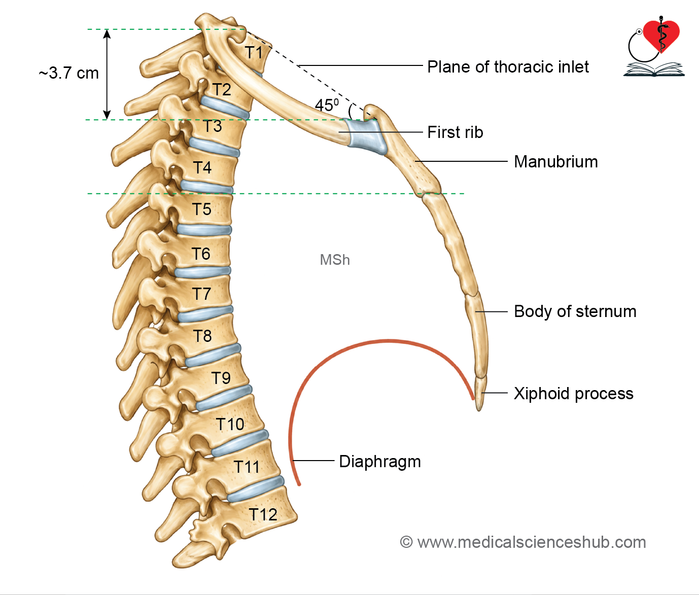
- The plane of the thoracic inlet is directed downward and forward and forms an angle of approximately 45° with the horizontal plane.
- Its anterior end lies about 3.7 cm lower than its posterior end.
Suprapleural Membrane (Sibson’s Fascia)
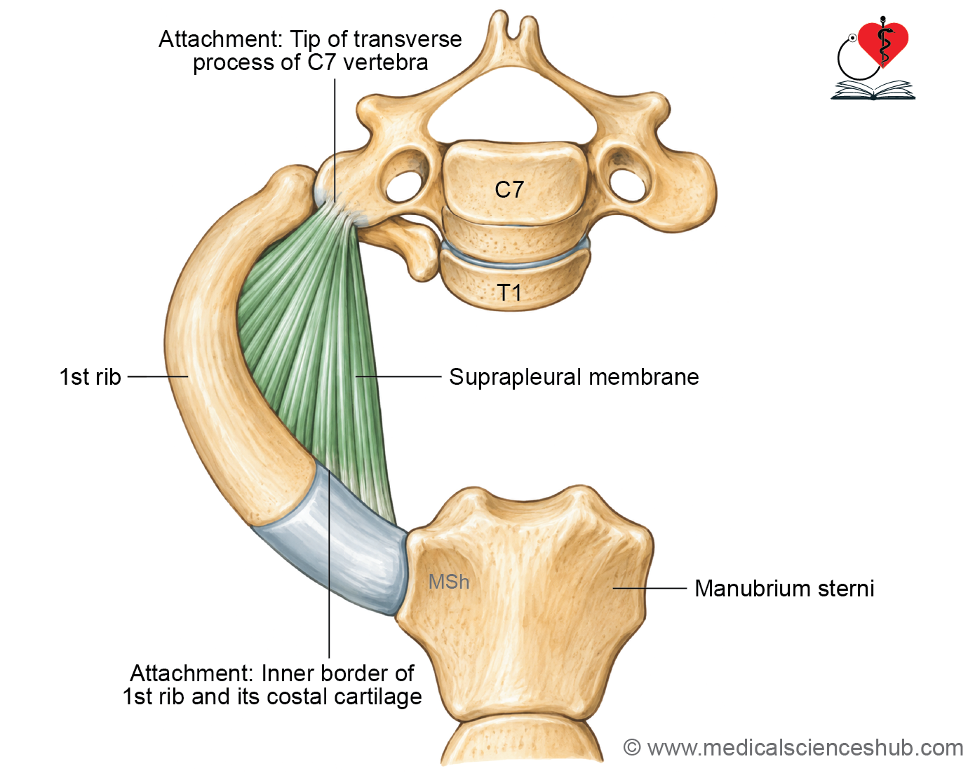
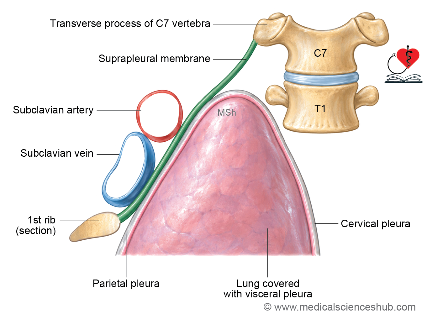
- The suprapleural membrane, or Sibson’s fascia, consists of a pair of fascial sheets that partially separate the thoracic inlet from the root of the neck.
- Each membrane is triangular and tent-shaped and covers the cervical pleura and apex of the lung.
- The space between the two membranes permits passage of structures between the thorax and neck.
Attachment
- Apex: The apex is attached to the tip of the transverse process of the C7 vertebra.
- Base: The base is attached to the inner border of the first rib and its costal cartilage.
Relations
- Superiorly: The subclavian vessels are related to the membrane.
- Inferiorly: It is related to the cervical pleura and apex of the lung.
Morphology
- Sibson’s fascia is considered a flattened and degenerated tendon of the scalenus minimus (pleuralis) muscle.
Functions
- It protects the cervical pleura and apex of the lung.
- It provides rigidity to the thoracic inlet, helping prevent excessive vertical movement of structures at the root of the neck during respiration.
Structures Passing through Thoracic Inlet
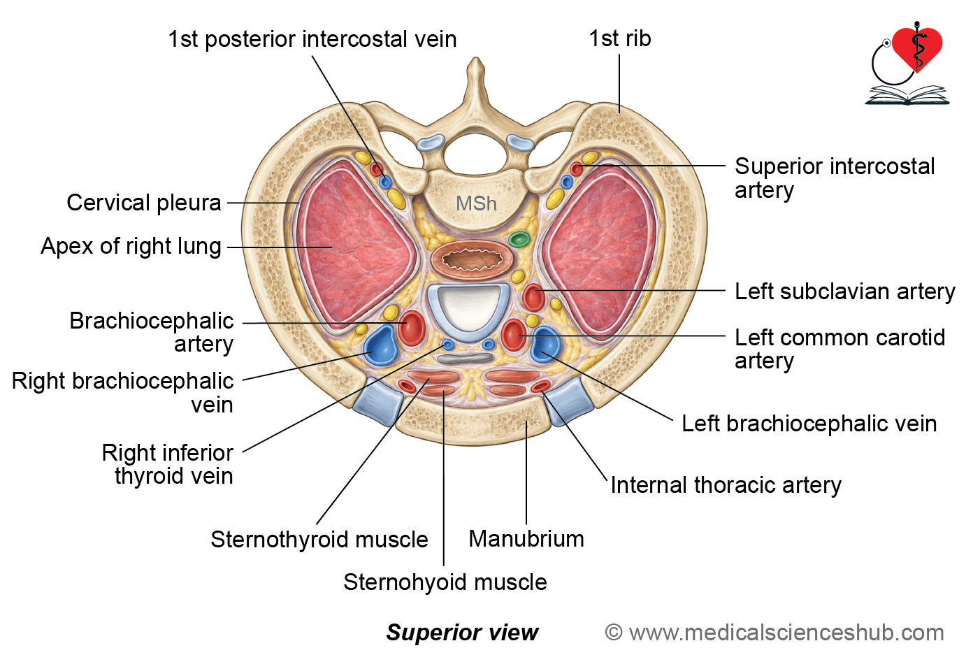
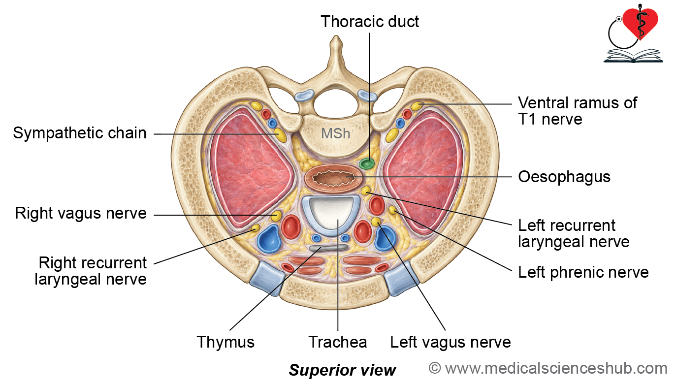
The thoracic inlet provides a passage between the thoracic cavity and root of the neck for several important structures.
Viscera
- Trachea and esophagus pass through the thoracic inlet.
- The thymus and apices of the lungs also extend through this region.
Blood Vessels
- The major arteries include the brachiocephalic artery, left common carotid artery, and left subclavian artery.
- Other vessels include the internal thoracic arteries, superior intercostal arteries, first posterior intercostal veins, and inferior thyroid vein.
Nerves
- The phrenic nerves, vagus nerves, sympathetic trunks, and T1 spinal nerves pass through the thoracic inlet.
Muscles
The sternohyoid, sternothyroid, and longus colli muscles are related to or pass through the region.
Clinical Anatomy
Thoracic Outlet Syndrome
- Clinicians commonly use the term thoracic outlet for the anatomical thoracic inlet; therefore, the condition is clinically termed thoracic outlet syndrome.
- It results from compression of the subclavian vessels and/or lower trunk of the brachial plexus.
- Common causes include a cervical rib and hypertrophy of the scalenus anterior muscle.
- Clinical Features
- Neural compression: Pain, tingling, and numbness may occur along the medial forearm and hand, with possible wasting of the hand muscles.
- Arterial compression: Reduced blood flow may cause pallor and coldness of the affected upper limb.
Inferior Thoracic Aperture
- The inferior thoracic aperture, also known anatomically as the thoracic outlet, forms the lower opening of the thoracic cage.
- It lies between the thoracic and abdominal cavities.
- The aperture is closed by the thoracoabdominal diaphragm, which separates the two cavities and contributes to respiration.
Boundaries
- Anteriorly: The infrasternal angle.
- Posteriorly: The body of the T12 vertebra.
- Laterally: The costal margin, formed by the 7th to 10th costal cartilages and the 11th and 12th ribs.
Thoracoabdominal Diaphragm
- The thoracoabdominal diaphragm separates the thoracic cavity from the abdominal cavity. It is the principal muscle of respiration and forms a dome-shaped musculoaponeurotic partition.
- It consists of right and left muscular domes that converge into a central tendon. The right dome is higher than the left, mainly because of the underlying liver.
Origin
The diaphragm has three muscular parts:
- Sternal part: It consists of two fleshy slips arising from the posterior surface of the xiphoid process.
- Costal part: It arises from the inner surfaces of the lower six ribs and their costal cartilages.
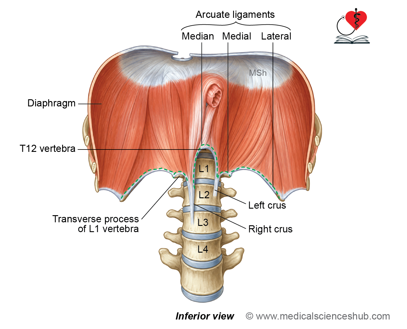
- Vertebral part: It is formed by the right and left crura and five arcuate ligaments.
Crura
- The right crus arises from the anterior surfaces of the right sides of L1–L3 vertebrae and their intervertebral discs.
- The left crus is shorter and arises from the anterior surfaces of L1 and L2 vertebrae and their intervertebral disc.
Arcuate Ligaments
- The median arcuate ligament connects the upper ends of the two crura.
- The medial arcuate ligaments extend from L2 to the transverse processes of L1.
- The lateral arcuate ligaments extend from the transverse processes of L1 to the 12th ribs.
Insertion
- All muscular fibers converge toward and attach to the central tendon.
- The central tendon is an aponeurotic, trifoliate sheet with one anterior leaflet and two posterior leaflets.
- Superiorly, it is fused with the fibrous pericardium.
Nerve supply
Motor Supply
- The diaphragm receives its motor supply from the phrenic nerves (C3, C4, and C5).
- The central part is supplied mainly by the phrenic nerve.
Sensory Supply
- The central part of the diaphragm receives sensory fibers from the phrenic nerves.
- The peripheral part receives sensory fibers from the lower six to seven intercostal nerves.
Blood supply and lymphatic drainage
Arterial supply
The diaphragm receives blood from branches of the internal thoracic artery and the aorta:
- Musculophrenic artery.
- Pericardiophrenic artery.
- Lower five posterior intercostal arteries.
- Subcostal arteries.
- Superior phrenic arteries.
- Inferior phrenic arteries.
Venous Drainage
- The veins generally accompany the corresponding arteries and drain into the systemic venous circulation.
Lymphatic Drainage
- Lymph from the diaphragm drains into several groups of diaphragmatic lymph nodes.
- The principal groups are:
- Anterior diaphragmatic lymph nodes.
- Posterior diaphragmatic lymph nodes.
- Right lateral diaphragmatic lymph nodes.
- Left lateral diaphragmatic lymph nodes.
Openings in the diaphragm
- The diaphragm contains several openings that allow important structures to pass between the thorax and abdomen.
- These openings are classified into major and minor openings.
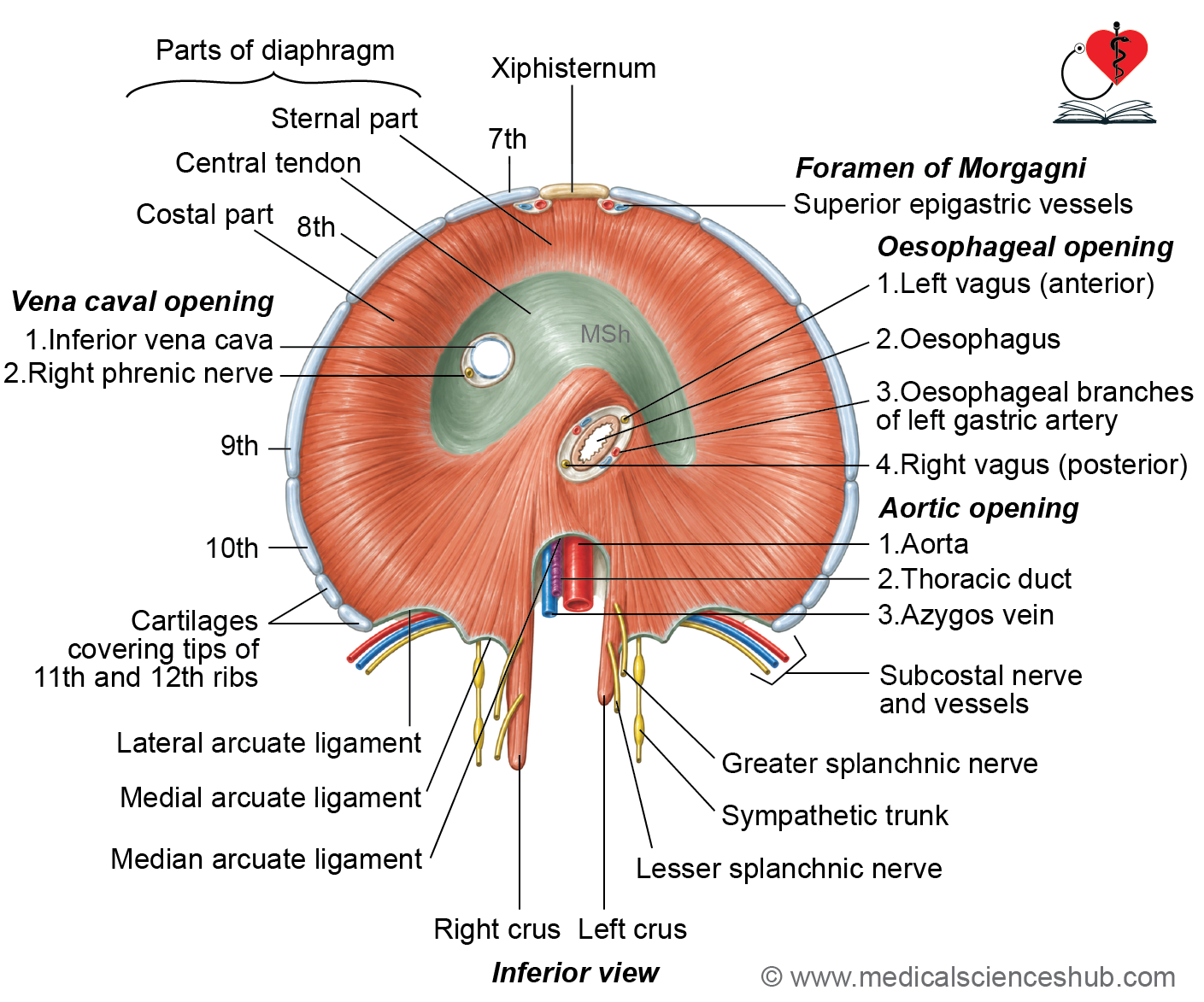
Table
Structures passing through minor openings
- Space of Larry: Transmits the superior epigastric vessels.
- Gap between the 7th and 8th costal slips: Transmits the musculophrenic artery.
- Deep to the lateral arcuate ligament: Transmits the subcostal nerves and vessels.
- Deep to the medial arcuate ligament: Transmits the sympathetic trunks.
- Through the crura: The greater, lesser, and least splanchnic nerves pass through the diaphragm.
- Left crus: Allows passage of the hemiazygos vein.
Actions of diaphragm
1. Respiration
- The diaphragm is the principal muscle of respiration.
- During contraction, it descends and flattens, increasing the vertical diameter of the thoracic cavity.
- This produces more negative intrathoracic pressure, allowing air to enter the lungs during inspiration.
2. Other Functions
- Expulsive actions: The diaphragm assists in micturition, defecation, vomiting, and parturition by increasing intra-abdominal pressure.
- Weight lifting: It helps stabilize the vertebral column by increasing intra-abdominal pressure during heavy lifting.
- Venous return: Diaphragmatic contraction facilitates venous return to the heart by lowering intrathoracic pressure and increasing intra-abdominal pressure, thereby compressing abdominal veins.
- Esophageal sphincteric action: Contraction of the arching fibers of the right crus around the esophageal opening contributes to the functional lower esophageal sphincter mechanism.
Clinical Anatomy
Paralysis of the Diaphragm
- Damage to the phrenic nerve can result in paralysis of the diaphragm.
- In unilateral paralysis, the affected dome remains elevated during inspiration and moves downward during expiration. This abnormal movement is called paradoxical movement and can be demonstrated by real-time fluoroscopy.
- Bilateral diaphragmatic paralysis can cause severe respiratory impairment and may lead to respiratory failure.
Hiccups
- Hiccups result from sudden, involuntary spasmodic contractions of the diaphragm, usually following diaphragmatic or phrenic nerve irritation.
Diaphragmatic Hernia
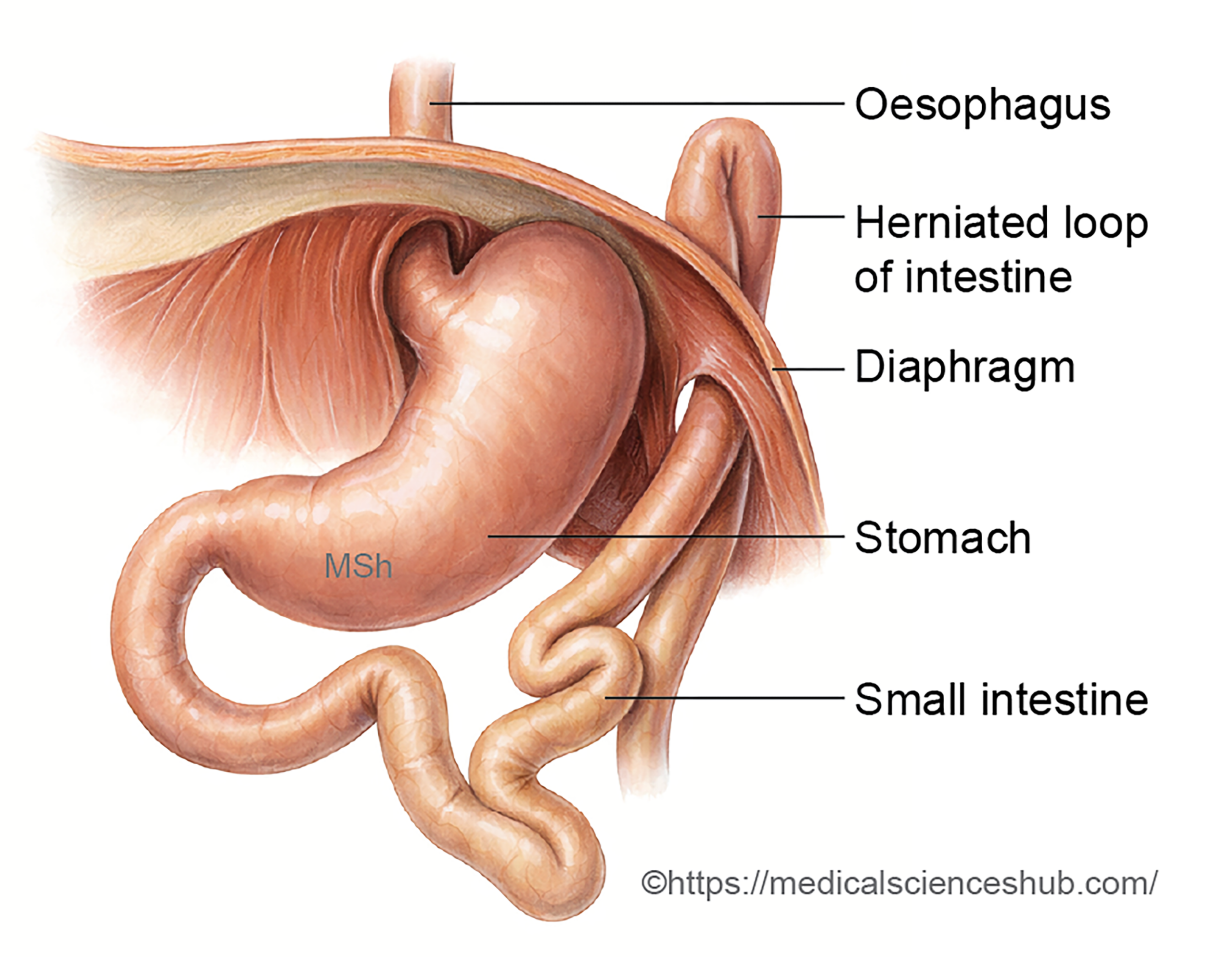
- A diaphragmatic hernia occurs when abdominal contents protrude into the thoracic cavity through a defect or abnormal opening in the diaphragm.
- It may be congenital or acquired.
Congenital Diaphragmatic Hernia
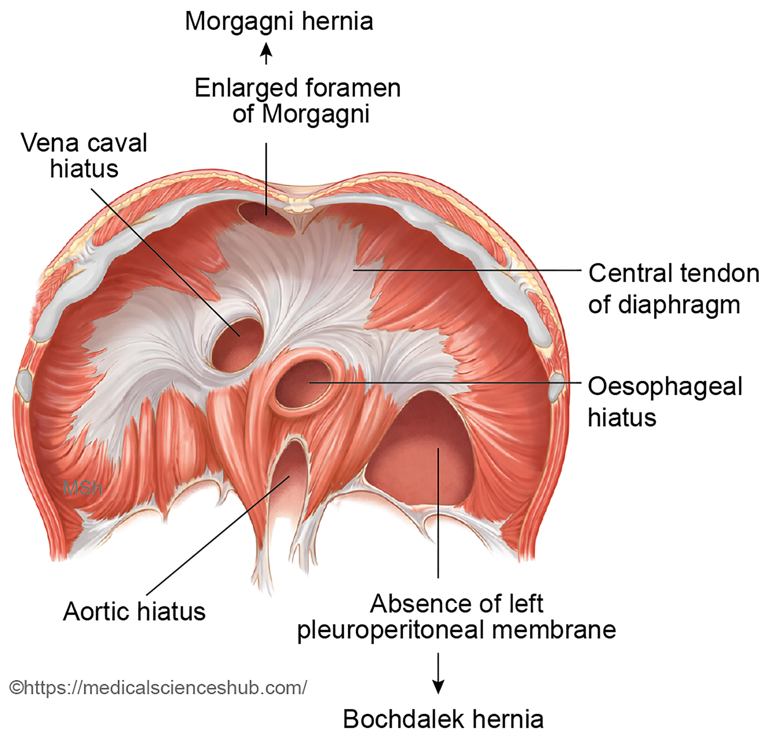
- Bochdalek hernia: This is a posterolateral hernia and accounts for most cases of congenital diaphragmatic hernia. It results from incomplete development of the pleuroperitoneal membrane.
- Morgagni hernia: This retrosternal or parasternal hernia occurs through the foramen of Morgagni (space of Larry).
- Diaphragmatic eventration: It refers to abnormal elevation or contour of a diaphragmatic dome due to varying degrees of muscle aplasia, atrophy, or paralysis.
Acquired Diaphragmatic Hernia
- Acquired hernias may result from trauma or occur as hiatal hernias.
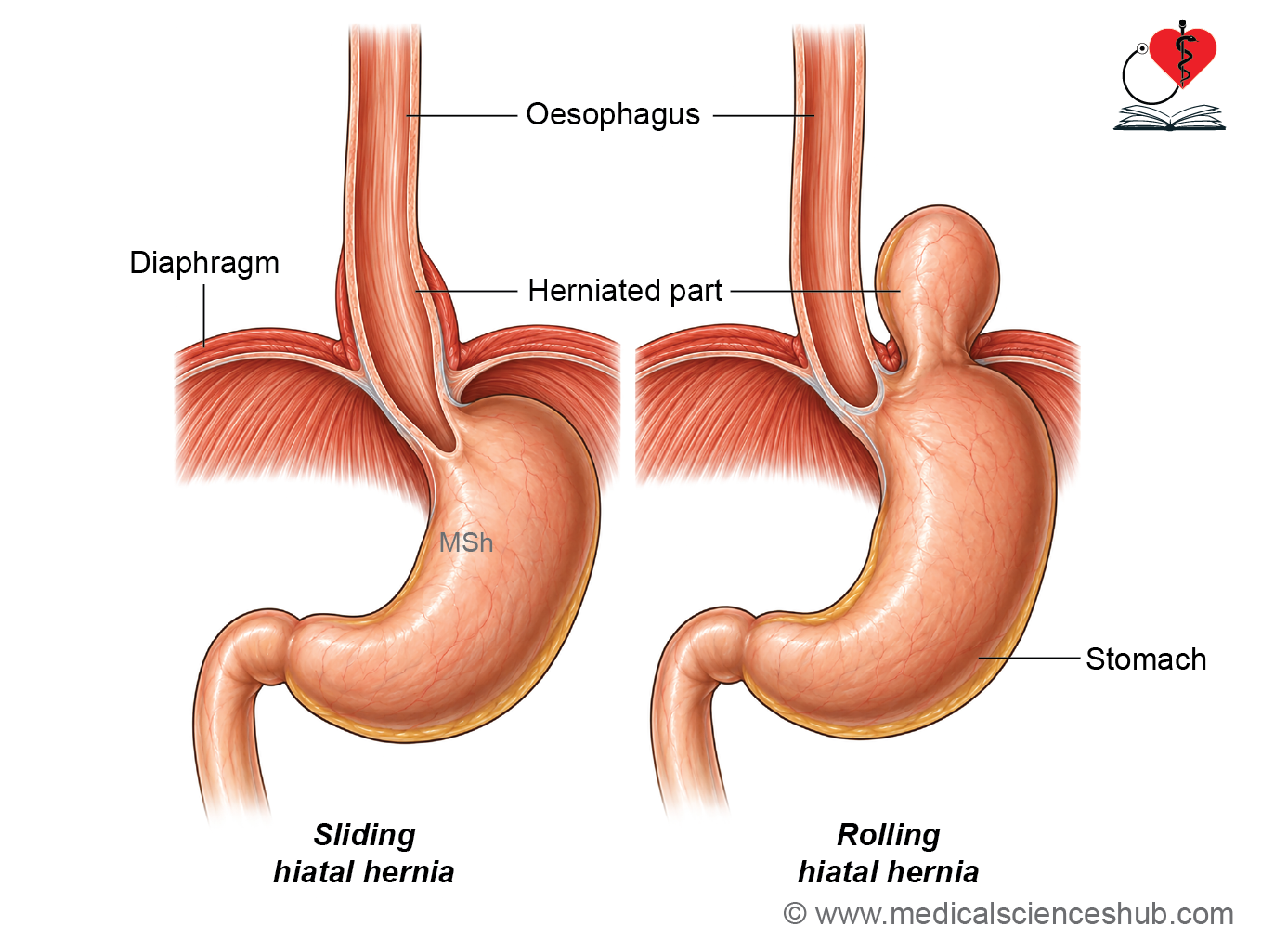
- Sliding hiatal hernia: The gastroesophageal junction and part of the stomach move into the thorax through the esophageal opening. It is associated with factors such as aging, obesity, and chronic cough and may contribute to GERD.
- Rolling (paraesophageal) hiatal hernia: The gastroesophageal junction remains in the abdomen, while the fundus of the stomach herniates into the thoracic cavity.
📝 Test Your Knowledge – Practice MCQs
Attempt the chapter MCQ quiz and assess your understanding of key concepts.
Click to see: Medical Disclaimer and Copyright Notice