
Competencies
- AN21.1: Identify and describe the salient features of sternum, typical rib, and typical thoracic vertebra.
- AN21.2: Identify and describe the features of 1st, 2nd, 11th, and 12th ribs, and 1st, 11th, and 12th thoracic vertebrae
This chapter includes sternum and ribs.
- The thoracic cage forms the skeletal framework of the thoracic wall.
- It consists of:
- Sternum.
- Twelve pairs of ribs.
- Twelve thoracic vertebrae.
- The thoracic cage protects the heart and lungs from external injury.
Sternum
- The sternum is a median bone situated anteriorly in the thoracic wall. It is an elongated flat bone that is convex anteriorly and concave posteriorly.
- It consists of three parts:
- Manubrium, which forms the upper part.
- Body, which forms the middle and longest part.
- Xiphoid process, which forms the lower part.
- The sternum is approximately 17 cm long.
- It articulates with both clavicles at the sternoclavicular joints. It also articulates with the costal cartilages of the first seven pairs of ribs at the sternocostal joints.
- The sternum contains highly vascular trabecular bone and retains hematopoietic activity even in adults.
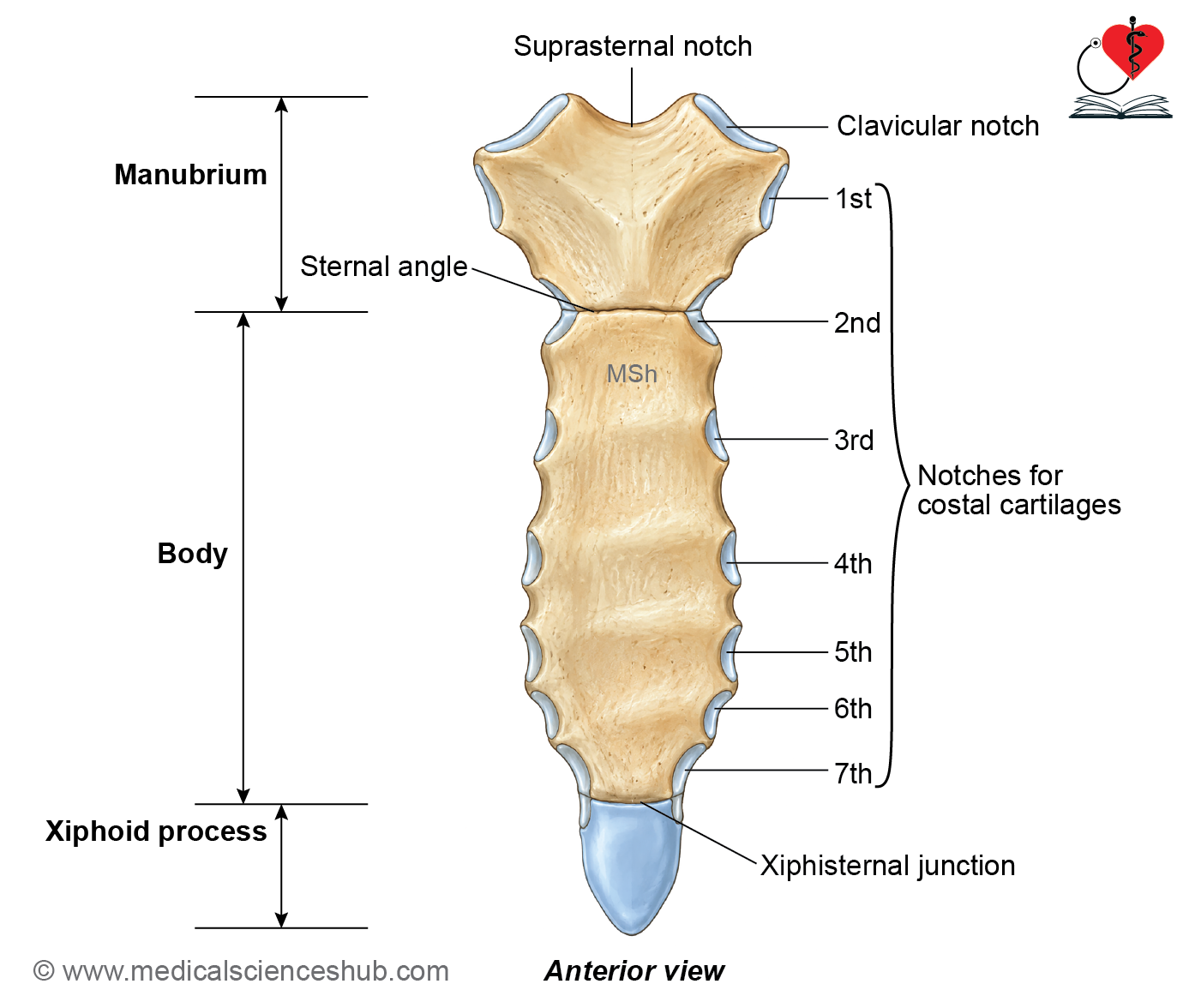
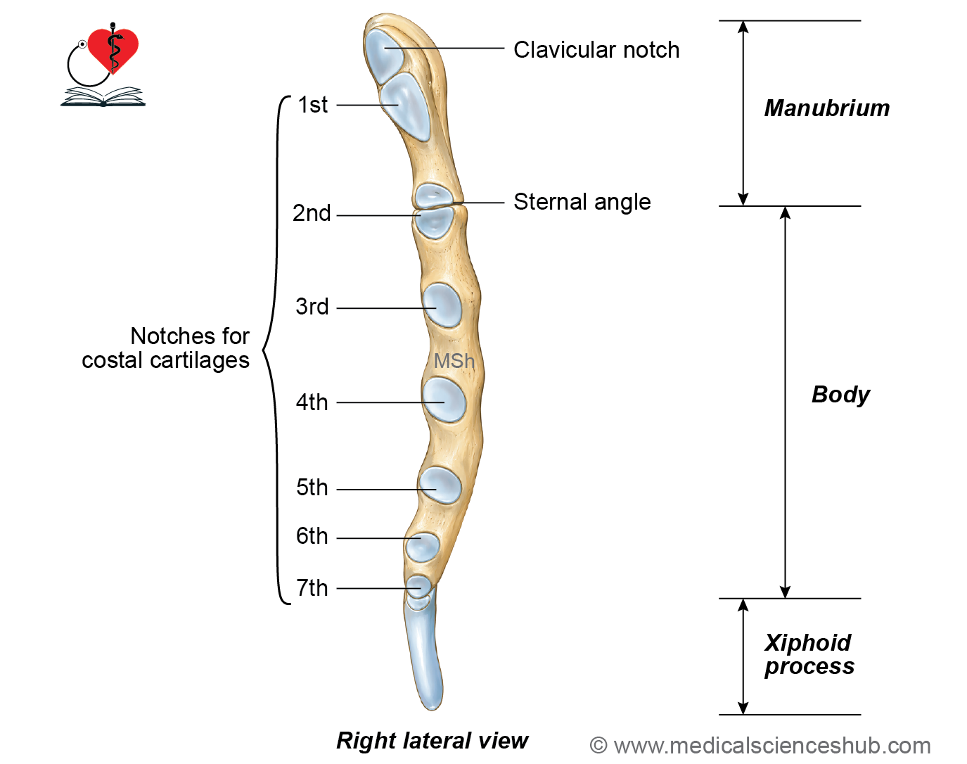
Anatomical Position
Hold the sternum in such a way so that:
- The rough, convex surface is directed anteriorly.
- The broad manubrium is positioned superiorly, while the pointed xiphoid process is directed inferiorly.
- The anterior surface is oriented slightly forward.
Features of Sternum
The sternum is divided into three parts: the manubrium, body, and xiphoid process.
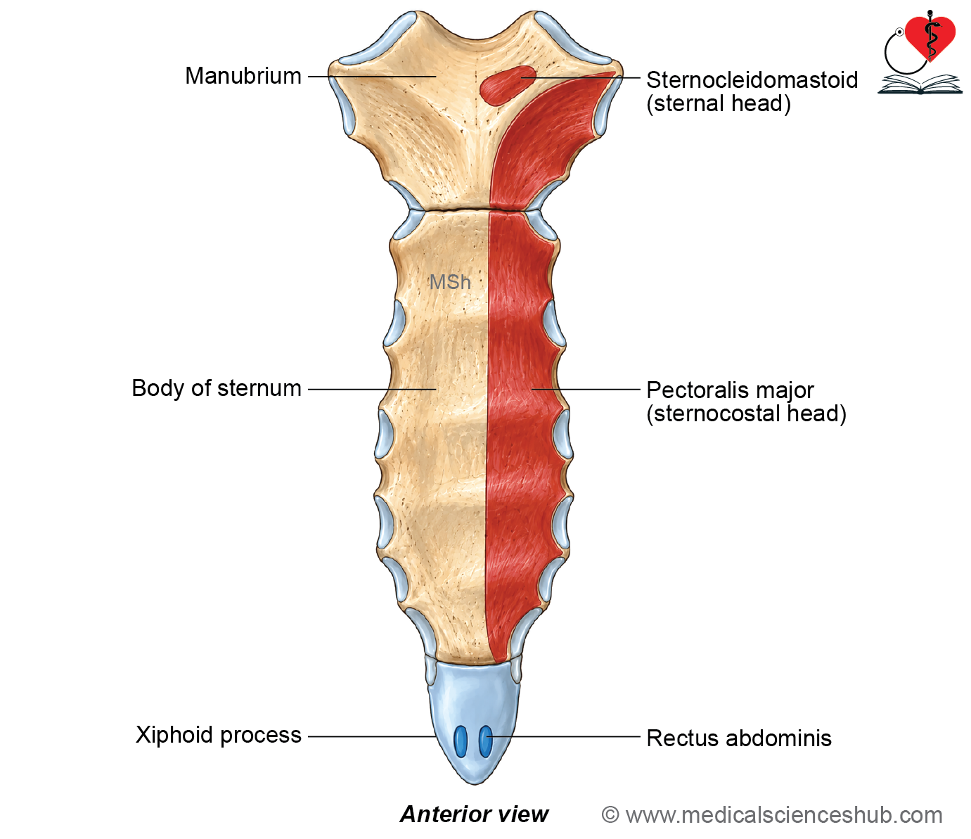
Manubrium
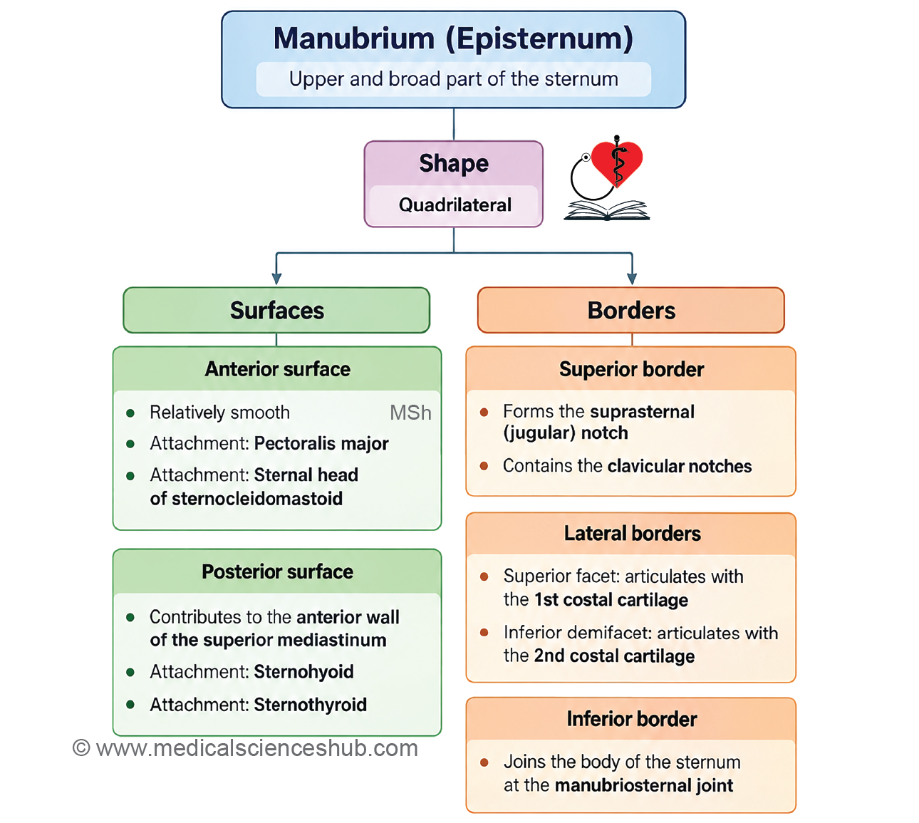
- The manubrium sterni, also called the episternum, is the upper part of the sternum. It is quadrilateral, broad superiorly and progressively narrower inferiorly.
- It has two surfaces, anterior and posterior, and four borders: superior, inferior, and two lateral borders.
Anterior surface
- The anterior surface is smooth, convex transversely, and slightly concave vertically.
- It provides attachment to the pectoralis major and sternal head of the sternocleidomastoid muscles on each side.
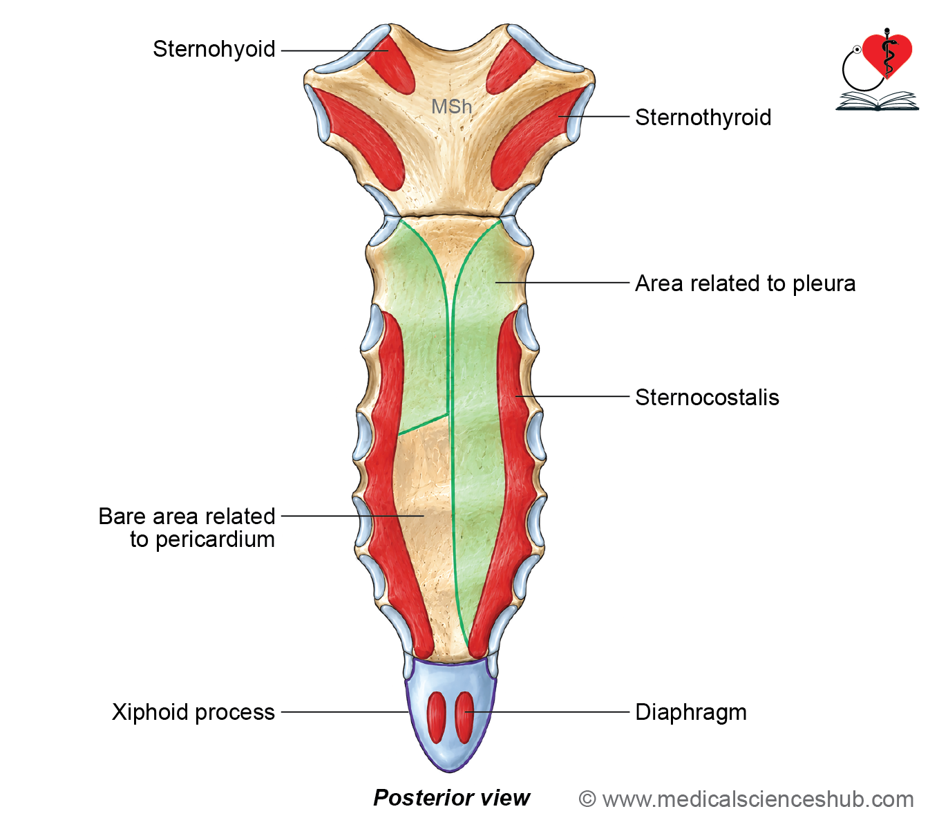
Posterior surface
- The posterior surface is smooth and concave.
- It provides attachment to the sternohyoid superiorly and sternothyroid inferiorly.
- It forms the anterior boundary of the superior mediastinum and is related to the arch of the aorta, major branches of the aortic arch, left brachiocephalic vein, lungs, and pleurae.
Superior border
- The superior border is thick, rounded, and concave, with a midline suprasternal (jugular) notch.
- A clavicular notch on each side articulates with the medial end of the clavicle to form the sternoclavicular joint.
- The suprasternal notch provides attachment to the interclavicular ligament and cervical fascia.
Lateral borders
- Each lateral border has two costal facets:
- The upper facet articulates with the first costal cartilage.
- The lower demifacet contributes to articulation with the second costal cartilage.
Inferior border
- The inferior border has an oval facet that articulates with the body of the sternum. It forms the manubriosternal joint and marks the sternal angle.
Body (Mesosternum)
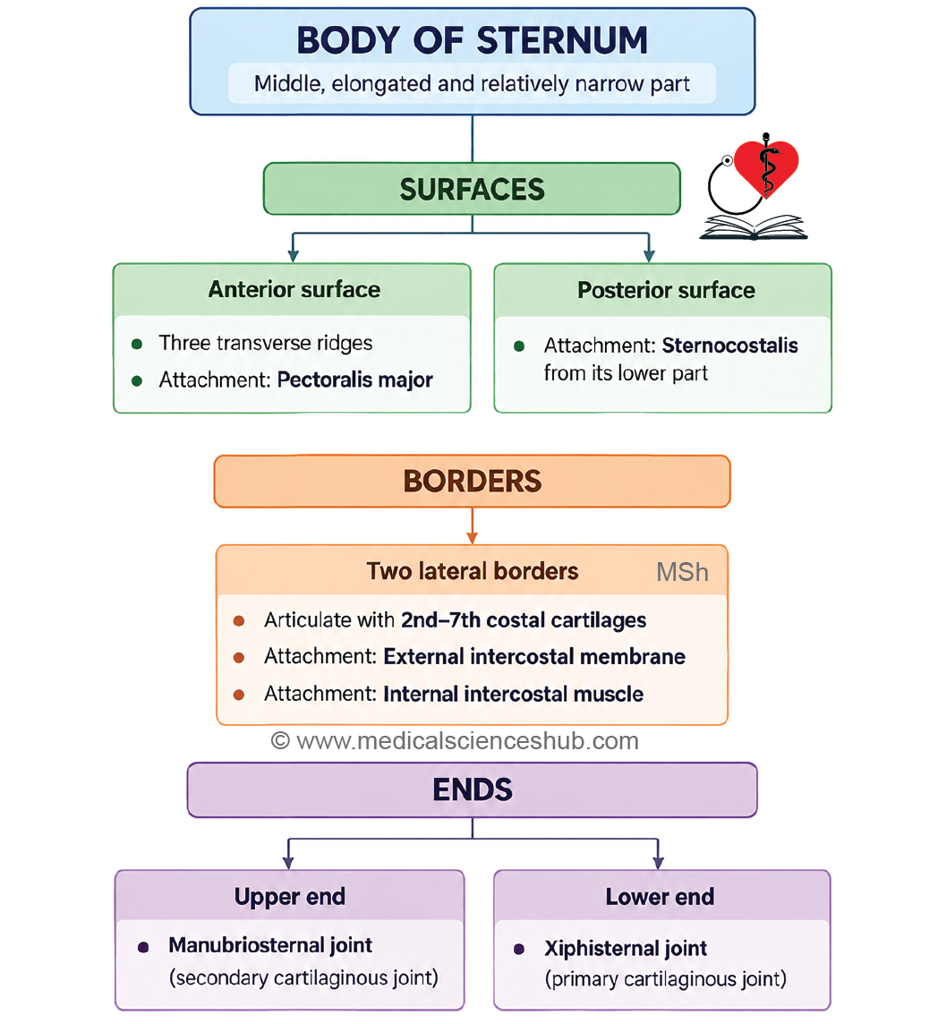
- The body of the sternum is the middle, elongated, relatively narrow and thin part of the sternum.
- It is widest at the level of the 5th costal cartilage.
- It has two surfaces, two lateral borders, and two ends.
Anterior surface
- The anterior surface is flat and faces forward with a slight upward inclination. It presents three transverse ridges, which indicate the lines of fusion between the four embryonic sternal segments, known as sternebrae.
- A sternal foramen may occasionally be present near the line of fusion between the third and fourth sternebrae.
- Muscle Attachment
- The pectoralis major takes origin from the anterior surface on either side.
Posterior surface
- The posterior surface is concave. It shows faint transverse ridges representing the fusion lines between the sternebrae.
- Muscle Attachment:
- The sternocostalis muscle arises from the lower part of the posterior surface.
Lateral borders
- Each lateral border bears four complete, cup-shaped articular facets for articulation with the third, fourth, fifth, and sixth costal cartilages.
- Each border also has two demifacets.
- The upper demifacet articulates with the second costal cartilage.
- The lower demifacet articulates with the seventh costal cartilage.
Attachments
- The lateral borders provide attachment to the external intercostal membranes.
- They also provide attachment to the internal intercostal muscles.
Ends
Upper end
- The upper end articulates with the manubrium to form the manubriosternal joint.
- The manubriosternal joint is a secondary cartilaginous joint (symphysis).
Lower end
- The lower end is narrow. It articulates with the xiphoid process to form the xiphisternal joint.
- The xiphisternal joint is a primary cartilaginous joint (synchondrosis).
Xiphoid Process (Metasternum)
- The xiphoid process is the smallest and most inferior part of the sternum.
- Its shape is variable, and it may occasionally be bifid or perforated by a foramen.
- It lies in the floor of the epigastric fossa.
- It has anterior and posterior surfaces, two lateral borders, and upper and lower ends.
- Attachments
- The anterior surface provides origin to the rectus abdominis and attachment to the aponeuroses of the external and internal oblique muscles.
- The posterior surface gives origin to the sternal fibres of the diaphragm.
- The lower end or tip provides attachment to the upper end of the linea alba.
Ossification
- Sternum develops by fusion of two cartilaginous sternal plates one on each side of the median · The sternum develops from two cartilaginous sternal plates, situated on either side of the median plane.
- The two sternal plates gradually unite in the midline, with fusion progressing in a craniocaudal direction.
- Incomplete fusion, particularly in the lower part, may result in developmental variations such as a bifid sternum or sternal foramen.
Ossification of the Sternum
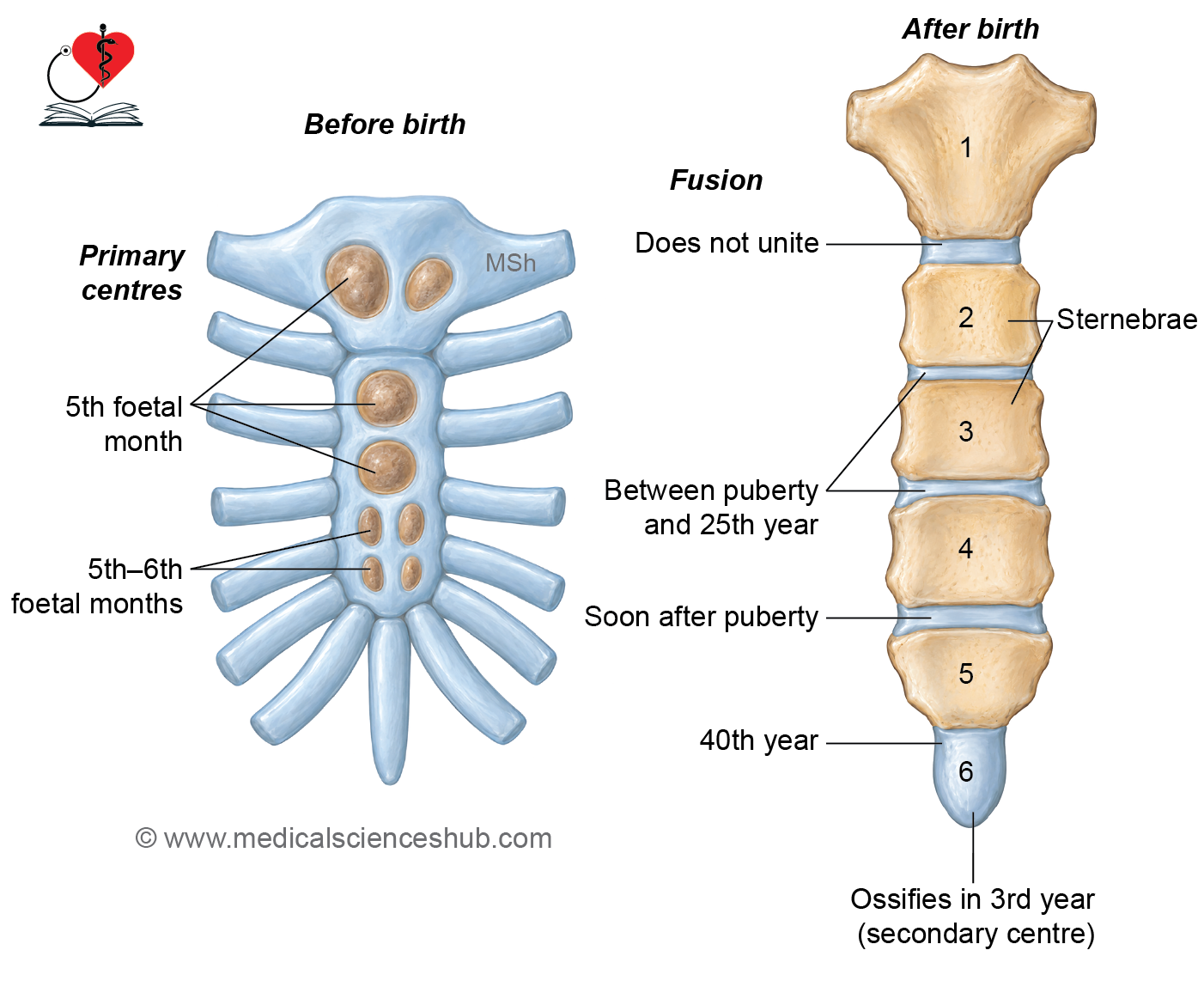
The sternum ossifies from several centers that appear at different stages of development.
Manubrium
- The manubrium usually ossifies from one primary ossification center. This center appears during the fifth month of intrauterine life.
Body of Sternum
- The body develops from four sternebrae, each having its own ossification center or centers.
- The first sternebra usually has one center, which appears during the fifth month of intrauterine life.
- The second sternebra usually has one center, which also appears during the fifth month of intrauterine life.
- The third sternebra usually has a pair of centers that appear during the fifth to sixth month of intrauterine life.
- The fourth sternebra usually has a pair of centers that appear during the fifth to sixth month of intrauterine life.
Xiphoid Process
- The xiphoid process usually ossifies from one center. Its ossification center appears around the third year of life.
Fusion of the Sternum
- Fusion between the sternebrae proceeds from below upward, which is opposite to the direction in which the two sternal plates unite during development.
- Fusion of the sternebrae generally begins around puberty and is usually completed by approximately 25 years of age.
- The xiphoid process usually fuses with the body of the sternum at about 40 years of age.
- The manubrium and body are connected by the manubriosternal joint, which is a secondary cartilaginous joint. Therefore, they usually remain separate rather than undergoing complete bony fusion during early adult life.
- The xiphoid process is the only part of the sternum described as ossifying from a secondary ossification center.
Clinical Anatomy
- Hematopoiesis: The sternum contains red bone marrow throughout life and therefore remains an important site of hematopoiesis in adults.
- Sternal Puncture: Sternal puncture may be performed to obtain a sample of red bone marrow for hematological examination. A suitable needle is introduced carefully into the sternum to aspirate bone marrow. Excessive penetration must be avoided because important mediastinal structures lie immediately behind the sternum. Sternal puncture is generally avoided in young children because their sternum is relatively thin.
- Funnel Chest (Pectus Excavatum): Pectus excavatum, also called funnel chest, cobbler’s chest, or sunken chest, is the most common congenital deformity of the anterior chest wall. It occurs in approximately 1 in 500 live births. The lower part of the sternum and adjoining costal cartilages are displaced posteriorly. This posterior displacement produces a decrease in the anteroposterior diameter of the chest.
- Pigeon Chest (Pectus Carinatum): Pectus carinatum, or pigeon chest, is characterized by forward protrusion of the sternum. It results from abnormal growth of the costal cartilages, which pushes the sternum anteriorly. The deformity alters the normal shape and dimensions of the thoracic cage.
- Median Sternotomy: During many cardiothoracic surgical procedures, the sternum is divided longitudinally along the midline to provide access to structures within the thoracic cavity. At the end of the operation, the divided parts of the sternum are approximated and commonly secured using stainless-steel wires.
- Sternal Fracture: Fracture of the sternum may occur following high-energy blunt trauma, particularly during road traffic accidents. A fractured sternum may be associated with injury to the underlying heart, lungs, or other thoracic structures.
- Ectopia Cordis: Ectopia cordis is a rare congenital anomaly in which the heart is located partially or completely outside the thoracic cavity. It is associated with failure of normal midline fusion of the developing anterior thoracic wall, including the sternal components.
RIBS
Ribs
- The ribs are also known as costae; the term costa means rib in Latin.
- They are flat, elongated, and curved bones that form a major part of the thoracic cage. Each rib is connected anteriorly to its corresponding costal cartilage. Together, a rib and its costal cartilage form a costal arch.
Number
- There are 12 pairs of ribs, making a total of 24 ribs. The ribs are arranged symmetrically on the two sides of the thoracic cage.
Arrangement
- The ribs are arranged one below another on either side of the thoracic cage. The gap between two adjacent ribs is called an intercostal space.
Obliquity
- The ribs are arranged obliquely, extending downward and forward from their posterior to anterior ends. The degree of obliquity generally increases from the upper ribs toward the lower ribs. The ninth rib is the most obliquely placed. Below the ninth rib, the degree of obliquity gradually decreases.
Length
- The length of the ribs progressively increases from the first to the seventh rib. From the seventh rib onward, their length gradually decreases. The seventh rib is the longest rib.
Anterior Ends
- The anterior ends of the ribs continue as costal cartilages. Through their costal cartilages, the upper ten pairs of ribs have direct or indirect connections with the sternum. The eleventh and twelfth ribs have no anterior attachment to the sternum, and their anterior ends remain free.
Classification
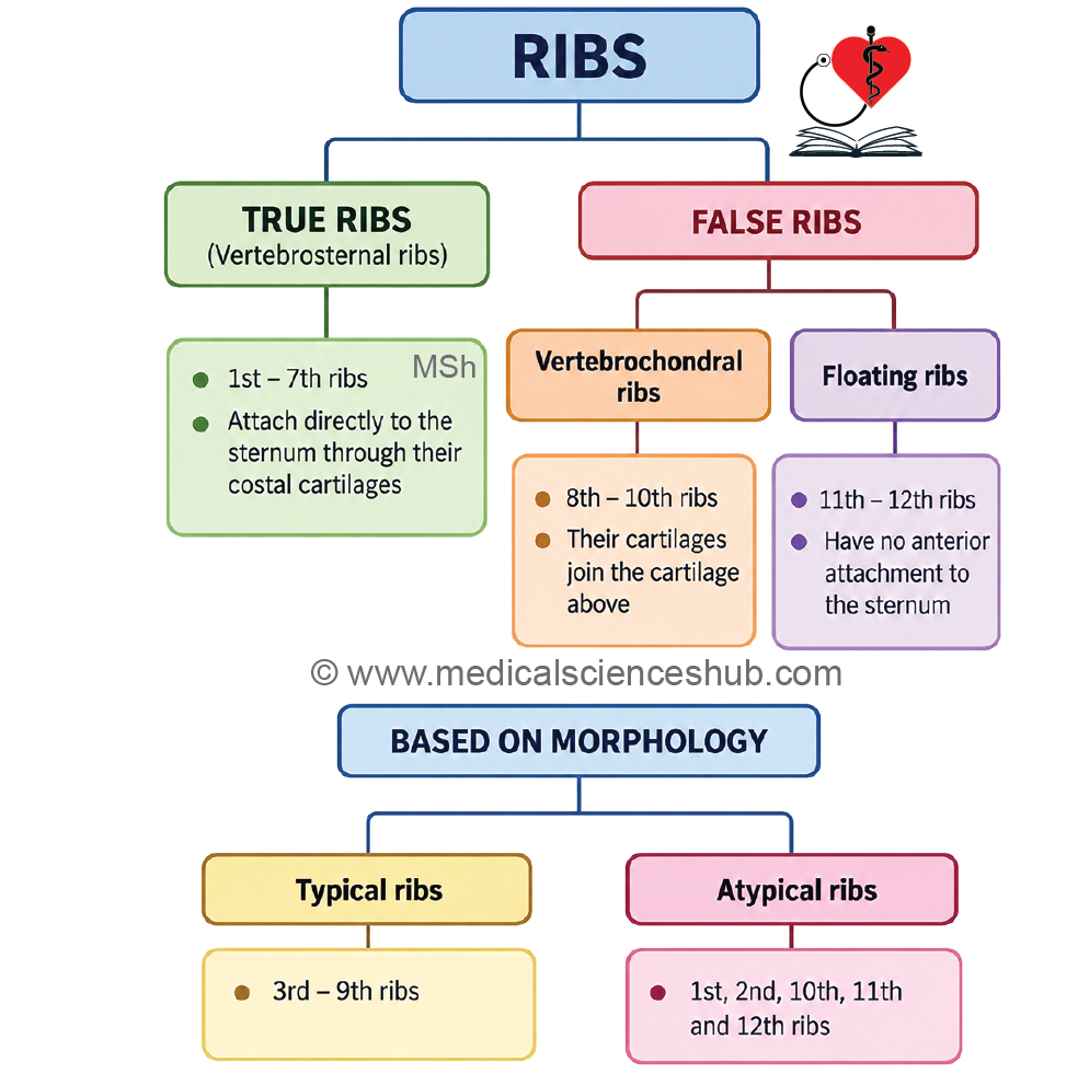
Ribs can be classified according to their relationship with the sternum and their morphological features.
According to Articulation with the Sternum
1. True Ribs (Vertebrosternal Ribs)
- The anterior ends of true ribs are connected directly to the sternum through their own costal cartilages. The first to seventh pairs of ribs are classified as true ribs.
2. False Ribs
- The anterior ends of false ribs do not articulate directly with the sternum. The eighth to twelfth pairs of ribs are classified as false ribs. False ribs are further divided into vertebrochondral ribs and floating ribs.
A. Vertebrochondral Ribs
- The costal cartilage of each vertebrochondral rib joins the costal cartilage of the rib immediately above it rather than attaching directly to the sternum. The eighth, ninth, and tenth pairs of ribs are vertebrochondral ribs.
B. Floating (Vertebral) Ribs
- The anterior ends of floating ribs remain free and have no attachment to the sternum or the costal cartilage of another rib. The eleventh and twelfth pairs of ribs are floating ribs.
According to Morphological Features
1. Typical Ribs
- The third to ninth ribs are considered typical ribs. They share a common pattern of anatomical features and are described using the same general characteristics.
2. Atypical Ribs
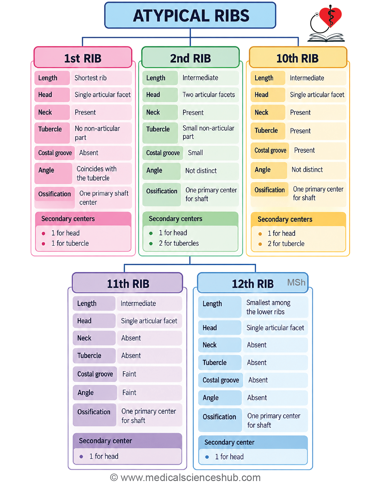
The first, second, tenth, eleventh, and twelfth ribs are classified as atypical ribs. These ribs possess distinctive anatomical features that differ from the general pattern seen in typical ribs.
Typical Ribs
- Typical ribs possess a common set of anatomical features.
- The third to ninth ribs are classified as typical ribs.
Side Determination and Anatomical Position
To determine the side of a typical rib and place it in its anatomical position:
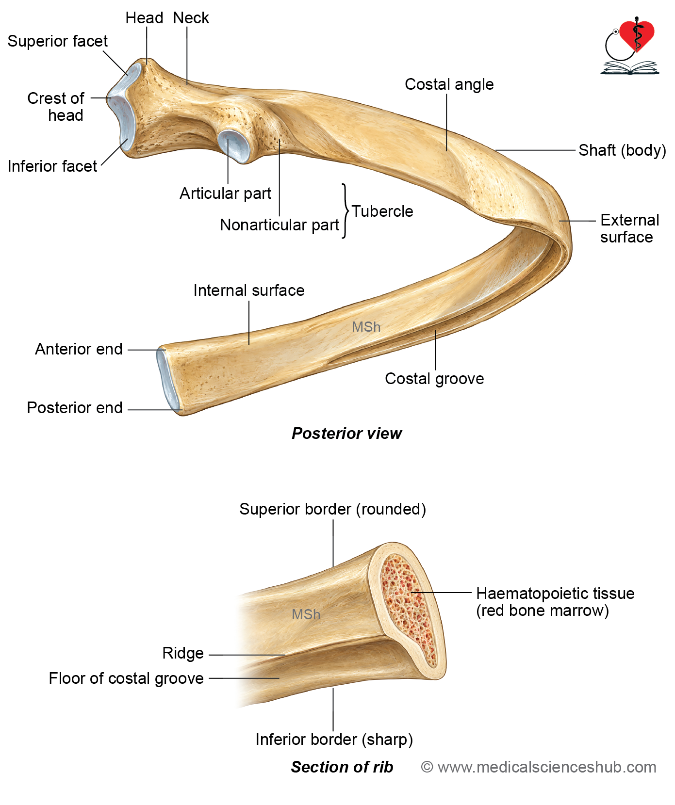
- Hold the rib so that the anterior end, which bears a shallow concave depression, is directed forward.
- Keep the posterior end, identified by its rounded head, directed backward.
- Position the shaft so that its convex surface faces outward.
- Ensure that the costal groove and sharp inferior border are directed downward.
- The convexity of the shaft should face toward the side to which the rib belongs.
- Tilt the rib so that the posterior end lies at a higher level than the anterior end.
Parts
Each typical rib consists of three main parts:
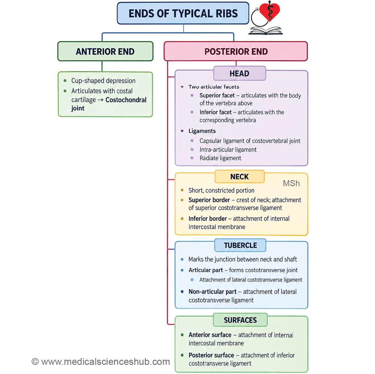
- Anterior end: The anterior end presents a small, cup-shaped depression. This depression receives the corresponding costal cartilage.
- Posterior end: The posterior end consists of the head, neck, and tubercle.
- Shaft: The shaft forms the middle and major part of the rib. It is flat, elongated, and curved.
Features
Anterior end
- The anterior end presents a small, cup-shaped depression or socket.
- This depression articulates with the corresponding costal cartilage to form the costochondral joint.
- The costochondral joint is a primary cartilaginous joint (synchondrosis).
Posterior end
- The posterior end consists of the head, neck, and tubercle.
Head
- The head of a typical rib bears two articular facets.
- The upper facet is smaller and articulates with the body of the vertebra immediately above.
- The lower facet is larger and articulates with the body of the numerically corresponding vertebra.
- The two articular facets are separated by a ridge known as the crest of the head.
Attachments to the Head
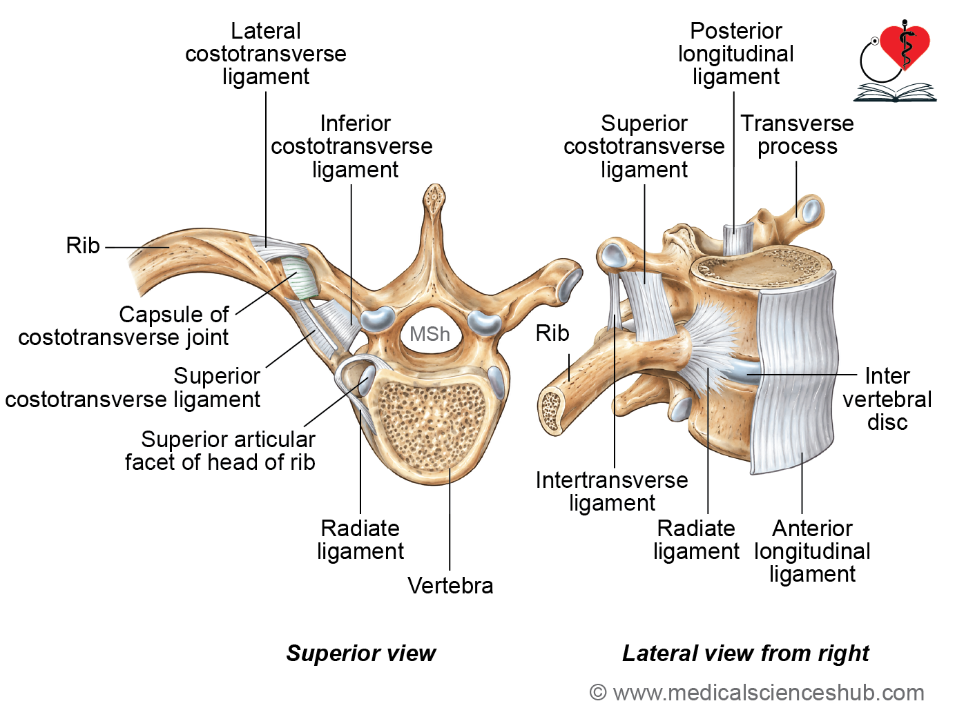
- The margins of the articular facets provide attachment to the capsular ligament of the costovertebral joint.
- The intra-articular ligament extends from the crest of the head of the rib to the corresponding intervertebral disc.
- The radiate ligament connects the anterior aspect of the head of the rib with the bodies of the corresponding vertebra and the vertebra above, as well as the intervening intervertebral disc.
Neck
- The neck is a narrow and flattened part of the rib situated between the head and tubercle. It lies anterior to the transverse process of the numerically corresponding thoracic vertebra. The neck has two borders and two surfaces.
Borders
- The superior border is sharp and forms the crest of the neck.
- The inferior border is smooth and rounded.
Surfaces
- The anterior surface is smooth.
- The posterior surface is rough and contains numerous vascular foramina.
Attachments
- The inferior costotransverse ligament connects the posterior surface of the neck with the transverse process of the corresponding vertebra.
- The superior costotransverse ligament consists of two laminae. It extends from the crest of the neck to the transverse process of the vertebra above.
- The internal intercostal membrane is attached to the anterior surface and inferior border of the neck of the rib forming the upper boundary of the intercostal space.
- The posterior intercostal membrane is also related to the posterior part of the intercostal space and should not be considered synonymous with the internal intercostal membrane.
Tubercle
- The tubercle is located on the posterolateral aspect of the neck of the rib.
- It marks the junction between the neck and shaft.
- The tubercle is divided into medial articular and lateral non-articular parts.
- Medial Articular Part
- The medial part bears a small articular facet.
- This facet articulates with the transverse process of the numerically corresponding thoracic vertebra.
- The articulation forms the costotransverse joint, which is a synovial joint.
- Lateral Non-articular Part
- The lateral part is rough and non-articular.
- It provides attachment to the lateral costotransverse ligament.
Attachment: The lateral costotransverse ligament extends from the rough, non-articular part of the tubercle to the tip of the transverse process of the corresponding vertebra.
Shaft/body
- The shaft, or body, is the flat, elongated, and curved part of a typical rib.
- It has two borders, superior and inferior.
- It also has two surfaces, outer and inner, and two angles, posterior and anterior.
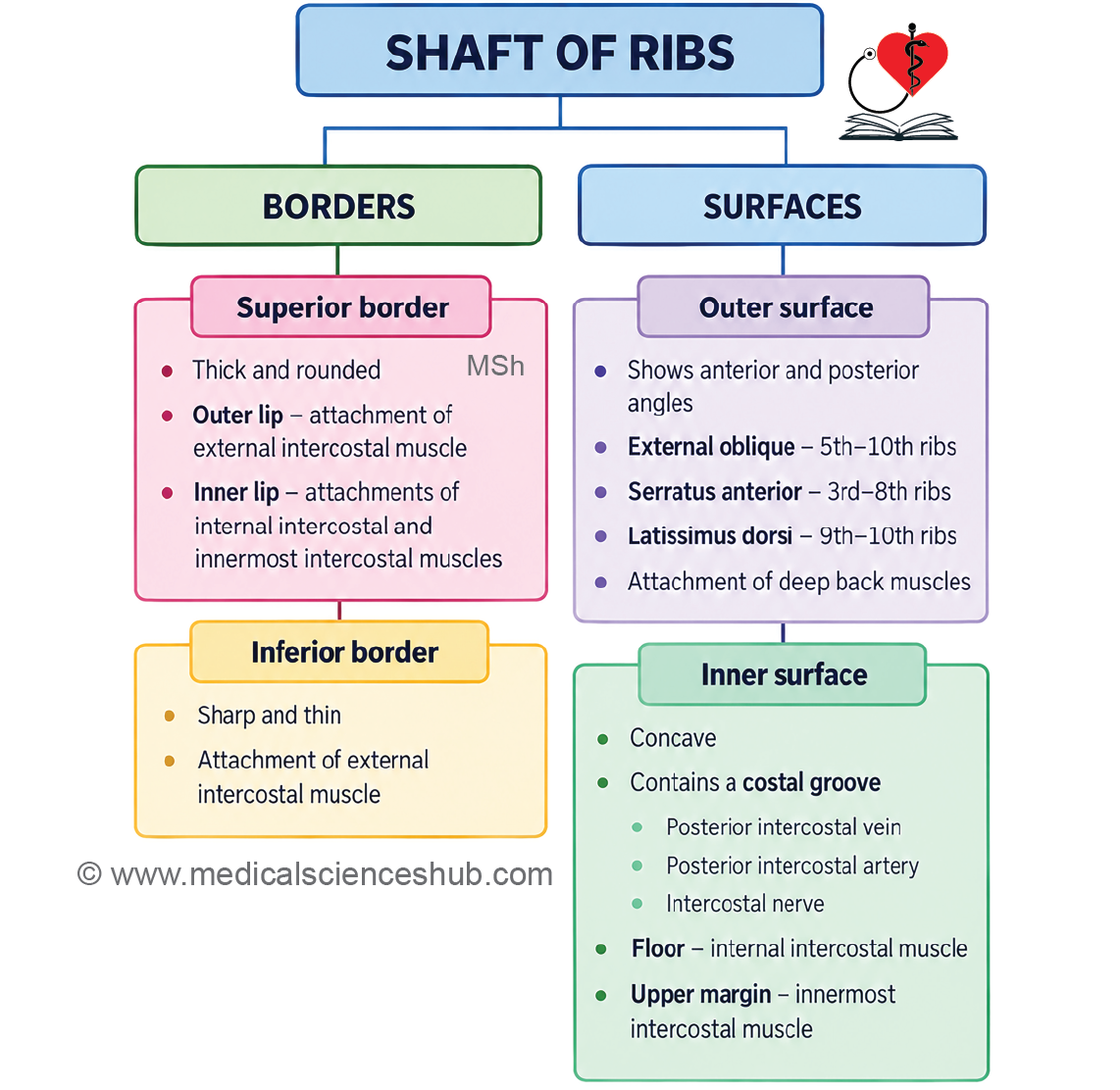
Superior border
- The superior border is thick and rounded.
- It is divided into an outer lip and an inner lip.
Attachments
- The external intercostal muscle is inserted into the outer lip of the superior border.
- The internal intercostal and intercostalis intimus muscles are inserted into the inner lip.
Inferior border
- The inferior border is thin and sharp.
- It forms the lower boundary of the costal groove.
Attachment
- The external intercostal muscle takes origin from the inferior border of the rib.
Outer surface
- The outer surface is smooth and convex.
Posterior Angle
- The posterior angle is indicated by an oblique ridge on the outer surface.
- It is located approximately 5 cm lateral to the tubercle.
Anterior Angle
- The anterior angle is represented by a faint oblique line.
- It is situated approximately 5 cm from the anterior end of the rib.
Attachments
- The thoracolumbar fascia is attached near the posterior angle.
- The levator costae and sacrospinalis (erector spinae) are inserted into the outer surface between the tubercle and posterior angle.
- The external oblique muscle takes origin from the fifth to tenth ribs, anterior to their anterior angles.
- The serratus anterior takes origin from the lateral surfaces of the upper eight ribs. On the third to eighth ribs, its attachment extends up to the region of the anterior angle.
- The latissimus dorsi takes origin from the ninth and tenth ribs lateral to their anterior angles.
Inner surface
- The inner surface of the shaft is smooth and concave.
- Near its inferior border, it presents the costal groove.
Costal Groove
- The costal groove has an upper margin, floor, and lower margin.
- The upper margin is marked by a ridge.
- The lower margin corresponds to the inferior border of the rib.
Contents of the Costal Groove
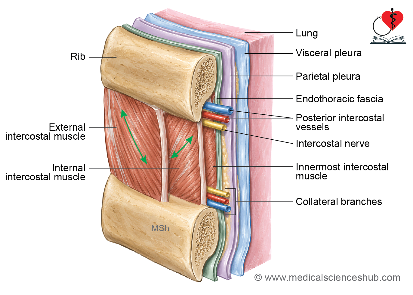
- The costal groove lodges the main intercostal neurovascular bundle between the internal intercostal and intercostalis intimus muscles.
- From above downward, the structures are arranged as:
- Posterior intercostal vein
- Posterior intercostal artery
- Intercostal nerve
- Their arrangement can be remembered by the mnemonic VAN: Vein, Artery, Nerve.
Muscle Attachments
- The external intercostal muscle arises from the lower margin of the costal groove.
- The internal intercostal muscle arises from the floor of the costal groove.
- The intercostalis intimus muscle arises from the upper margin of the costal groove.
Ossification of Ribs
- The third to tenth ribs usually ossify from one primary ossification center for the shaft.
- The primary center appears around the eighth week of intrauterine life.
- Secondary Ossification Centers: In a typical rib, three secondary ossification centers appear around puberty:
- One center for the head.
- One center for the articular part of the tubercle.
- One center for the non-articular part of the tubercle.
- These secondary centers usually fuse with the rest of the rib between 20 and 25 years of age.
Ossification of Atypical Ribs
First Rib
- The first rib has one secondary center for the tubercle, unlike a typical rib, which has two secondary centers for the tubercle.
Eleventh and Twelfth Ribs
- The eleventh and twelfth ribs each have one primary center for the shaft. Each also has one secondary center for the head. They do not have secondary ossification centers for a tubercle because the tubercle is absent in these ribs.
First Rib
Identification Features
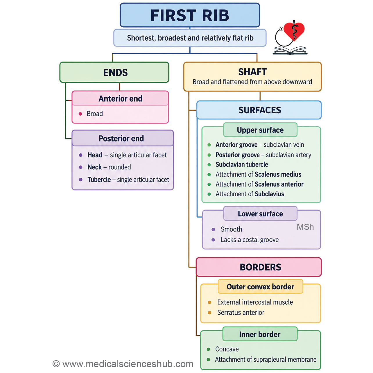
- The first rib is the shortest, broadest, and most strongly curved of all the ribs. Its shaft is flattened from above downward and therefore has superior and inferior surfaces and inner and outer borders.
- The head bears a single articular facet for articulation with the body of the first thoracic vertebra. The tubercle has an articular facet, but a distinct non-articular part is absent.
- A costal groove is absent.
- The anterior end is broad and thick and joins the first costal cartilage. The superior surface presents two vascular grooves separated by a prominent scalene tubercle.
Side Determination and Anatomical Position
To determine the side of the first rib and place it in its correct anatomical position:
- Hold the rib so that the superior surface, identified by its two grooves, faces upward and forward at approximately 45° to the horizontal plane.
- Keep the rounded head posteriorly and the broad anterior end anteriorly.
- The anterior end should lie at a lower level than the posterior end.
- The convex outer border should face toward the side to which the rib belongs.
Features of First Rib
Anterior end
- The anterior end is broad and thick.
- It articulates with the first costal cartilage to form the first costochondral joint, which is a primary cartilaginous joint.
Posterior end
- The posterior end consists of the head, neck, and tubercle.
Head
- The head is small and rounded.
- It bears a single articular facet.
- This facet articulates with the body of the first thoracic vertebra.
Neck
- The neck is rounded and elongated.
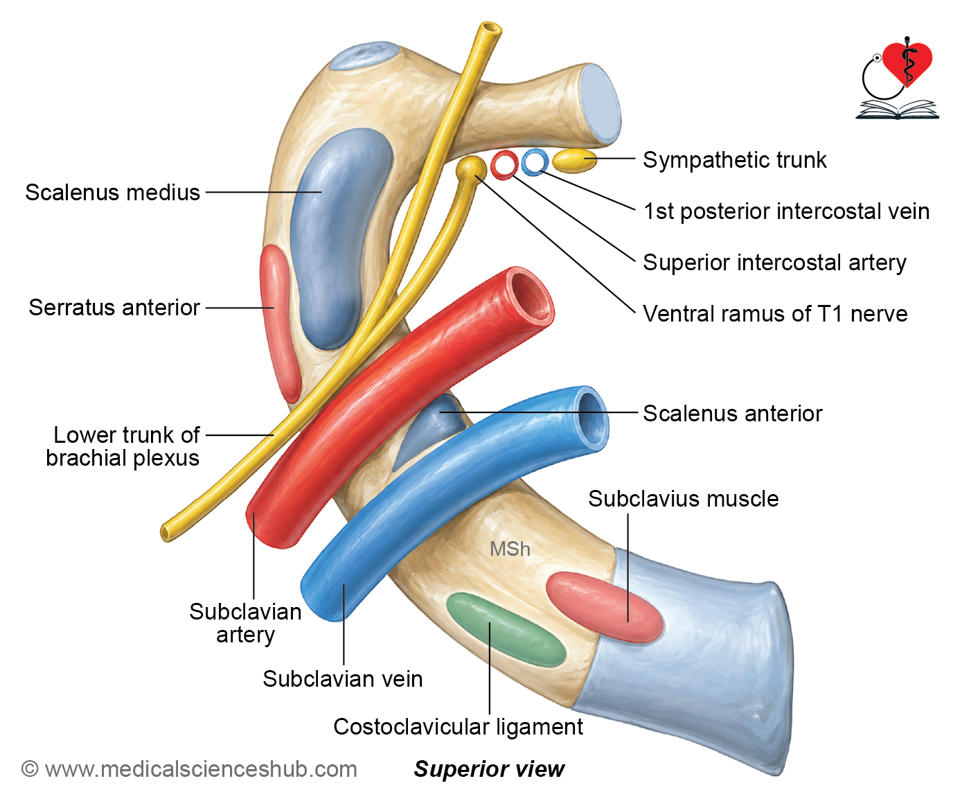
- Anteriorly, the neck is related to the following structures from medial to lateral:
- Sympathetic chain
- First posterior intercostal vein
- Superior intercostal artery
- Ventral ramus of the first thoracic nerve
Tubercle
- The tubercle is large and prominent and coincides with the posterior angle of the rib.
- It has only an articular part, which articulates with the transverse process of the first thoracic vertebra.
- The lateral costotransverse ligament is attached to the tubercle.
Shaft
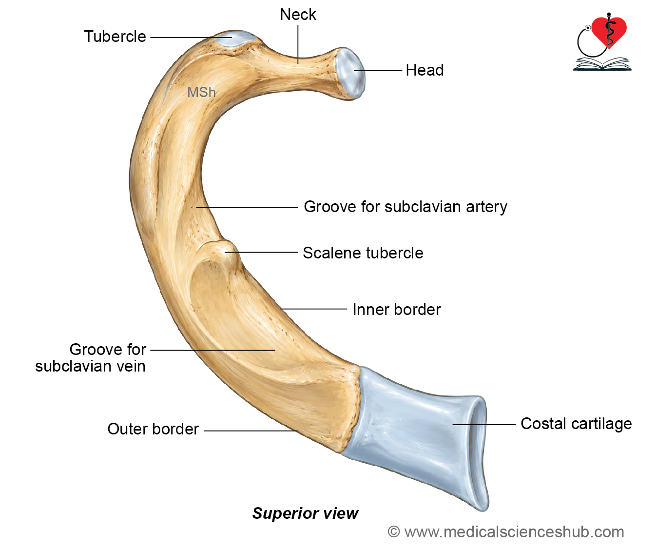
- The shaft is flattened from above downward. It has superior and inferior surfaces and outer and inner borders.
Superior Surface
- The superior surface presents two shallow grooves separated by the scalene tubercle, which lies close to the inner border.
Relations
- The anterior groove lodges the subclavian vein. The posterior groove lodges the subclavian artery and is related to the lower trunk of the brachial plexus.
Attachments
- The scalenus anterior is inserted into the scalene tubercle. The scalenus medius is inserted into an elongated rough area behind the groove for the subclavian artery.
- The subclavius takes origin from the lateral part of the anterior end near its junction with the shaft. The costoclavicular ligament extends from the medial part of the anterior end to the medial end of the clavicle.
Inferior Surface
- The inferior surface is smooth. Unlike a typical rib, the first rib has no costal groove.
- The inferior surface is related to the costal pleura. It provides attachment to the internal intercostal and intercostalis intimus muscles.
Outer Border
- The outer border is convex. The external intercostal muscle is attached to the outer border.
- The first digitation of the serratus anterior takes origin from the outer border, just behind the groove for the subclavian artery.
Inner Border
The inner border is concave. The scalene tubercle lies close to this border on the superior surface. The inner border provides attachment to the suprapleural membrane.
Second Rib
Features
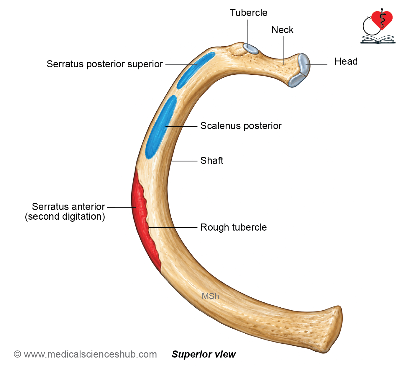
- The second rib is approximately twice the length of the first rib. Its shaft is sharply curved but not twisted.
- The head is small and bears two articular facets for articulation with the bodies of the first and second thoracic vertebrae. The tubercle has a relatively small non-articular part.
Shaft
- The outer surface of the shaft faces upward and outward. The inner surface faces downward and inward.
- A prominent rough tuberosity is present near the middle of the outer surface and provides attachment to the serratus anterior. A short costal groove is present on the posterior part of the inner surface.
Muscle Attachments
- The serratus anterior takes origin from the rough tuberosity on the outer surface. The scalenus posterior is inserted into the upper border of the second rib. The serratus posterior superior is also inserted into the upper border in its posterior part.
Last Three Ribs
Tenth Rib
Identification features
- The head bears a single articular facet, which articulates with the body of the tenth thoracic vertebra.
- The tenth rib is shorter than the typical ribs.
Eleventh Rib
Identification features
- The head bears a single large articular facet for articulation with the eleventh thoracic vertebra.
- The neck is absent. The tubercle is absent.
- The anterior end is pointed and free. A faint angle can be identified on the outer surface.
Twelfth Rib
Identification features
- The head bears a single articular facet for articulation with the twelfth thoracic vertebra.
- The anterior end is pointed and free.
- The neck, tubercle, angle, and costal groove are absent.
Side Determination and Anatomical Position
To determine the side of the twelfth rib:
- Keep the pointed anterior end directed forward and laterally.
- The sharp border should face downward.
- The concave inner surface should face inward and slightly upward.
- The convex outer surface faces toward the side to which the rib belongs and therefore helps in determining its side.
Attachments and Relations
Outer Surface
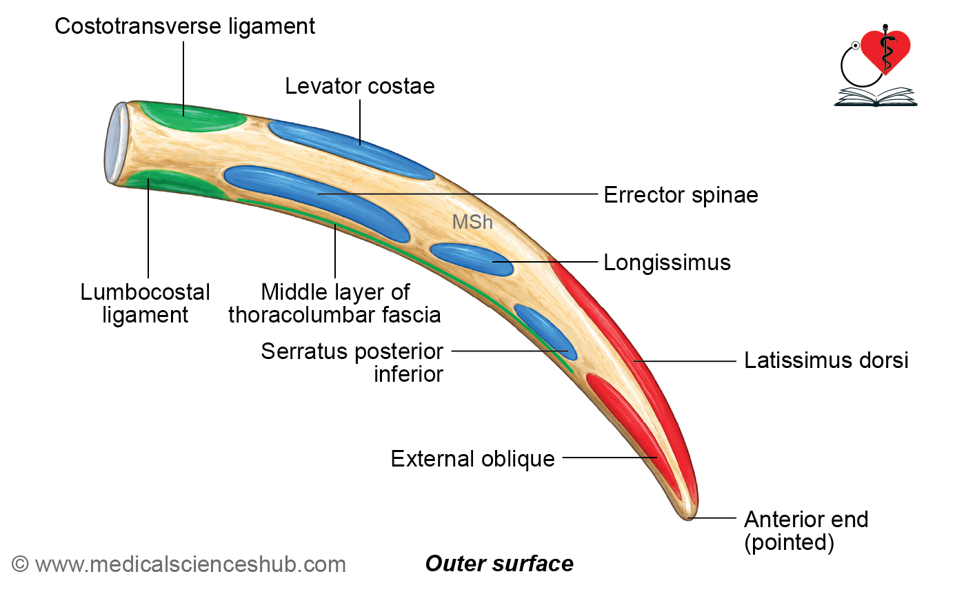
- The latissimus dorsi takes origin from the upper part of the area near the anterior end of the rib. The external oblique takes origin from the lower part of the area near the anterior end.
- The erector spinae and levator costae are inserted into the medial half of the outer surface. The serratus posterior inferior is inserted into the lateral half of the outer surface.
Inner Surface
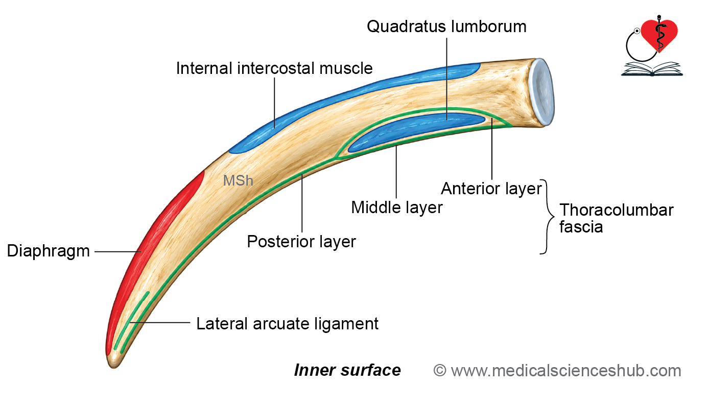
- The quadratus lumborum is inserted into the lower part of the medial half of the inner surface. The internal intercostal muscle is attached near the upper border. The diaphragm takes origin from the anterior part of the inner surface.
Upper Border
- The upper border provides attachment to the external and internal intercostal muscles.
Lower Border
- The lumbocostal ligament extends from the twelfth rib to the transverse process of the first lumbar vertebra. The lateral arcuate ligament extends from the twelfth rib to the transverse process of the first lumbar vertebra. The middle layer of the thoracolumbar fascia is also attached to the lower border.
Important Relations
The inner surface of the twelfth rib is related to the costodiaphragmatic recess of the pleura. An oblique line on the middle part of the inner surface indicates the line of pleural reflection.
Costal Cartilages
- Costal cartilages form the unossified anterior extensions of the ribs. There are 12 pairs of costal cartilages, one corresponding to each pair of ribs. They are composed of hyaline cartilage and contribute to the flexibility and elasticity of the thoracic wall.
Articulations
- The lateral end of each costal cartilage articulates with the anterior end of its corresponding rib. The first to seventh costal cartilages articulate with the sternum and form the sternocostal (chondrosternal) joints.
- The eighth to tenth costal cartilages join the costal cartilage immediately above. The eleventh and twelfth costal cartilages are short and have free anterior ends.
General Features
- Each costal cartilage has two ends, medial and lateral. It also has two borders, upper and lower, and two surfaces, anterior and posterior.
Anterior Surface
- The anterior surface of the first costal cartilage provides attachment to the articular disc of the sternoclavicular joint and the costoclavicular ligament. It also gives origin to the subclavius muscle.
- The anterior surfaces of the second to sixth costal cartilages provide origin to the pectoralis major muscle.
Posterior Surface
- The posterior surface of the first costal cartilage provides origin to the sternothyroid muscle.
The posterior surfaces of the second to sixth costal cartilages provide insertion to the sternocostalis (transversus thoracis) muscle. The lower costal cartilages provide attachments to the diaphragm and transversus abdominis, contributing to the muscular attachments along the lower thoracic margin.
📝 Test Your Knowledge – Practice MCQs
Attempt the chapter MCQ quiz and assess your understanding of key concepts.
Click to see: Medical Disclaimer and Copyright Notice