
Competencies
- AN10.1: Identify and describe boundaries and contents of axilla.
- AN10.2: Identify, describe, and demonstrate the origin, extent, course, parts, relations and branches of axillary artery and tributaries of axillary vein.
- AN10.4: Describe the anatomical groups of axillary lymph nodes and specify their areas of drainage.
- AN10.7: Explain anatomical basis of enlarged axillary lymph nodes.
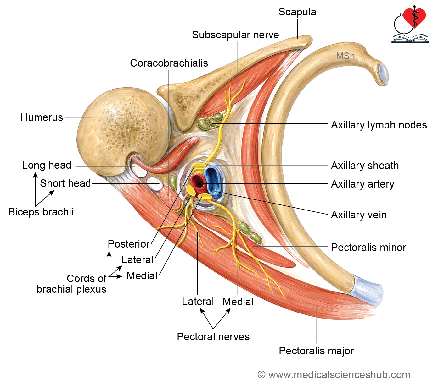
- The axilla (armpit) is a pyramidal-shaped space located between the upper arm and the lateral wall of the thorax.
- It serves as an important passage for blood vessels, nerves, and lymphatics traveling between the neck, upper limb, and thoracic region.
Axillary folds
- The axillary folds are skin folds formed by the margins of underlying muscles.
- The anterior axillary fold is formed by the lateral border of the pectoralis major muscle.
- The posterior axillary fold is formed by the latissimus dorsi and teres major muscles.
- The axillary pit is the depression located between the anterior and posterior axillary folds.
Boundaries of Axilla
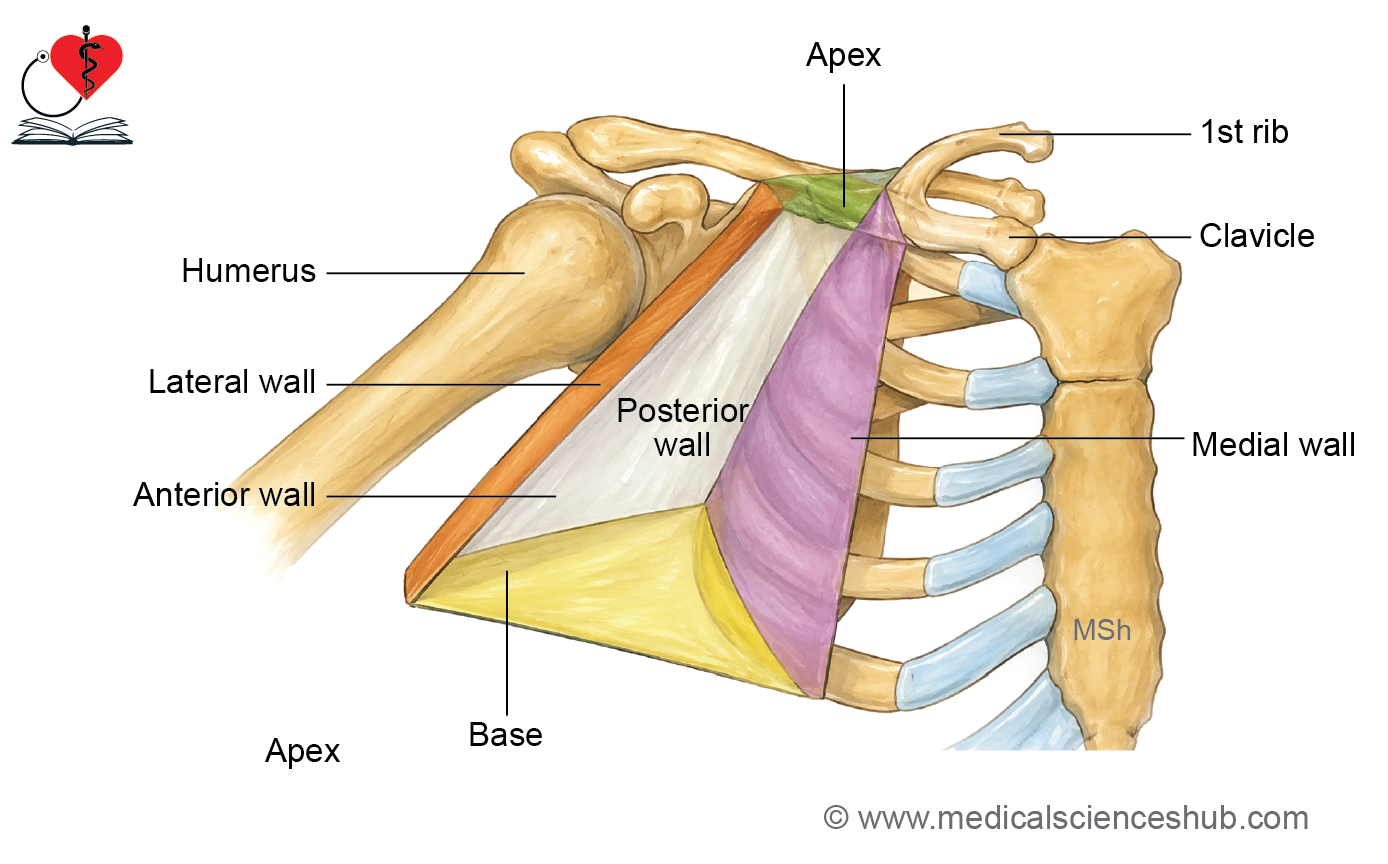
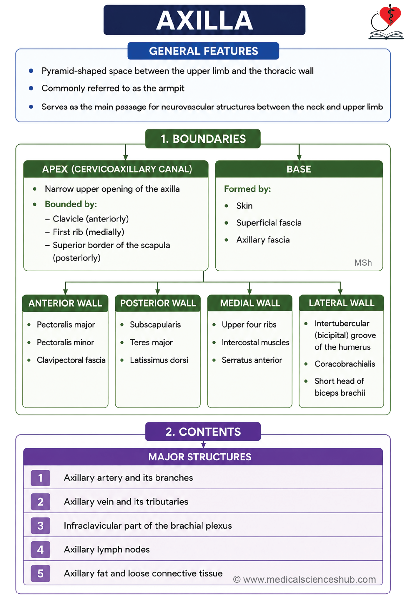
- The axilla is a pyramidal-shaped space with an apex, a base, and four walls.
- The apex is directed upward and medially toward the root of the neck.
- The base is directed downward and is formed by the skin and superficial fascia of the armpit.
- The four walls of the axilla are the anterior wall, posterior wall, medial wall, and lateral wall.
Apex (cervicoaxillary canal)
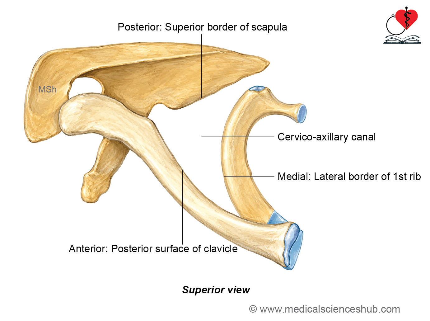
- The apex of the axilla is directed upward and medially toward the root of the neck.
- It serves as a passage for structures traveling between the neck and the axilla and is therefore called the cervicoaxillary canal. The apex is blunt (truncated) in shape.
Boundaries of apex
- It is a triangular opening bounded by:
- Anteriorly: Posterior surface of the middle third of the clavicle.
- Posteriorly: Superior border of the scapula.
- Medially: Outer border of the first rib.
Base (floor)
- The base (floor) of the axilla is directed downward. It is formed by the skin and the underlying axillary fascia. The base is dome-shaped, with its convexity directed upward. It extends between the anterior and posterior axillary folds.
- Medially, it is continuous with the chest wall, and laterally, it continues with the medial aspect of the arm.
Anterior wall
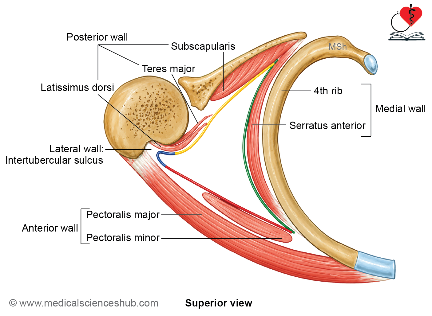
The anterior wall of the axilla is formed by the following structures arranged from superficial to deep:
- Pectoralis major muscle.
- Clavipectoral fascia.
- Pectoralis minor muscle.
- Subclavius muscle.
Posterior wall
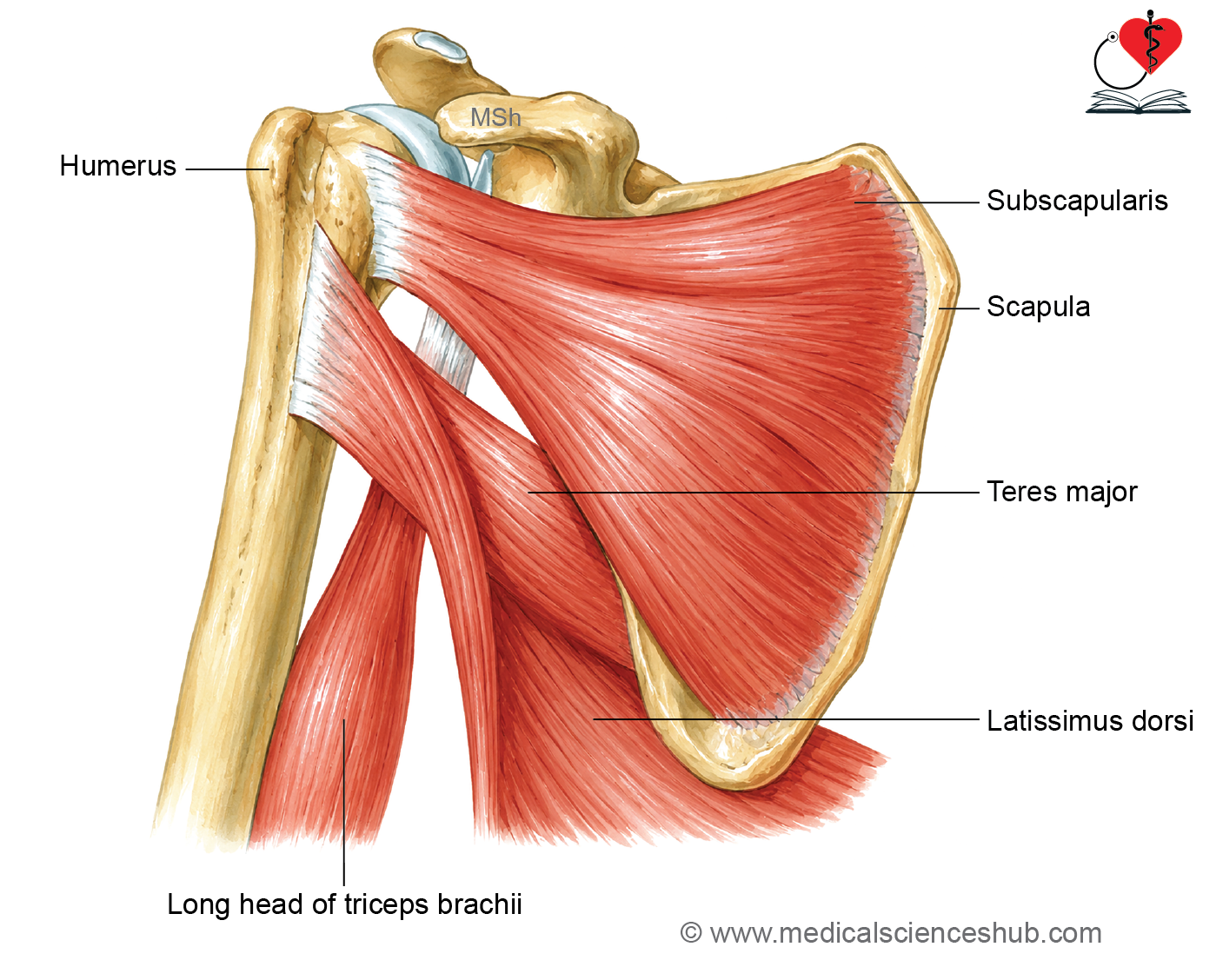
The posterior wall of the axilla is formed by the following structures from above downward:
- Subscapularis muscle.
- Teres minor and teres major muscles.
- Latissimus dorsi muscle.
H5: Medial wall
- The medial wall of the axilla is convex laterally.
- It is formed by the thoracic wall, including:
- The upper four ribs and their intercostal muscles.
- The upper four to five digitations of the serratus anterior muscle.
Lateral wall
- The lateral wall of the axilla is the narrowest of its four walls.
- It is formed by:
- The coracobrachialis muscle and the short head of the biceps brachii, which arise from the tip of the coracoid process.
- The intertubercular (bicipital) groove of the humerus containing the tendon of the long head of the biceps brachii.
Contents of Axilla
- The axilla contains the following important structures:
- Axillary artery and its branches.
- Axillary vein and its tributaries.
- Infraclavicular part of the brachial plexus.
- Axillary lymph nodes.
- Axillary fat and connective tissue.
- Axillary tail of the breast (axillary process of the mammary gland).
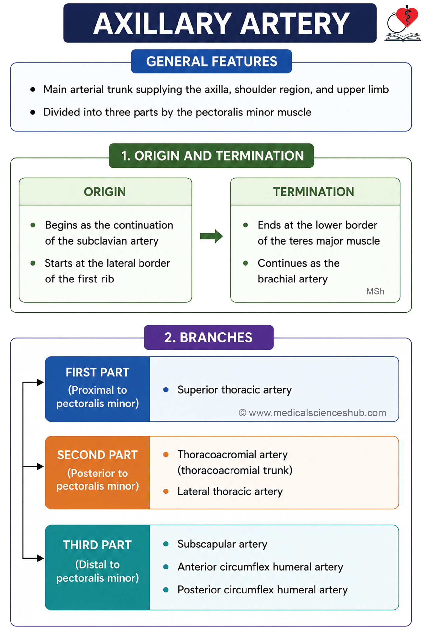
Axillary Artery
- The axillary artery is the main artery supplying the upper limb.
Beginning
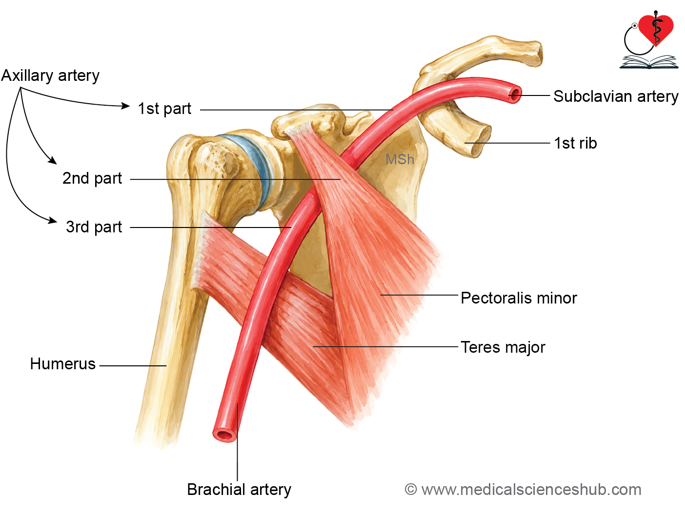
- It is the continuation of the subclavian artery.
- It begins at the outer border of the first rib.
Termination
- It continues as the brachial artery at the lower border of the teres major muscle.
Course
- The artery runs downward, forward, and laterally with a lateral convexity. In an outstretched arm, it follows a nearly straight horizontal course.
Parts of axillary artery
- The pectoralis minor muscle crosses the artery anteriorly and divides it into three parts:
- First part: Proximal to the muscle.
- Second part: Posterior (deep) to the muscle.
- Third part: Distal to the muscle.
- The third part is the largest content of the lower axilla and lies inferomedial to the coracobrachialis muscle, short head of the biceps brachii, and coracoid process.
Relation of Axillary Artery
- The axillary artery is related to the walls of the axilla throughout its course.
- In addition, its anatomical relations are described separately for the first, second, and third parts because the surrounding structures differ in each segment.
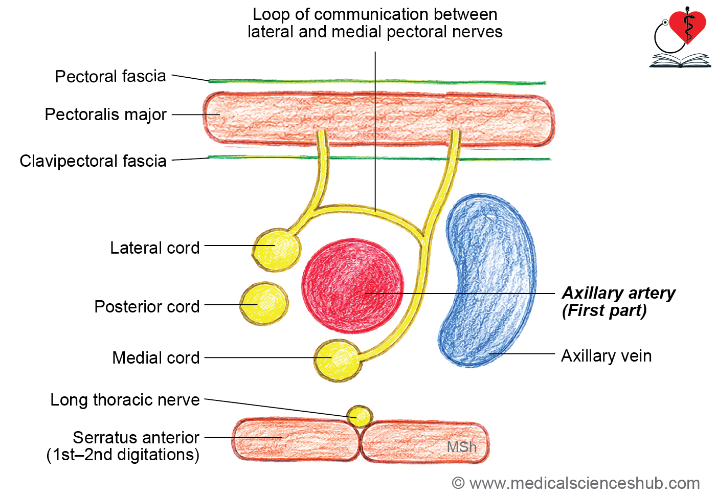
Relations of First part
The first part of the axillary artery has the following relations:
- Anteriorly:
- Pectoralis major (clavicular part).
- Clavipectoral fascia.
- The anterior wall of the axilla, except the pectoralis minor.
- The communication loop between the lateral and medial pectoral nerves.
- Posteriorly:
- Medial cord of the brachial plexus.
- Long thoracic nerve.
- First and second digitations of the serratus anterior muscle.
- First intercostal space.
- Medially:
- Axillary vein.
- Laterally:
- Lateral and posterior cords of the brachial plexus.
Relations of Second part
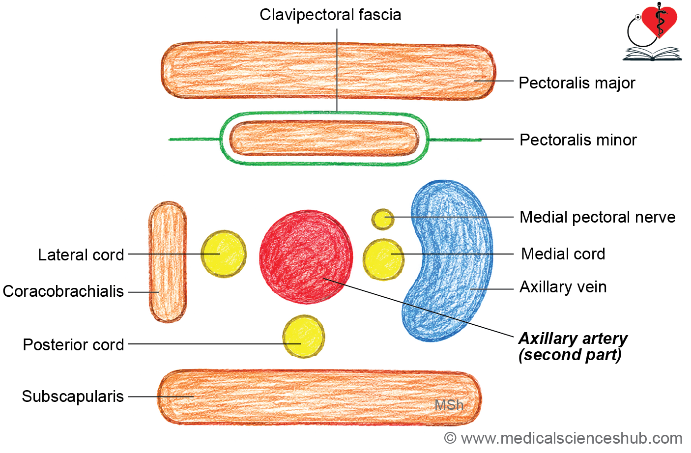
he second part of the axillary artery has the following relations:
- Anteriorly:
- Anterior wall of the axilla, including the pectoralis minor muscle.
- Posteriorly:
- Posterior cord of the brachial plexus.
- Subscapularis muscle.
- Medially:
- Medial cord of the brachial plexus.
- Axillary vein.
- Laterally:
- Lateral cord of the brachial plexus.
- Coracobrachialis muscle.
Relations of Third part
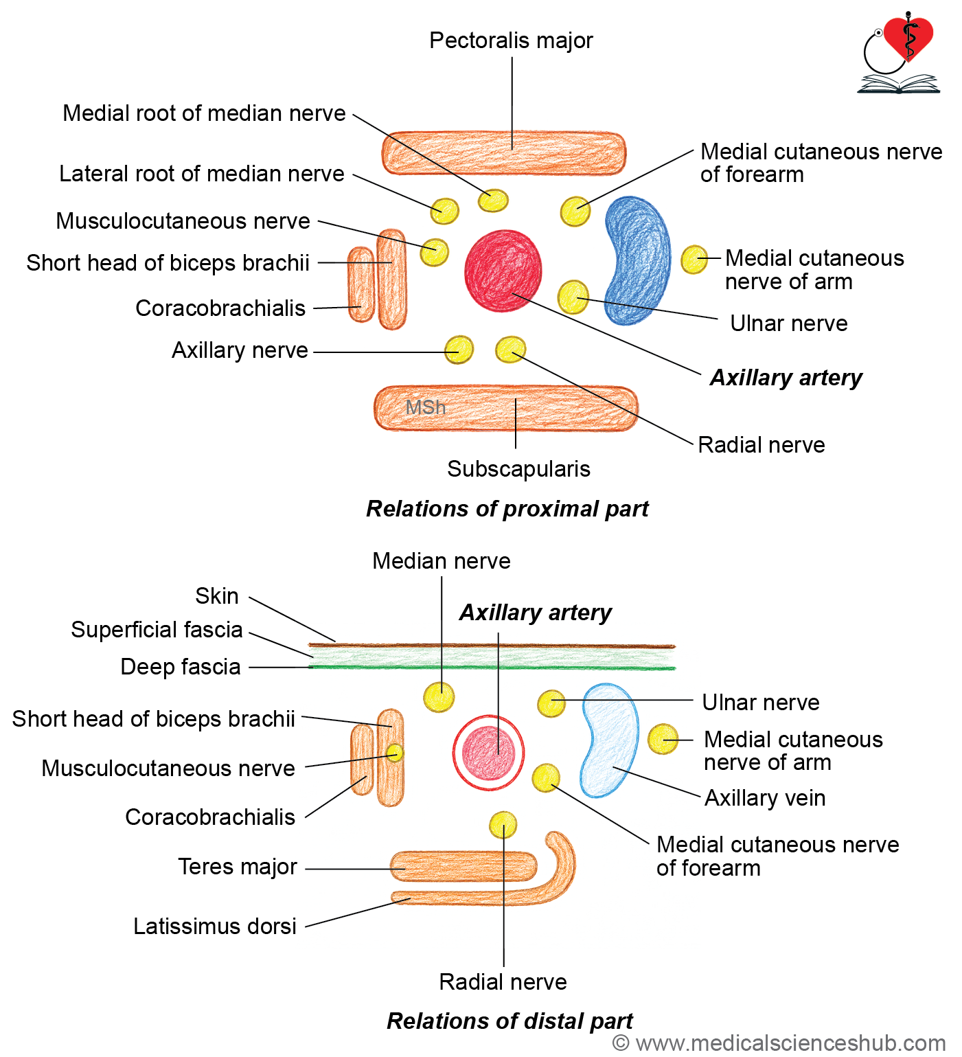
- The third part of the axillary artery has the following relations:
- Anteriorly:
- Pectoralis major muscle.
- Medial root of the median nerve.
- Posteriorly:
- Axillary nerve (upper part).
- Radial nerve.
- Subscapularis muscle (upper part).
- Latissimus dorsi and teres major muscles (lower part).
- Medially:
- Axillary vein.
- Medial cutaneous nerve of the arm.
- Laterally:
- Musculocutaneous nerve.
- Anteriorly:
Branches
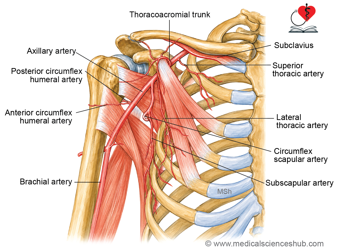
The axillary artery gives six branches, distributed according to its three parts:
- First part:
- Superior thoracic artery.
- Second part:
- Thoracoacromial trunk, which divides into the pectoral, acromial, clavicular, and deltoid branches.
- Lateral thoracic artery.
- Third part:
- Subscapular artery.
- Anterior circumflex humeral artery.
- Posterior circumflex humeral artery.
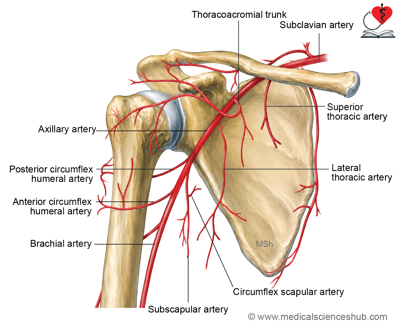
- Superior thoracic artery is a small branch of the first part of the axillary artery. It runs along the superior border of the pectoralis minor muscle.
- Thoracoacromial artery arises from the second part of the axillary artery. It emerges at the superior border of the pectoralis minor, pierces the clavipectoral fascia, and divides into four branches:
- Pectoral branch: Supplies the pectoral muscles.
- Acromial branch: Participates in the acromial anastomosis.Deltoid branch: Runs in the deltopectoral groove.
- Clavicular branch: Supplies the sternoclavicular joint and subclavius muscle.
- Lateral thoracic artery arises from the second part of the axillary artery and runs along the lateral border of the pectoralis minor. In females, it gives lateral mammary branches.
- Subscapular artery is the largest branch of the third part of the axillary artery. It runs along the lower border of the subscapularis to the inferior angle of the scapula and supplies the latissimus dorsi and serratus anterior muscles. It gives the circumflex scapular artery, which passes through the triangular space to reach the infraspinous fossa.
- Anterior circumflex humeral artery passes anterior to the surgical neck of the humerus, anastomoses with the posterior circumflex humeral artery, and gives an ascending branch in the intertubercular (bicipital) groove.
- Posterior circumflex humeral artery accompanies the axillary nerve through the quadrangular space and winds around the surgical neck of the humerus.
CLINICAL INTEGRATION
- The pulsation of the axillary artery can be felt in the lower part of the axilla by pressing it against the shaft of the humerus.
- During injuries, surgical procedures, or amputation, the axillary artery can be compressed against the humerus to temporarily control bleeding.
- An aneurysm of the axillary artery is an abnormal dilatation that most commonly affects its first part. An enlarged aneurysm may compress the trunks of the brachial plexus, producing neurological symptoms in the upper limb.
Axillary Vein
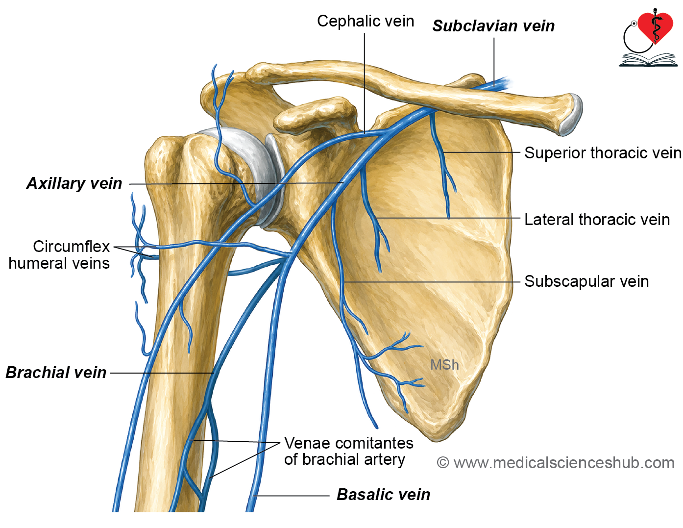
- The axillary vein is the main vein that drains deoxygenated blood from the upper limb. It is enclosed by a thin axillary sheath, allowing it to expand during increased venous return. In contrast, the sheath around the axillary artery is thicker.
Formation
- The axillary vein is formed by the union of the basilic vein and brachial veins at the lower border of the teres major muscle. The brachial veins are formed by the union of the venae comitantes of the brachial artery.
- It passes through the axilla medial to the axillary artery.
Termination
It continues as the subclavian vein at the lateral border of the first rib.
Tributaries
- Its tributaries include:
- Cephalic vein.
- Veins accompanying the branches of the axillary artery (except the thoracoacromial trunk).
- Thoracoepigastric vein.
CLINICAL INTEGRATION
- The axillary vein is prone to injury because of its large size and thin wall, which may lead to air embolism.
Axillary Lymph Nodes
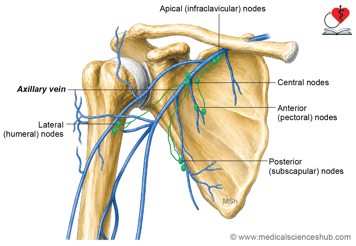
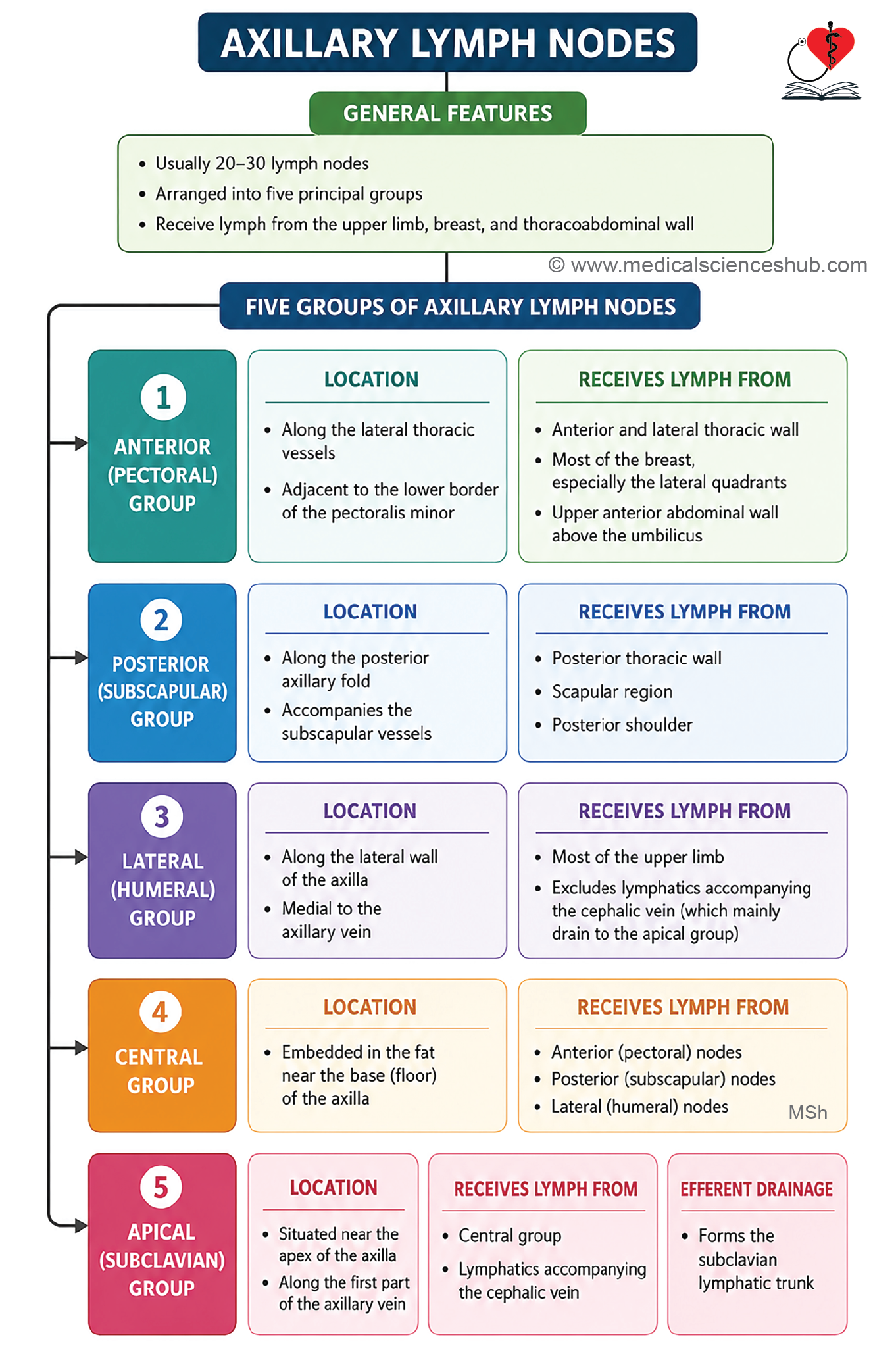
- The axillary lymph nodes consist of 20 to 30 lymph nodes located within the axilla.
- Based on their anatomical location, they are divided into five groups:
- Anterior (pectoral) group.
- Posterior (subscapular) group.
- ateral (humeral) group.
- Central group.
- Apical group.
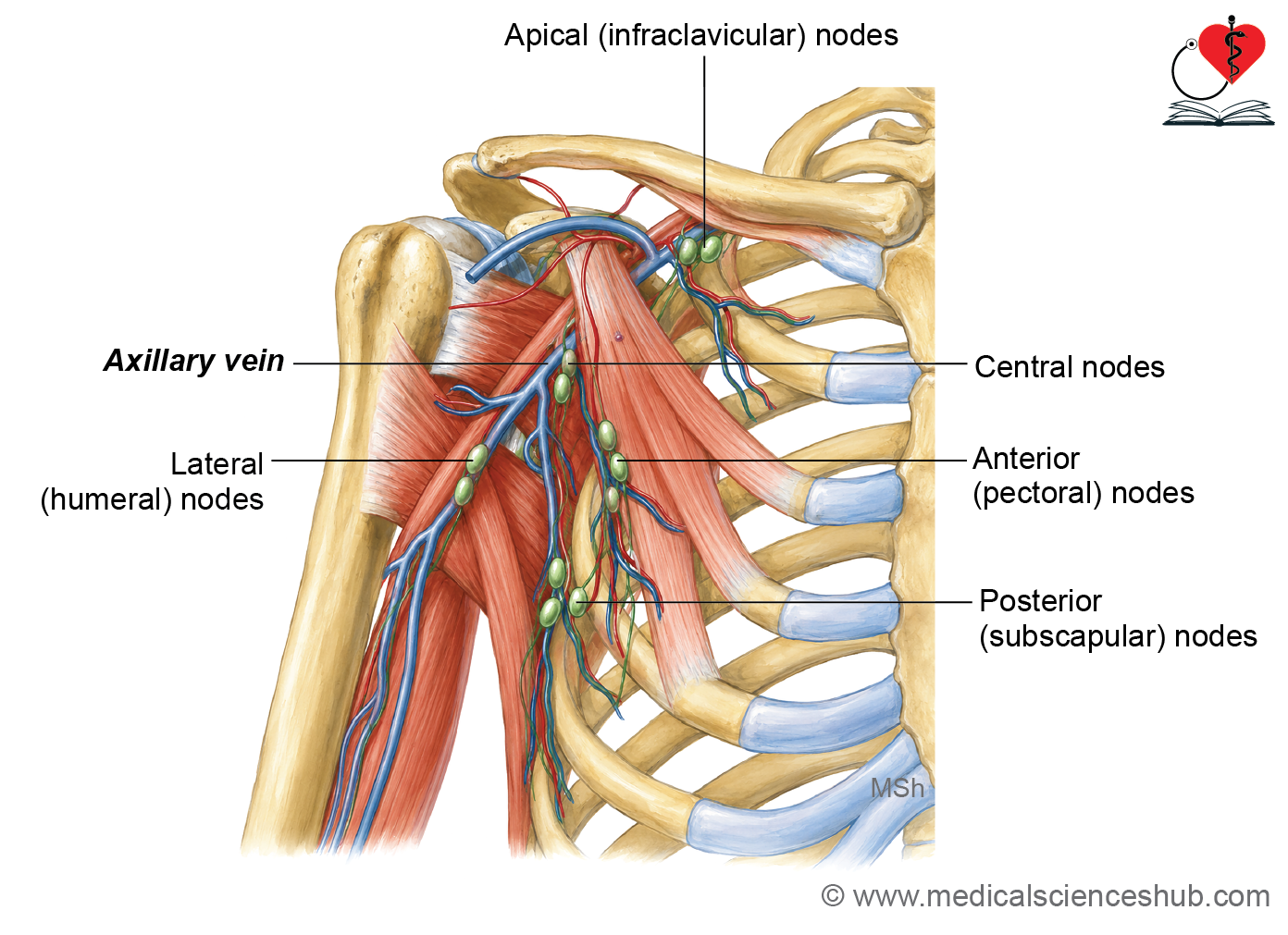
- Anterior (pectoral) nodes: These nodes lie along the lateral thoracic vein near the inferior border of the pectoralis minor. They receive lymph from the upper half of the trunk, most of the breast, and the subareolar plexus. The axillary tail (tail of Spence) of the breast is closely related to these nodes.
- Posterior (subscapular) nodes: These nodes are located along the posterior axillary fold with the subscapular vessels. They drain the posterior thoracic wall and scapular region.
- Lateral (humeral) nodes: These nodes lie medial to the axillary vein along the lateral wall of the axilla. They receive lymph from the upper limb, except lymphatics accompanying the cephalic vein.
- Central nodes: These nodes are embedded in the axillary fat near the base of the axilla, deep to the pectoralis minor. They receive lymph from the anterior, posterior, and lateral groups.
- Apical (infraclavicular) nodes: These nodes are situated near the apex of the axilla along the axillary vessels. They receive lymph from the central nodes, the upper part of the breast, and lymphatics accompanying the cephalic vein, including those from the thumb.
CLINICAL INTEGRATION
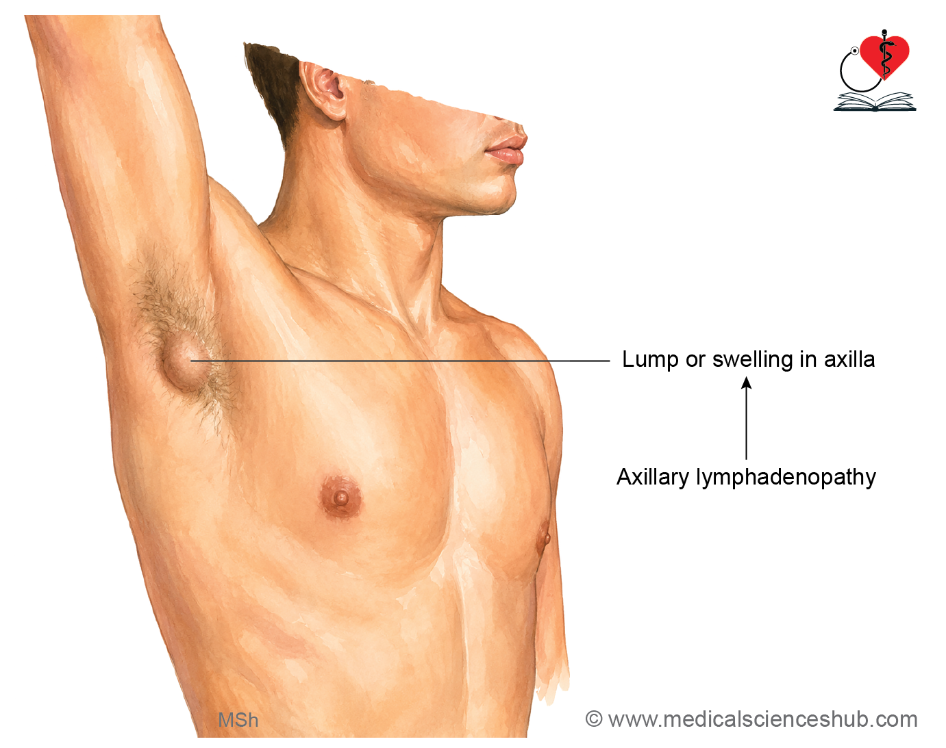
- Lymphadenopathy is the enlargement of lymph nodes due to infection, inflammation (lymphadenitis), or metastatic cancer.
- Axillary lymphadenopathy may result from infections of the upper limb, breast, or thoracic wall, or from the spread of breast cancer.
- Palpation of the axillary lymph nodes is an important part of the clinical examination for breast carcinoma, as enlarged nodes may be palpable.
- The central group of axillary lymph nodes lies close to the intercostobrachial nerve. Enlargement of these nodes may compress the nerve, causing pain along the medial side of the arm.
- An axillary abscess develops when infected axillary lymph nodes accumulate pus. The abscess is drained through an incision in the floor of the axilla, midway between the anterior and posterior axillary folds and close to the medial wall, to minimize injury to major vessels and nerves.
- Boils in the axilla are infections of the hair follicles of the axillary hairs.
Important Questions
- Describe the boundaries and content of axilla.
- List the contents of axilla.
- Describe the axillary artery under the following heads: beginning, termination, parts, relations, branches, and applied aspects.
- List the branches of axillary artery.
- Write a short note on axillary vein.
- Write a short note on axillary lymph nodes.
📝 Test Your Knowledge – Practice MCQs
Attempt the chapter MCQ quiz and assess your understanding of key concepts.
Click to see: Medical Disclaimer and Copyright Notice