
Competencies
- AN8.1: Features of individual bones (upper limb): Identify the given bone, its side, important features, and keep it in anatomical position.
- AN8.2: Features of individual bones (upper limb): Identify and describe joints formed by the given bone.
- AN8.3: Enumerate peculiarities of clavicle.
- AN8.4: Features of individual bones (upper limb): Demonstrate important muscle attachment on the given bone. This chapter includes description of scapula and clavicle.
- Posterior axio-appendicular muscles connect the upper limb to the axial skeleton.
- These muscles attach the pectoral girdle and humerus to the vertebral column.
- They stabilize the shoulder girdle and produce movements of the upper limb.
- A sound understanding of the anatomical structures on the back is essential for accurate clinical examination and diagnosis.
Surface Landmarks
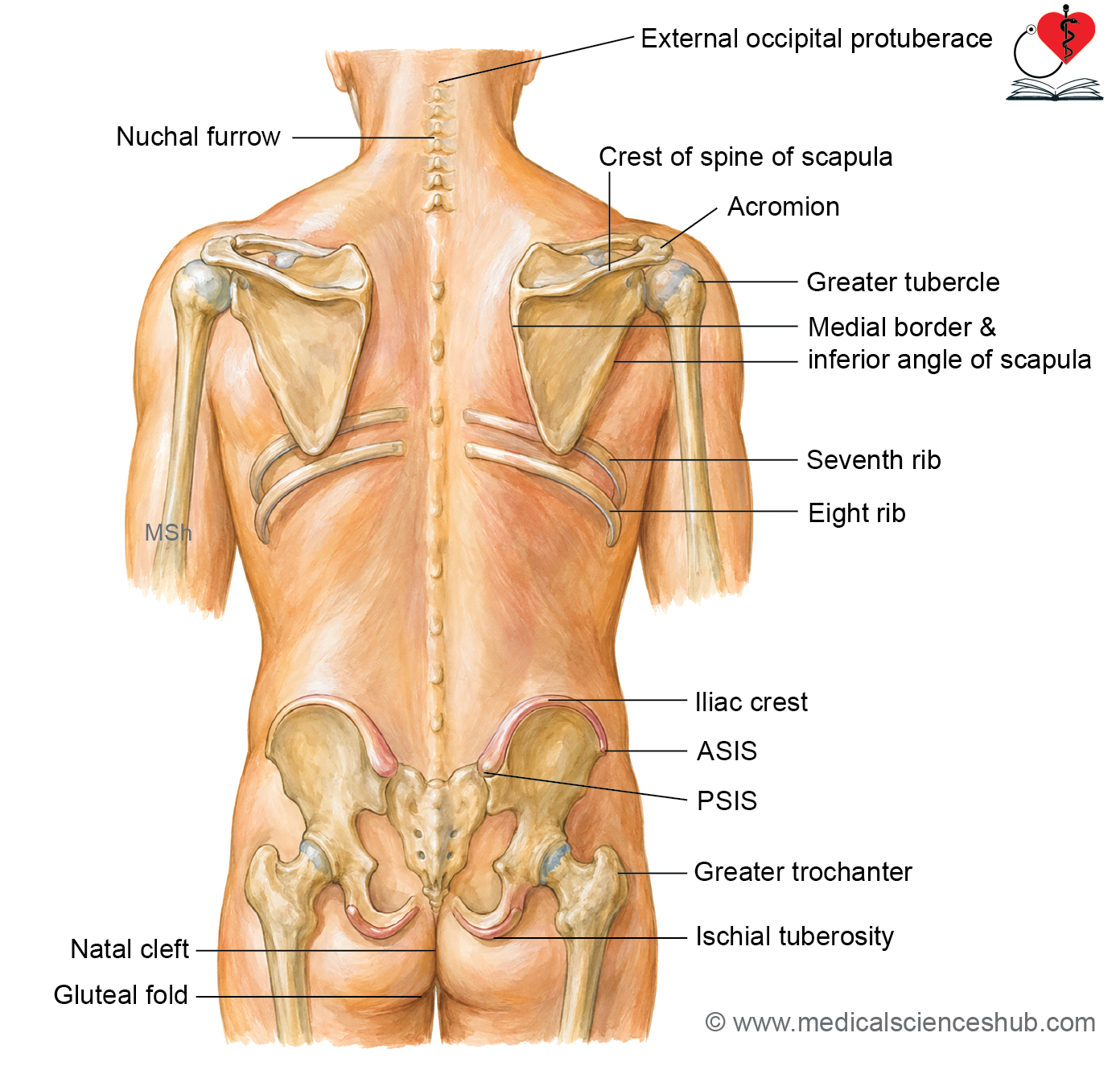
- Scapula lies on the posterolateral chest wall and extends from the 2nd to 7th ribs. The inferior angle of the scapula lies opposite the 7th rib and the T7 vertebral spine. The medial border extends from the T2 to T7 vertebral levels. The root of the scapular spine is opposite the T3 vertebral spine. The acromion forms the highest point of the shoulder and is easily palpable. The crest of the scapular spine extends downward and medially from the acromion to the root of the spine. The 8th rib lies immediately below the inferior angle of the scapula.
- The iliac crest is a curved bony ridge of the hip bone located below the waist.
- The anterior superior iliac spine (ASIS) forms the anterior end of the iliac crest.
- The posterior superior iliac spine (PSIS) forms the posterior end of the iliac crest and is palpable as a shallow dimple about 5 cm from the midline.
- The external occipital protuberance is a palpable midline bony prominence on the back of the skull.
- The nuchal groove extends from the external occipital protuberance to the C7 vertebral spine.
- The C7 spine is palpable at the root of the neck. The T2 spine corresponds to the superior angle of the scapula. The L4 spine lies at the level of the highest point of the iliac crest. The S2 spine lies at the level of the posterior superior iliac spine.
Cutaneous Innervation
- The skin of the back is supplied mainly by the posterior (dorsal) primary rami of the spinal nerves.
- The posterior primary rami of C1, C7, C8, L4, and L5 do not provide cutaneous innervation.
- From C2 to T6, the medial branches of the posterior primary rami supply the skin of the back.
- Below the T6 level, the lateral branches of the posterior primary rami provide cutaneous innervation.
- The posterior primary rami of L1, L2, L3, and S1 to S3 supply the skin of the gluteal region.
Muscles of the Back
- Posterior axio-appendicular muscles are the muscles of the back that connect the upper limb (appendicular skeleton) to the axial skeleton.
- These muscles primarily move and stabilize the scapula and contribute to movements of the upper limb.
- They are arranged into two anatomical layers:
- Superficial layer:
- Trapezius
- Latissimus dorsi
- Deep layer:
- Levator scapulae
- Rhomboid minor
- Rhomboid major.
- Superficial layer:
Superficial Posterior Axio-Appendicular Muscles
- The superficial posterior axio-appendicular muscles form the outermost layer of the back muscles.
- These muscles connect the upper limb to the axial skeleton and assist in movements of the scapula, shoulder, and upper limb.
- The superficial posterior axio-appendicular muscles are:
- Trapezius
- Latissimus dorsi
Trapezius Muscle
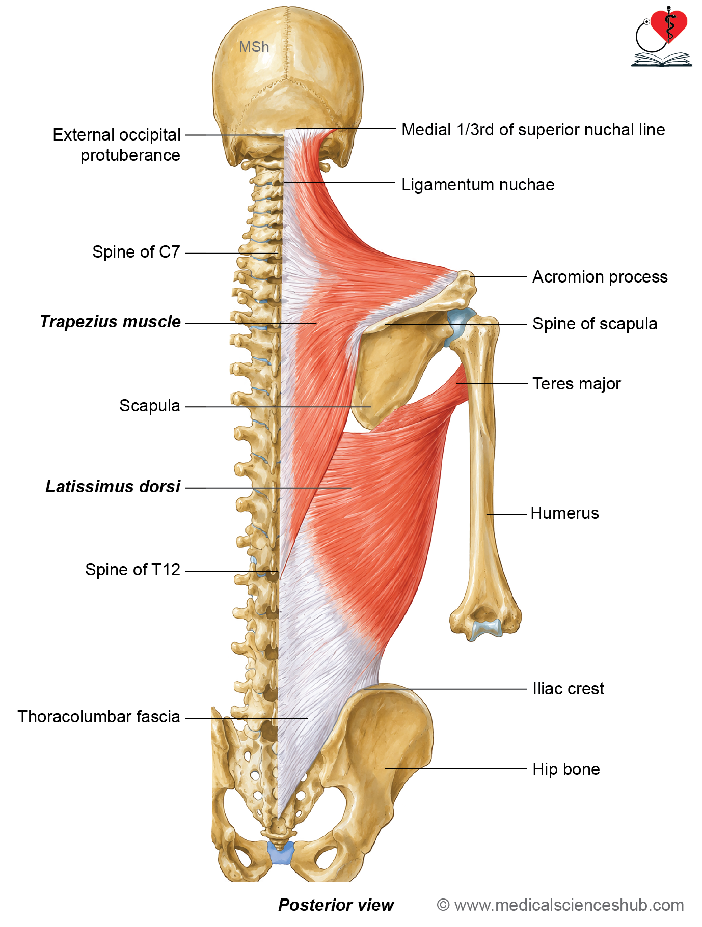
- Trapezius is a large, flat, triangular muscle located in the superficial layer of the back.
- The right and left trapezius muscles together form a trapezium-shaped outline across the upper back.
- It extends over the posterior aspect of the neck and the upper thoracic region.
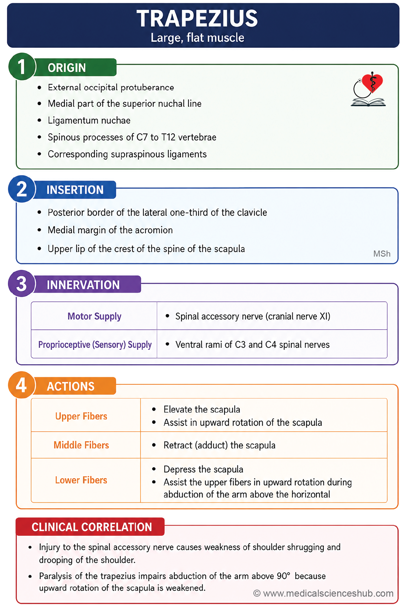
Origin
The trapezius arises from:
- Medial one-third of the superior nuchal line.
- External occipital protuberance.
- Ligamentum nuchae.
- Spine of the C7 vertebra.
- Spinous processes of the T1 to T12 vertebrae.
Insertion
The trapezius is inserted as follows:
- Upper fibers attach to the posterior border of the lateral one-third of the clavicle.
- Middle fibers attach to the medial margin of the acromion and the upper lip of the crest of the spine of the scapula.
- Lower fibers attach to the triangular area at the medial end of the spine of the scapula.
Nerve supply
- Motor innervation is provided by the spinal accessory nerve (cranial nerve XI).
- Proprioceptive sensory fibers are carried by the anterior (ventral) rami of C3 and C4 spinal nerves.
Actions
- The trapezius stabilizes the scapula during movements of the shoulder girdle and upper limb.
- Its actions vary according to the muscle fibers:
- Upper fibers: Elevate the scapula with the levator scapulae, producing shoulder shrugging.
- Middle fibers: Retract (adduct) the scapula with the rhomboid major and rhomboid minor.
- Lower fibers: Rotate the scapula upward with the serratus anterior, enabling overhead abduction of the arm.
Clinical testing
Trapezius function is assessed by asking the patient to shrug the shoulders against resistance.
CLINICAL ANATOMY
- The trapezius and sternocleidomastoid muscles develop from a common embryological source and are both supplied by the spinal accessory nerve (cranial nerve XI).
- Paralysis of the trapezius results in shoulder drooping, impaired scapular stabilization, and difficulty elevating the shoulder or abducting the arm above the horizontal plane.
Latissimus Dorsi
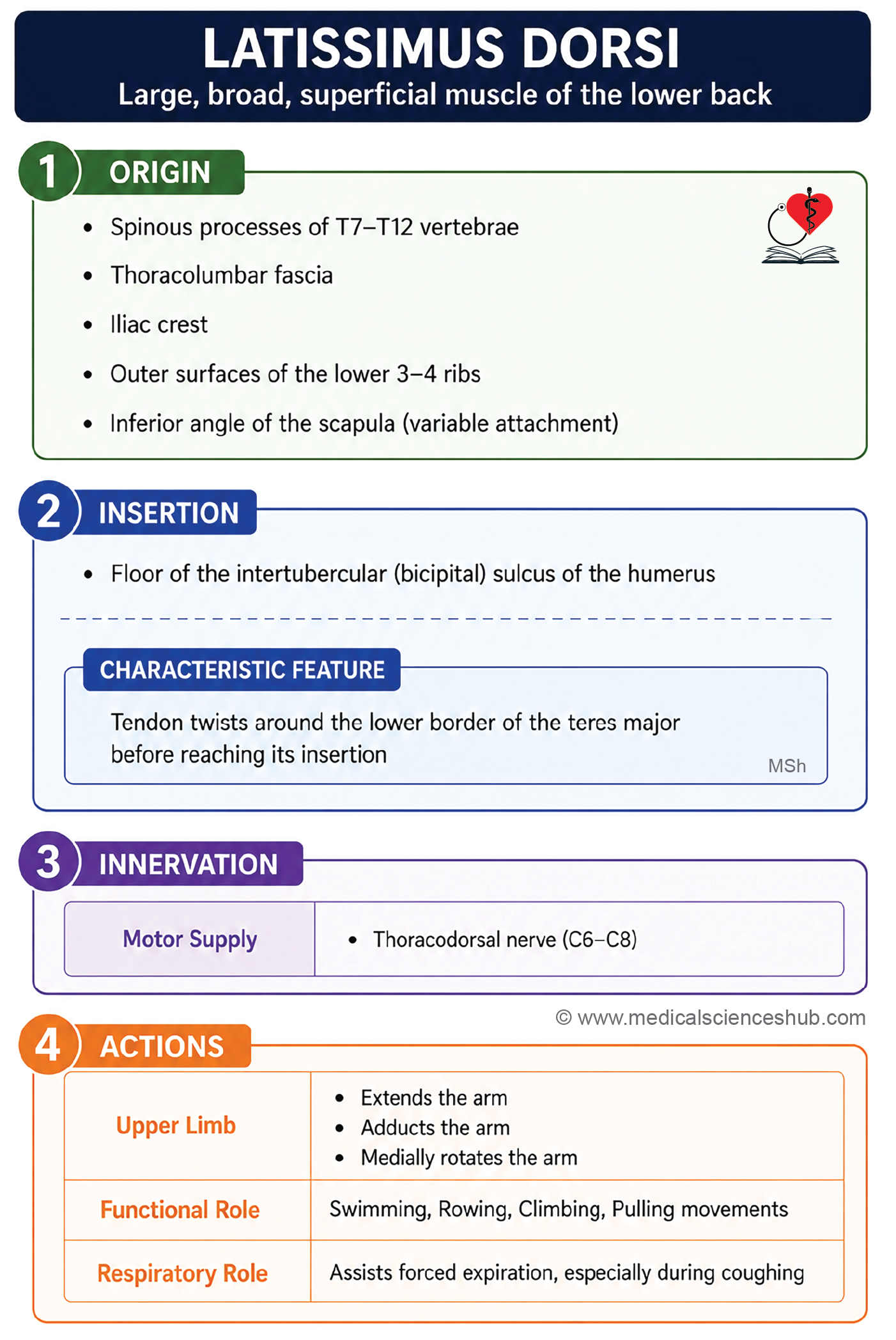
- Latissimus dorsi is a broad, thin, sheet-like muscle that covers a large part of the lower back.
- It is a large fan-shaped muscle that connects the humerus to the trunk and forms part of the superficial posterior axio-appendicular muscles.
Origin
- The latissimus dorsi arises from:
- Spinous processes of the lower six thoracic vertebrae (T7–T12).
- Thoracolumbar fascia.
- Outer lip of the iliac crest.
- Outer surfaces of the lower four ribs (9th–12th).
- Inferior angle of the scapula (this attachment is variable and may be absent in some individuals).
- The latissimus dorsi fibers pass upward and laterally toward the humerus.
- The lower fibers run almost vertically, whereas the upper fibers are nearly horizontal.
- The fibers wind around the inferolateral border of the teres major to form the posterior axillary fold.
Insertion
- The tendon inserts into the floor of the intertubercular sulcus (bicipital groove) of the humerus.
- During insertion, the fibers undergo a twist so that the lower fibers insert superiorly, while the upper fibers insert inferiorly.
Nerve supply
- The muscle is supplied by the thoracodorsal nerve (C6–C8), a branch of the posterior cord of the brachial plexus.
Actions
- The latissimus dorsi produces extension, adduction, and medial rotation of the arm at the shoulder joint.
- It plays an important role in activities such as swimming, rowing, climbing, and pulling, which require these shoulder movements.
- The muscle also acts as an accessory muscle of forced expiration, assisting during vigorous actions such as coughing and sneezing.
- During rope climbing, the humerus remains fixed while the muscle elevates the trunk toward the upper limb. The pectoralis major also contributes to this movement.
Clinical testing
The muscle is assessed by asking the patient to adduct an abducted arm (90°) against resistance. Contraction of the muscle makes the posterior axillary fold more prominent and can be palpated.
CLINICAL INTEGRATION
- The latissimus dorsi musculocutaneous flap is commonly used for breast reconstruction following mastectomy for breast carcinoma. The flap includes the latissimus dorsi muscle, thoracodorsal nerve, thoracodorsal artery (a branch of the subscapular artery), and accompanying veins (venae comitantes) to preserve its blood supply and function. In selected patients with severe heart failure who are unsuitable for heart transplantation, the latissimus dorsi can be wrapped around the heart and electrically stimulated with a pacemaker. This procedure is known as cardiomyoplasty.
- Thoracodorsal nerve injury may occur during axillary dissection in mastectomy because of its close relationship to the axillary tail of the breast. Injury to this nerve causes paralysis of the latissimus dorsi, resulting in difficulty raising the trunk during climbing and weakness of shoulder extension, adduction, and medial rotation.
- The latissimus dorsi is also widely used to reconstruct soft-tissue defects of the back because of its large size and reliable vascular supply.
Deep Posterior Axio-Appendicular Muscles
- The deep posterior axio-appendicular muscles lie beneath the superficial muscles of the back.
- These muscles connect the scapula to the vertebral column and help stabilize and move the scapula during upper limb movements.
- The deep posterior axio-appendicular muscles are:
- Levator scapulae
- Rhomboid minor
- Rhomboid major
Levator Scapulae
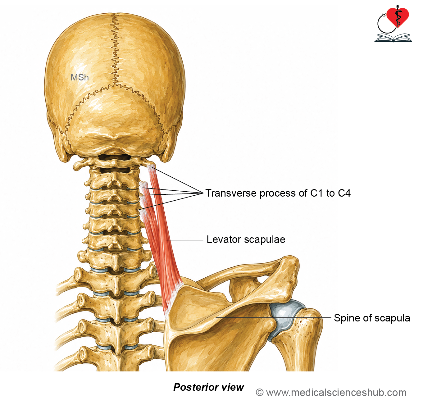
- Levator scapulae is a narrow, strap-like muscle located at the posterior aspect of the neck. Its primary function is to elevate the scapula, which gives the muscle its name.
Origin
- The muscle arises from:
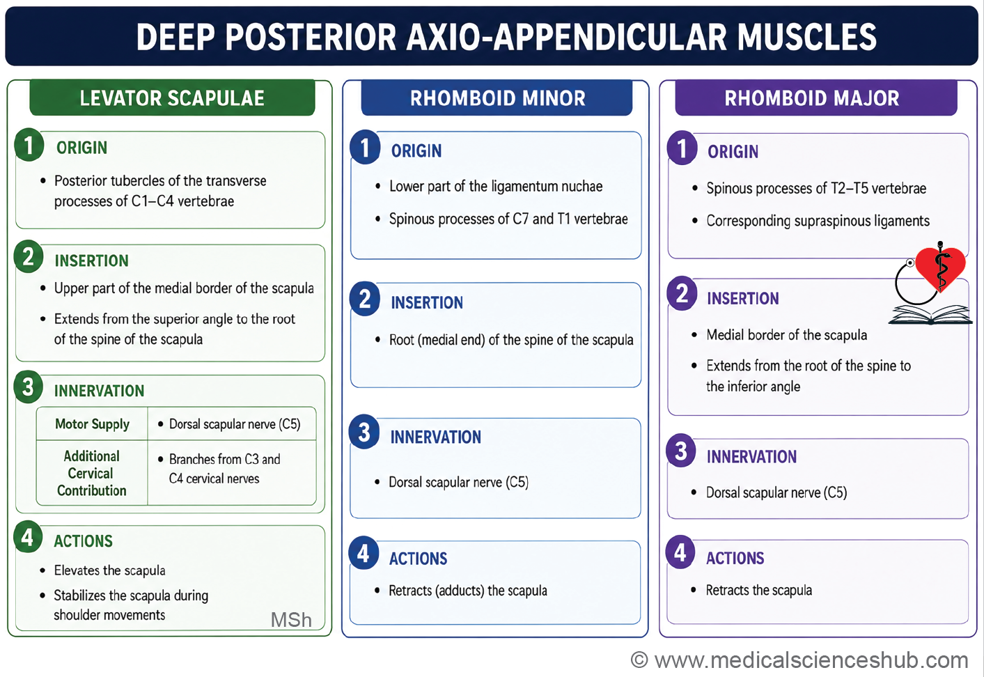
- Transverse processes of the C1 and C2 vertebrae.
- Posterior tubercles of the transverse processes of the C3 and C4 vertebrae.
- Direction of fibers: The fibers run obliquely downward and laterally toward the scapula.
Insertion
The muscle attaches to:
- Superior angle of the scapula.
- Posterior surface of the medial border of the scapula, between the superior angle and the root of the spine.
Nerve supply
- Dorsal scapular nerve (C5).
- Direct branches from the anterior (ventral) rami of C3 and C4 spinal nerves.
Action
The levator scapulae elevates and stabilizes the scapula during movements of the shoulder girdle.
Rhomboid Minor
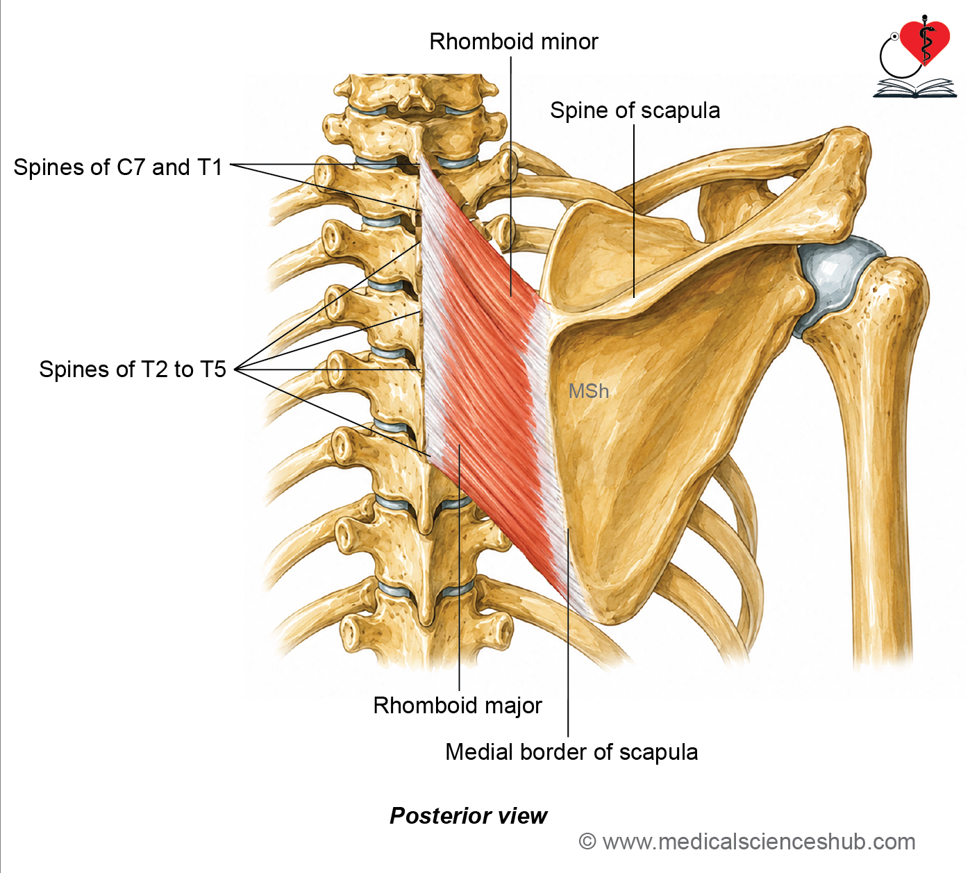
Rhomboid minor is a small, rhomboid-shaped muscle located deep to the trapezius.
Origin
The muscle arises from:
- Lower part of the ligamentum nuchae.
- Spinous processes of the C7 and T1 vertebrae.
- Supraspinous ligament between the C7 and T1 vertebrae.
The fibers pass downward and laterally toward the scapula.
Insertion
The muscle attaches to the triangular area at the root of the spine on the medial border of the scapula.
Nerve supply
- Dorsal scapular nerve (C5)
Action
The rhomboid minor retracts (adducts) and stabilizes the scapula, helping maintain its position against the thoracic wall.
Rhomboid Major
- Rhomboid major is a broad, quadrilateral muscle situated deep to the trapezius and inferior to the rhomboid minor.
Origin
The muscle arises from the spinous processes of T2 to T5 vertebrae and the intervening supraspinous ligaments.
Insertion
It attaches to the posterior surface of the medial border of the scapula, below the root of the spine.
Nerve supply
- Dorsal scapular nerve (C5).
Actions
- The rhomboid major, rhomboid minor, and middle fibers of the trapezius work together to retract (adduct) the scapula.
- The rhomboid muscles stabilize the scapula against the thoracic wall in coordination with the serratus anterior, providing a stable base for upper limb movements.
Dorsal Scapular Nerve (Nerve to Rhomboids)
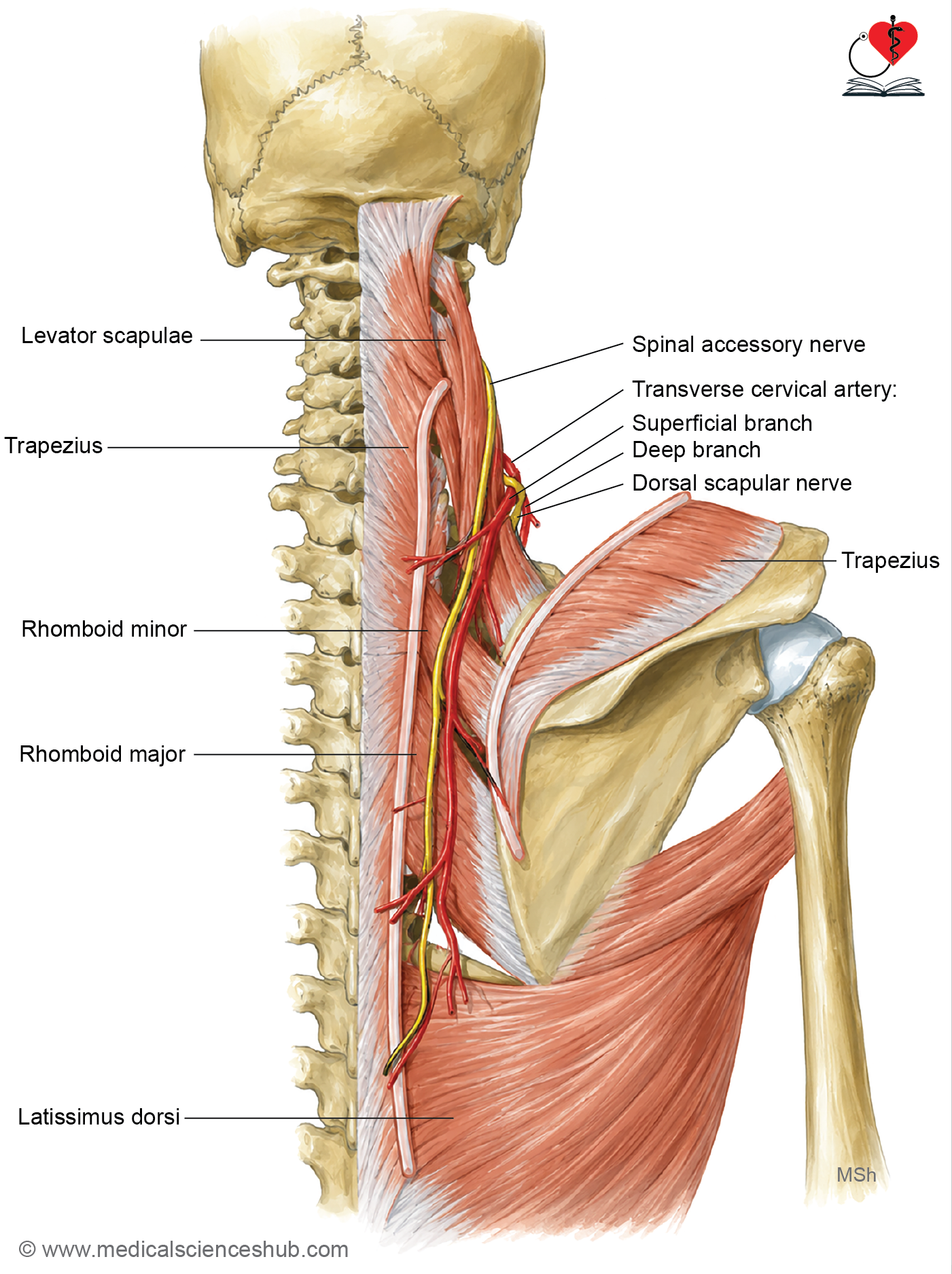
- The dorsal scapular nerve (nerve to the rhomboids) arises from the ventral primary ramus of the C5 spinal nerve in the neck.
- It enters the posterior triangle of the neck by passing between the scalenus anterior and scalenus medius muscles.
- The nerve runs laterally for a short distance and then pierces the scalenus medius muscle.
- It descends on the anterior surface of the levator scapulae muscle.
- During posterior triangle dissection, the spinal accessory nerve and the superficial branch of the transverse cervical artery are located between the trapezius and levator scapulae muscles.
- The dorsal scapular nerve and the deep branch of the transverse cervical artery lie deep to the levator scapulae muscle.
Structures supplied
The dorsal scapular nerve provides motor innervation to
- Levator scapulae
- Rhomboid minor
- Rhomboid major.
Triangle of auscultation
The triangle of auscultation is a small triangular gap in the muscles of the back, present on both sides.
- Boundaries
- Medial: Lateral border of the trapezius muscle.
- Lateral: Medial border of the scapula.
- Inferior: Superior border of the latissimus dorsi muscle.
- Floor
- 6th and 7th ribs.
- 6th intercostal space.
- Pleura.
- Lung.
- Some lower fibers of the rhomboid major muscle.
- The triangle of auscultation provides a relatively muscle-free area of the thoracic wall.
- It is an important clinical landmark because breath sounds can be heard more clearly through this region during auscultation.
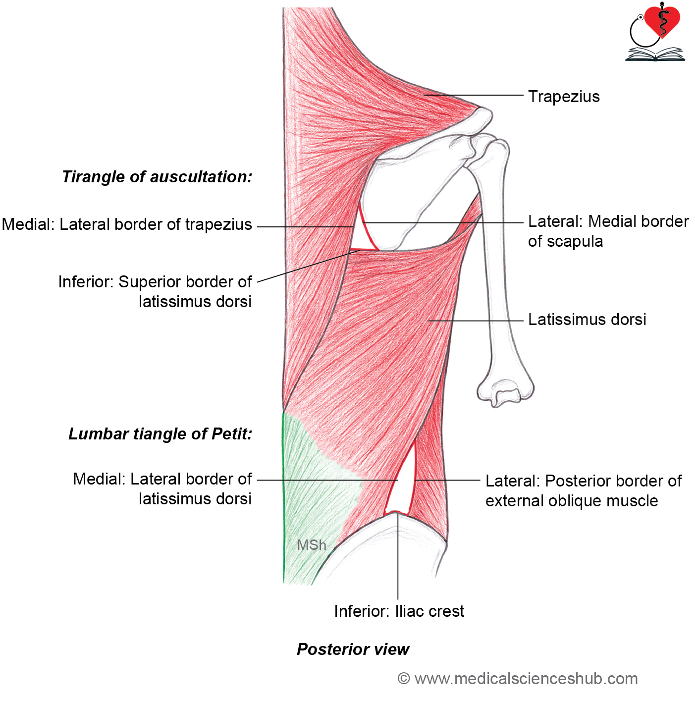
Lumbar triangle of Petit
- The lumbar triangle of Petit is a small triangular gap located in the lower part of the back.
- Boundaries:
- Medial: Lateral border of the latissimus dorsi muscle.
- Lateral: Posterior border of the external oblique muscle.
- Inferior (base): Iliac crest.
- Floor:
Internal oblique muscle of the anterior abdominal wall.
- The lumbar triangle is a potential weak area of the posterior abdominal wall.
- Clinical importance:
Protrusion of abdominal contents through this weak area results in a Petit’s (inferior lumbar) hernia, which is a rare type of lumbar hernia.
Important Questions
- Write a short note on triangle of auscultation.
- Write a short note on triangle of Petit.
- Write a short note on dorsal scapular nerve.
📝 Test Your Knowledge – Practice MCQs
Attempt the chapter MCQ quiz and assess your understanding of key concepts.
Click to see: Medical Disclaimer and Copyright Notice