
Competencies
- AN10.3: Describe, identify, and demonstrate formation, branches,relations, area of supply of branches, course and relations, and terminal branches of brachial plexus.
- AN10.5: Explain variations in formation of brachial plexus.
- AN10.6: Explain the anatomical basis of clinical features of Erb’s palsy and Klumpke’s paralysis.
Brachial Plexus
- The brachial plexus is a network of nerves formed by the ventral (anterior) primary rami of the C5, C6, C7, C8, and T1 spinal nerves.
- It provides the motor and sensory innervation to the upper limb, except for a few areas supplied by other nerves.
Components of Brachial Plexus
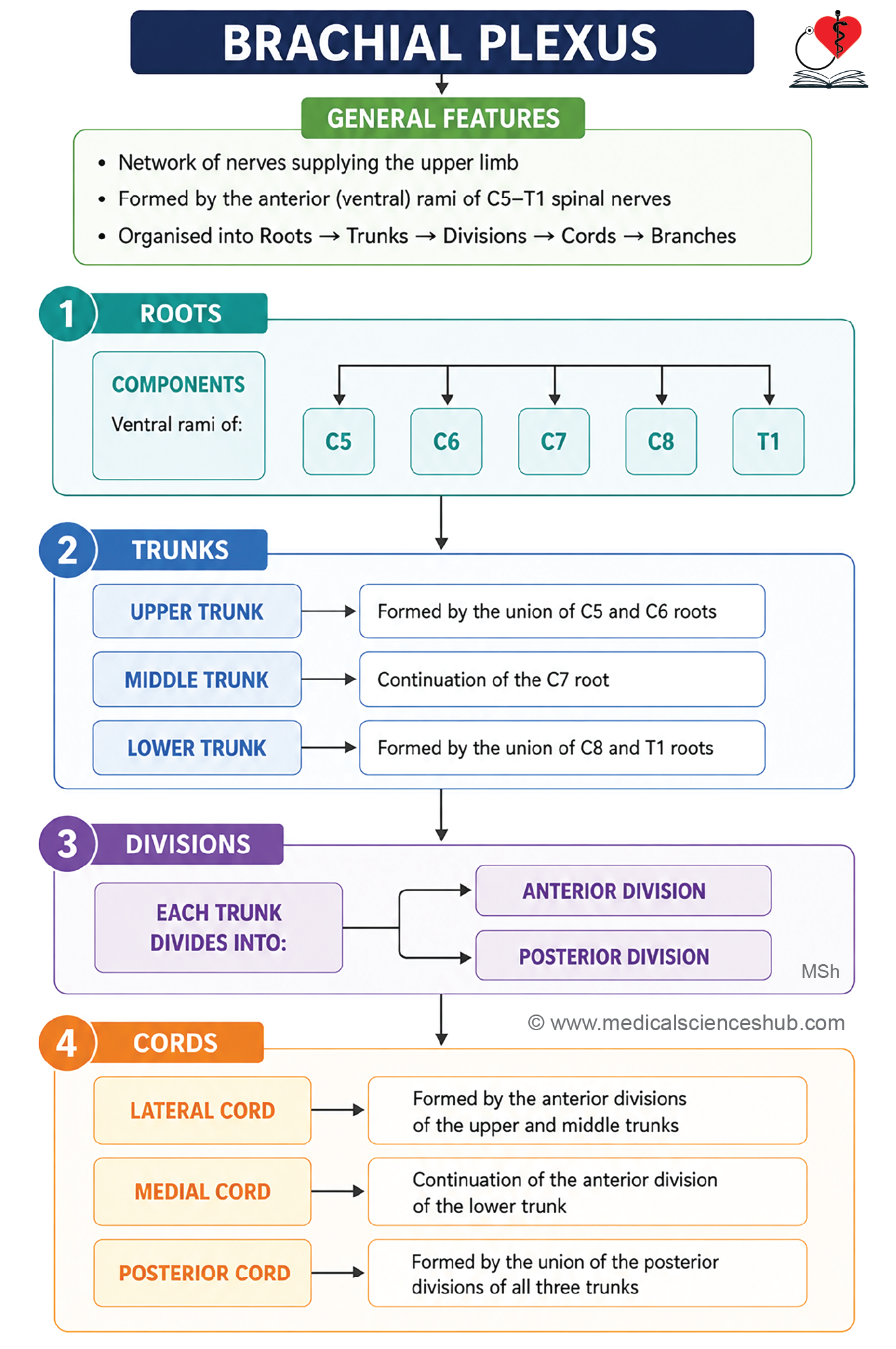
The brachial plexus is organized into five sequential components:
- Roots: The ventral primary rami of spinal nerves C5 to T1 form the roots.
- Trunks: The roots unite to form three trunks: upper (superior), middle, and lower (inferior).
- Divisions: Each trunk divides into one anterior and one posterior division.
- Cords: The divisions recombine to form three cords: lateral, medial, and posterior, named according to their relationship to the second part of the axillary artery.
- Branches: The terminal and collateral branches arise from the cords to supply the muscles and skin of the upper limb.
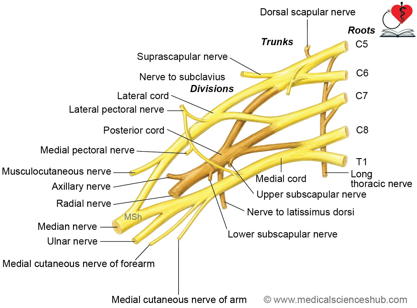
Roots
- The roots of the brachial plexus are formed by the ventral (anterior) primary rami of the C5, C6, C7, C8, and T1 spinal nerves.
- These five nerve roots constitute the normal brachial plexus and give rise to its trunks.
- A prefixed brachial plexus occurs when the ventral ramus of the C4 spinal nerve contributes to the plexus. In this variation, the contribution from T1 is usually reduced.
- A postfixed brachial plexus occurs when the ventral ramus of the T2 spinal nerve contributes to the plexus. In this variation, the contribution from C5 is usually reduced.
- Prefixed and postfixed brachial plexuses are normal anatomical variations and should be recognized during clinical examination and surgical procedures involving the neck and upper limb.
Trunks
- The roots of the brachial plexus unite to form three trunks:
- The upper (superior) trunk is formed by the union of the C5 and C6 roots.
- The middle trunk is the direct continuation of the C7 root.
- The lower (inferior) trunk is formed by the union of the C8 and T1 roots.
Relations
- The trunks are located in the posterior triangle of the neck.
- They emerge between the anterior scalene and middle scalene muscles.
- The trunks descend inferolaterally toward the root of the upper limb.
- The upper and middle trunks pass posterior to the subclavian artery, while the lower trunk lies posterior and slightly superior to the artery as it enters the cervicoaxillary canal.
Divisions
- Each of the three trunks of the brachial plexus divides into an anterior and a posterior division, producing a total of six divisions.
- The divisions pass through the cervicoaxillary canal to enter the axilla.
- Within the axilla, the anterior and posterior divisions recombine in a characteristic pattern to form the lateral, medial, and posterior cords of the brachial plexus.
Cords
- The divisions of the brachial plexus unite to form three cords: lateral, medial, and posterior.
- The lateral cord is formed by the union of the anterior divisions of the upper and middle trunks.
- The medial cord is the continuation of the anterior division of the lower trunk.
- The posterior cord is formed by the union of the posterior divisions of all three trunks.
Relations
- The cords and their branches constitute the infraclavicular part of the brachial plexus.
- In relation to the first part of the axillary artery, the lateral cord lies lateral to the artery, whereas the medial and posterior cords lie posterior to it.
- In relation to the second part of the axillary artery, the lateral, medial, and posterior cords lie lateral, medial, and posterior to the artery, respectively. These relationships give the cords their names.
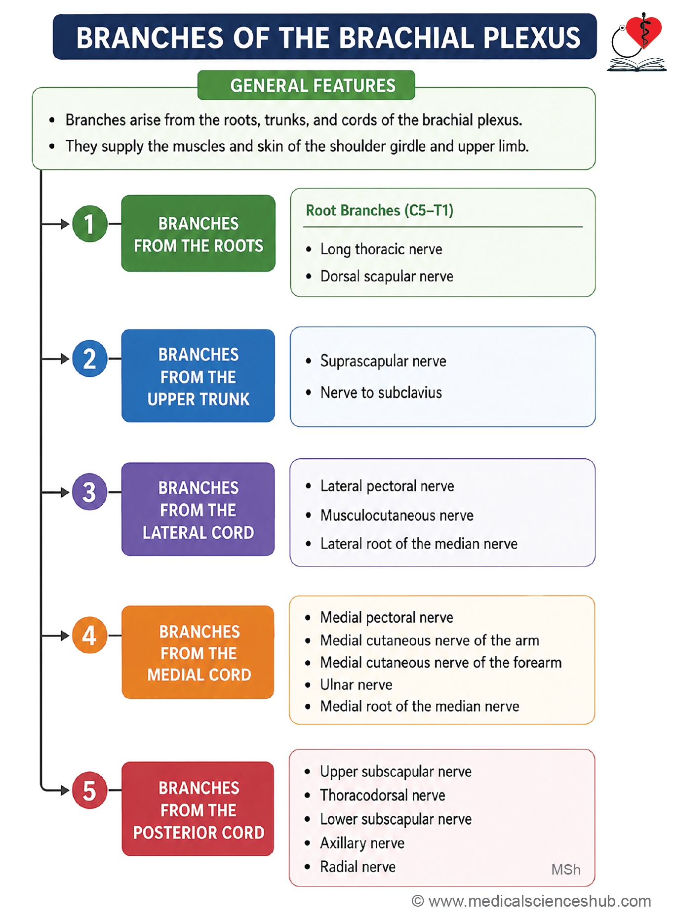
Branches of Brachial Plexus
Branches of Roots
- Long thoracic nerve (C5–C7): Supplies the serratus anterior muscle.
- Dorsal scapular nerve (C5): Supplies the rhomboid major, rhomboid minor, and levator scapulae muscles.
- Muscular branches (C5–C8): Supply the longus colli and scalene muscles.
- Contribution to the phrenic nerve (C3–C5): Fibres from C5 may contribute to the phrenic nerve.
Branches of Trunks
From upper trunk
- Suprascapular nerve (C5–C6): Supplies the supraspinatus and infraspinatus muscles.
- Nerve to subclavius (C5–C6): Supplies the subclavius muscle.
Branches of Cords
Branches of lateral cord
- Lateral pectoral nerve
- Musculocutaneous nerve
- Lateral root of median nerve
Branches of medial cord
- Medial pectoral nerve
- Medial cutaneous nerve of arm
- Medial cutaneous nerve of forearm
- Ulnar nerve
- Medial root of median nerve
Branches of posterior cord
- Upper subscapular nerve
- Nerve to latissimus dorsi
- Lower subscapular nerve
- Axillary neve
- Radial nerve
Key description
- The long thoracic nerve (C5-C7) arises from the ventral rami of C5, C6, and C7 spinal nerves in the neck. It passes posterior to the trunks of the brachial plexus and the subclavian artery, and anterior to the scalenus medius muscle. The nerve enters the axilla behind the axillary artery and the cords of the brachial plexus. It descends along the lateral surface of the serratus anterior in the midaxillary line and provides its motor supply.
- The dorsal scapular nerve (C5) originates from the C5 root of the brachial plexus. It pierces the scalenus medius, passes deep to the levator scapulae, and supplies the levator scapulae and both rhomboid muscles.
- The suprascapular nerve (C5-C6) arises from the upper trunk and passes beneath the inferior belly of the omohyoid to the suprascapular notch. It passes below the superior transverse scapular ligament and supplies the supraspinatus and infraspinatus muscles.
- The nerve to subclavius (C5-C6) arises from the upper trunk, passes anterior to the brachial plexus and subclavian vessels, and supplies the subclavius muscle and the sternoclavicular joint.
- The lateral pectoral nerve (C5-C7) arises from the lateral cord of the brachial plexus. It supplies the pectoralis major muscle and often communicates with the medial pectoral nerve through the ansa pectoralis. Through this communication, fibers may also reach the pectoralis minor muscle. The nerve pierces the clavipectoral fascia and enters the deep surface of the pectoralis major muscle. During dissection, reflect the pectoralis major superiorly to identify the lateral pectoral nerve.
- The musculocutaneous nerve (C5-C7) is a terminal branch of the lateral cord and lies lateral to the axillary artery. It pierces the coracobrachialis muscle, then descends between the biceps brachii and brachialis muscles. It provides motor innervation to the coracobrachialis, biceps brachii, and brachialis muscles.
- The nerve continues beyond the elbow as the lateral cutaneous nerve of the forearm, supplying sensation to the lateral aspect of the forearm.
- The lateral root of the median nerve (C5-C7) is the continuation of the lateral cord of the brachial plexus. It joins the medial root of the median nerve anterior to the third part of the axillary artery. The union of the two roots forms a characteristic Y-shaped configuration during dissection. The medial root of the median nerve (C8-T1) arises from the medial cord of the brachial plexus. It crosses anterior to the axillary artery to unite with the lateral root, completing the median nerve. The median nerve therefore contains fibers from all five roots of the brachial plexus (C5-T1). It supplies most muscles of the anterior compartment of the forearm, except the flexor carpi ulnaris and the medial half of the flexor digitorum profundus. In the hand, it innervates the thenar muscles (except adductor pollicis and the deep head of flexor pollicis brevis) and the lateral two lumbricals. It also provides sensory innervation to the lateral palm and the lateral 3½ digits.
- The medial pectoral nerve (C8-T1) arises from the medial cord of the brachial plexus. It pierces the pectoralis minor muscle and then enters the deep surface of the pectoralis major, supplying both muscles. During dissection, reflect the pectoralis major laterally to identify the medial pectoral nerve.
- The medial cutaneous nerve of the arm (C8-T1) supplies the skin on the medial side of the arm. It is a thin nerve that runs along the medial side of the axillary vein and may communicate with the intercostobrachial nerve.
- The medial cutaneous nerve of the forearm (C8-T1) provides sensory innervation to the skin on the medial side of the forearm. It descends between the axillary artery and vein, anterior to the thicker ulnar nerve.
- The ulnar nerve (C8-T1, occasionally C7) is the continuation of the medial cord of the brachial plexus. It supplies the flexor carpi ulnaris and the medial half of the flexor digitorum profundus in the forearm, and most intrinsic muscles of the hand. In the axilla, the ulnar nerve is thicker than the medial cutaneous nerve of the forearm and lies between the axillary artery and vein.
- The upper subscapular nerve (C5-C6) arises from the posterior cord and supplies the subscapularis muscle.
- The thoracodorsal (middle subscapular) nerve (C6-C8) arises from the posterior cord and supplies the latissimus dorsi muscle.
- The lower subscapular nerve (C5-C6) arises from the posterior cord and supplies the lower part of the subscapularis and the teres major muscles. These three nerves descend posterior to the axillary artery and vein on the anterior surface of the subscapularis muscle beneath fibrofatty tissue. The thoracodorsal nerve is the largest of the three and lies between the upper and lower subscapular nerves.
- The axillary nerve (C5-C6) arises from the posterior cord and supplies the deltoid and teres minor muscles. It passes posterolateral to the axillary artery and leaves the axilla through the quadrangular space. Retracting the axillary artery inferomedially helps identify it during dissection.
- The radial nerve (C5-T1) is the largest terminal branch and continuation of the posterior cord. It supplies the triceps brachii, all muscles of the posterior compartment of the forearm, and the skin of the posterior arm, forearm, and part of the dorsum of the hand. In the axilla, the radial nerve lies posterior to the axillary artery and is exposed by retracting the artery and the branches of the medial cord laterally.
CLINICAL INTEGRATION
- A cervical rib is an additional rudimentary rib that arises from the seventh cervical vertebra. Although often asymptomatic, it may compress the T1 nerve root or the lower trunk of the brachial plexus, producing neurological symptoms.
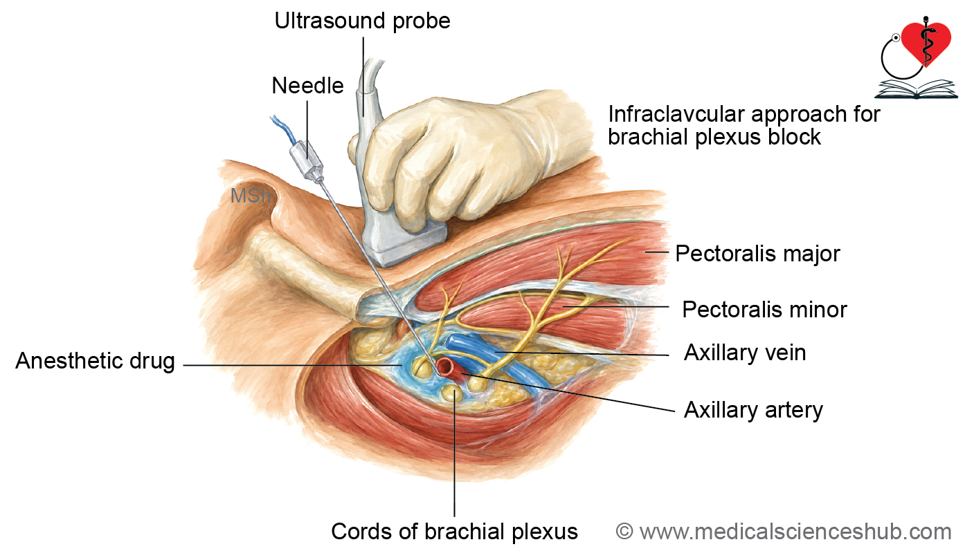
- A brachial plexus block is a regional anesthetic technique used to provide anesthesia and muscle relaxation for surgical procedures on the upper limb. The anesthetic is injected around the brachial plexus under ultrasound guidance, which improves accuracy and reduces the risk of injury to adjacent nerves and blood vessels.
- The upper trunk of the brachial plexus is the most common site of brachial plexus injury, typically resulting from excessive separation of the head and shoulder.
Erb’s paralysis
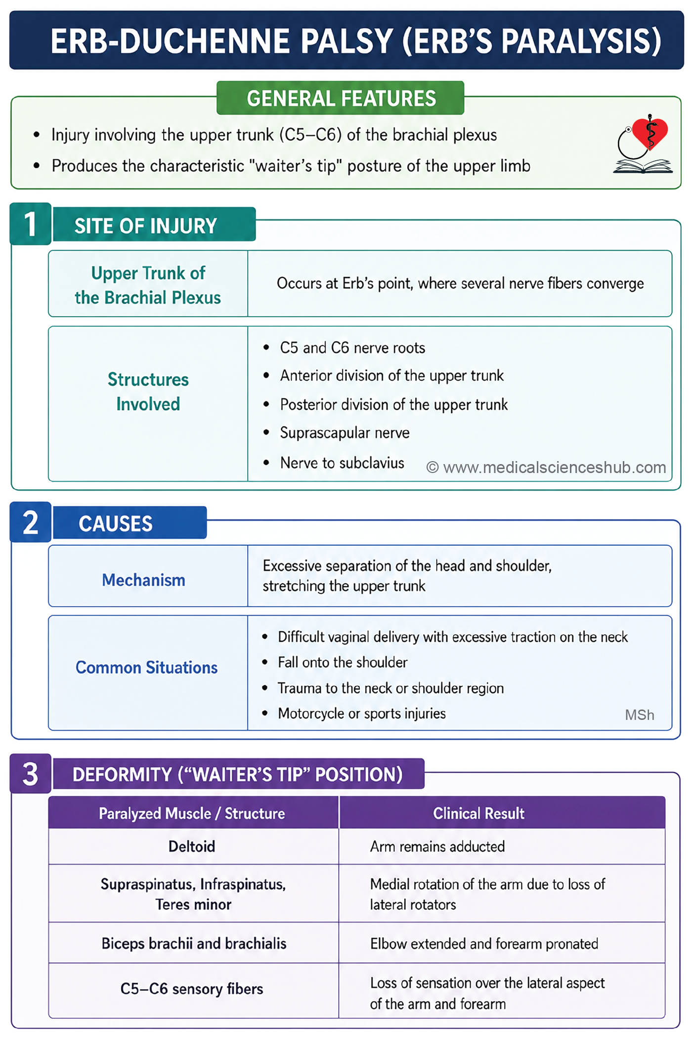
- Erb’s paralysis (Erb’s palsy) is paralysis of the upper limb caused by injury to the upper trunk (C5-C6) of the brachial plexus.
Site of injury
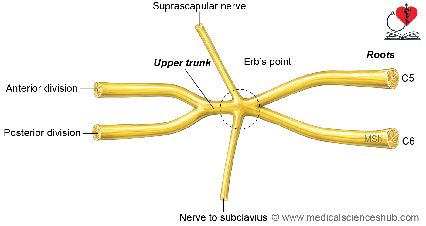
- The injury commonly occurs at Erb’s point, where six structures meet: the C5 and C6 roots, the anterior and posterior divisions of the upper trunk, the suprascapular nerve, and the nerve to subclavius.
Cause
- It is usually caused by excessive separation of the head and shoulder, as may occur during difficult childbirth or after a fall on the shoulder.
Deformity and muscles involved
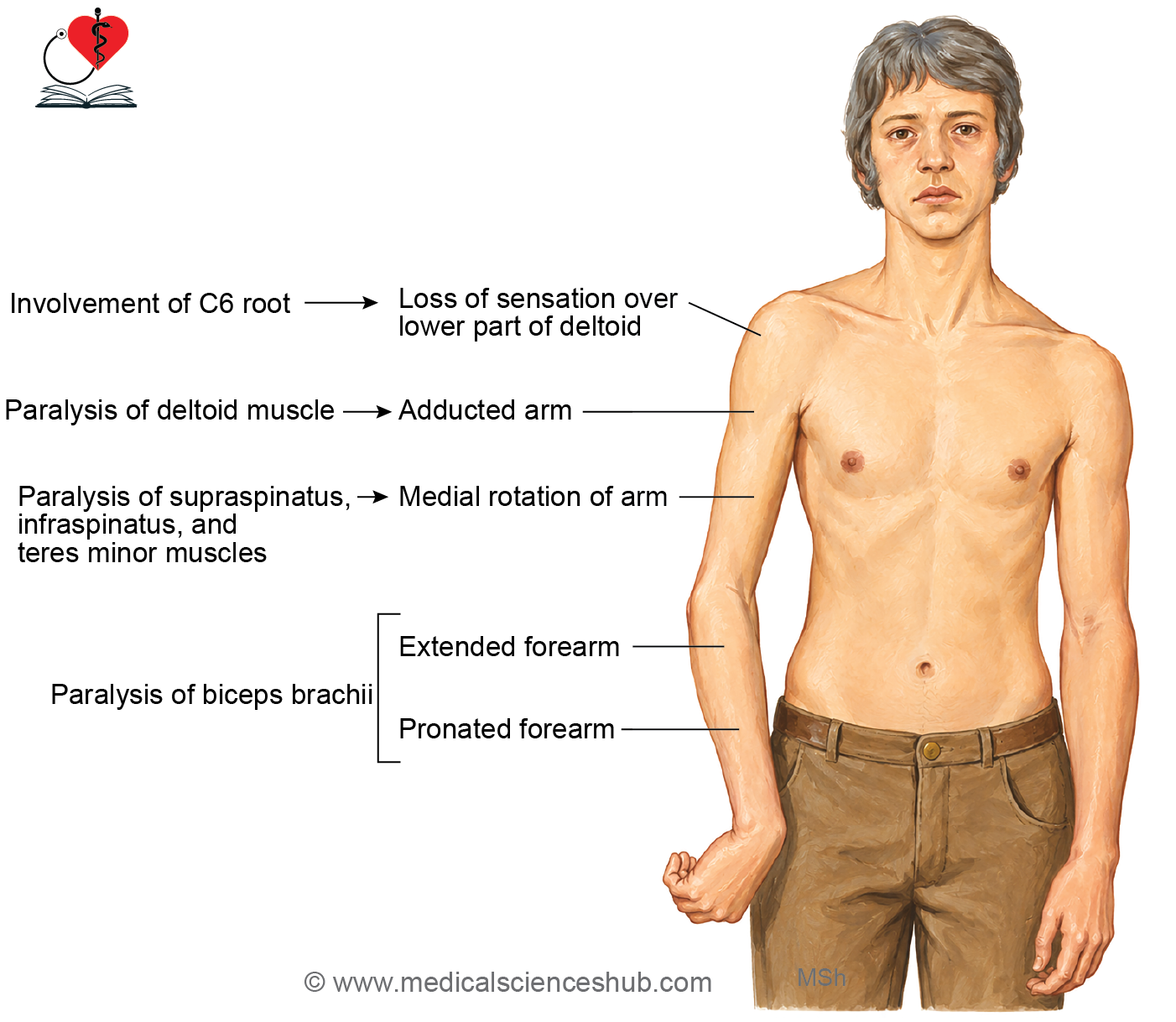
- The characteristic deformity is the “waiter’s tip” (policeman’s tip) posture, with the arm hanging by the side, adducted and medially rotated, and the forearm extended and pronated.
Disability Cause Result
- Paralysis of the deltoid causes loss of shoulder abduction, resulting in an adducted arm.
- Paralysis of the supraspinatus, infraspinatus, and teres minor causes loss of lateral rotation, producing medial rotation of the arm.
- Paralysis of the biceps brachii and brachialis prevents elbow flexion, leading to an extended forearm.
- Paralysis of the biceps brachii also abolishes forearm supination, resulting in a pronated forearm.
- Involvement of the C6 dermatome causes sensory loss over the lateral aspect of the arm, including the lower part of the deltoid region.
Klumpke’s paralysis
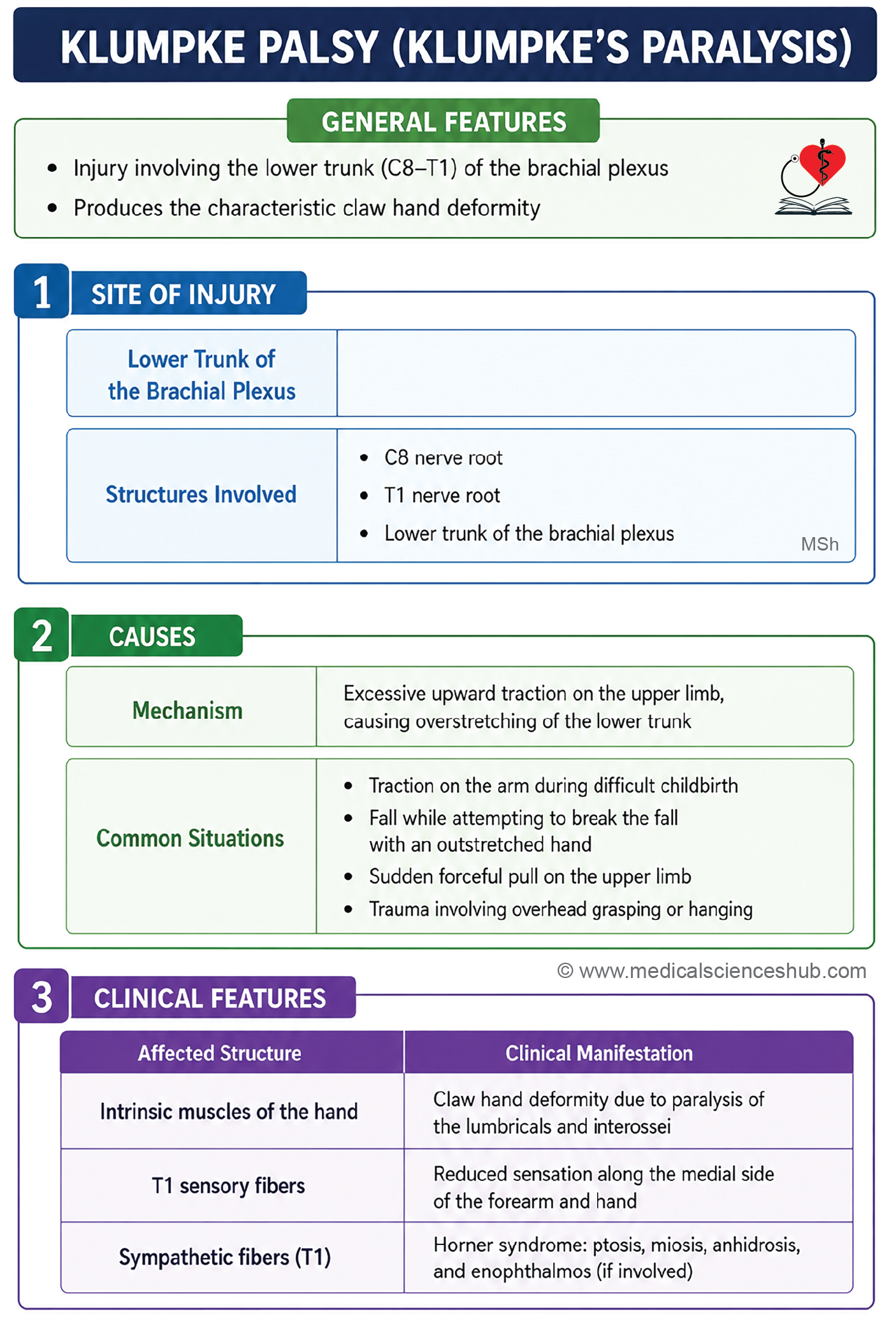
- Klumpke’s paralysis (Klumpke’s palsy) results from injury to the lower trunk (C8-T1) of the brachial plexus and primarily affects the muscles of the hand.
Site of injury
- The lesion involves the C8 and T1 nerve roots.
Cause
- It is commonly caused by excessive upward traction on the upper limb, such as a fall on an outstretched hand or forceful traction on the upper limb during difficult childbirth.
Deformity and muscles involved
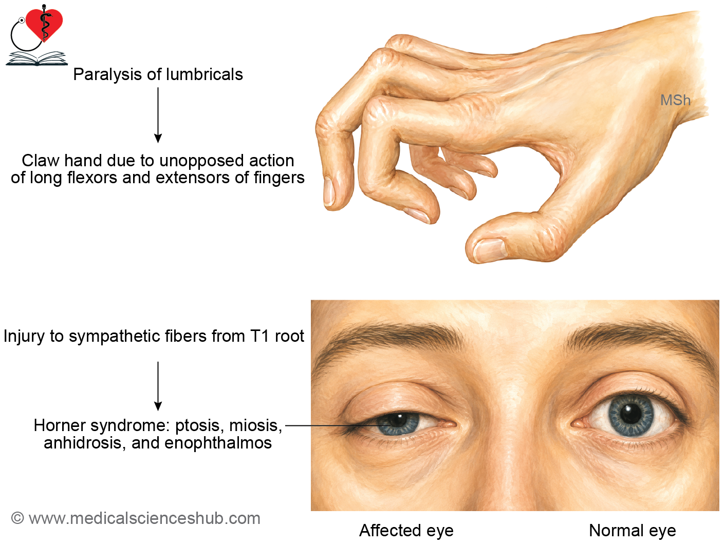
- The characteristic deformity is a claw hand, produced by paralysis of the intrinsic muscles of the hand, especially the lumbricals and interossei.
Disability Cause Result
- Paralysis of the lumbricals causes hyperextension at the metacarpophalangeal joints and flexion at the interphalangeal joints due to the unopposed action of the long extensor and flexor muscles.
- Involvement of the T1 dermatome results in sensory loss along the medial side of the forearm and hand.
- If the sympathetic fibers arising from the T1 spinal segment are damaged, Horner syndrome may occur.
- Horner syndrome is characterized by ptosis, miosis, anhidrosis, and enophthalmos on the affected side.
Important Questions
- Describe the formation and branches of brachial plexus.
- Draw a well-labeled diagram of brachial plexus.
- List the branches of brachial plexus.
- List the branches of medial cord of brachial plexus.
- List the branches of posterior cord of brachial plexus.
- Write a short note on Erbs’s palsy.
- Write a short note on Horner’s syndrome.
📝 Test Your Knowledge – Practice MCQs
Attempt the chapter MCQ quiz and assess your understanding of key concepts.
Click to see: Medical Disclaimer and Copyright Notice