
Competencies
- AN9.2: Breast: Describe the location, extent, deep relations,structure, age changes, blood supply, lymphatic drainage, microanatomy,and applied anatomy of breast.
- AN9.3: Describe development of breast.
- The mammary gland is a characteristic feature of mammals and is commonly known as the breast.
- The breast is located in the pectoral region on the anterior chest wall.
- Breasts are present in both males and females.
- The female breast undergoes significant development after puberty under the influence of hormones.
- In males and prepubertal females, the breast remains rudimentary and is minimally developed.
- After childbirth, the female breast produces milk to provide nutrition for the newborn through lactation.
- The anatomy described here refers specifically to the female breast.
Location
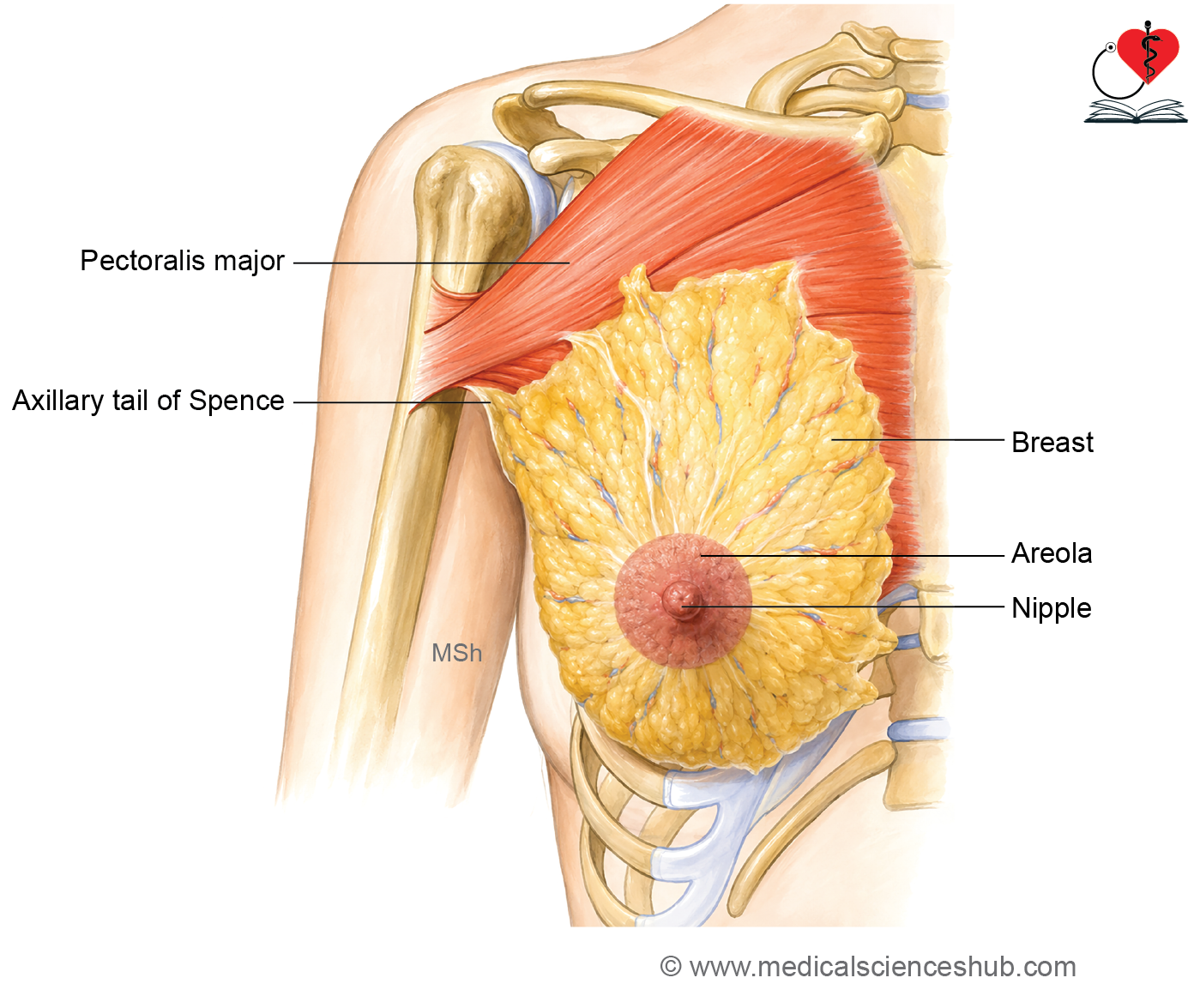
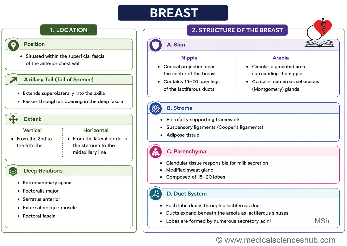
- The breast lies within the superficial fascia of the pectoral region on the anterior chest wall.
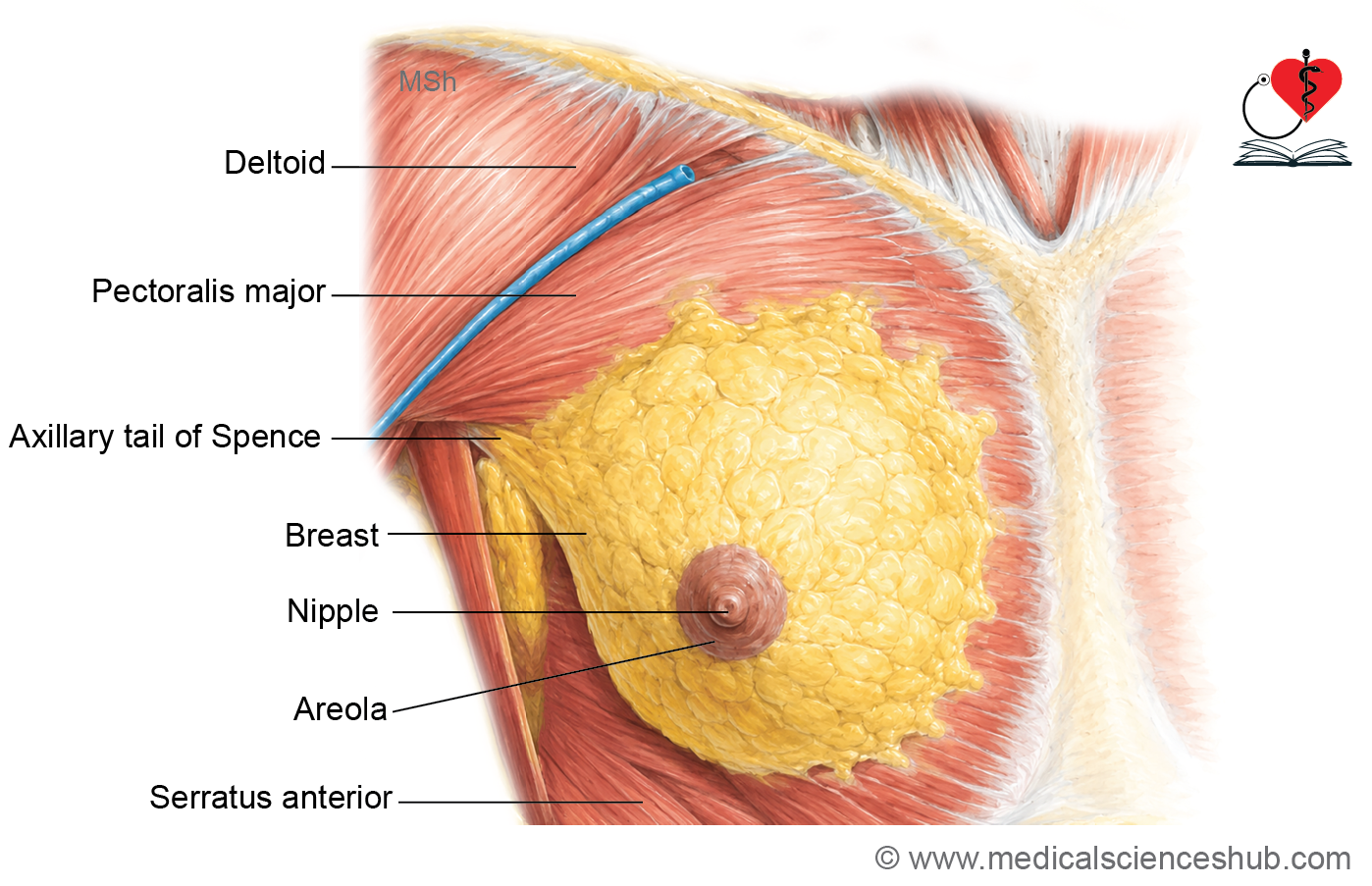
- A small extension from its superolateral part passes through the deep fascia into the axilla and is called the axillary tail.
- The opening in the deep fascia that transmits the axillary tail is known as the foramen of Langer.
- Breast carcinoma frequently develops in the axillary tail because glandular tissue extends into this region.
Shape
The shape of the breast varies among individuals and may be hemispherical, conical, pear-shaped, pendulous, or flat.
Extent
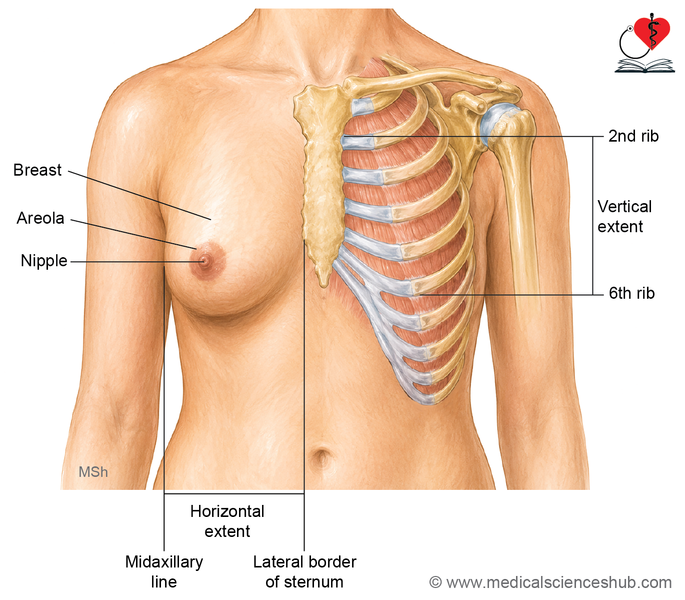
- Vertically, the breast usually extends from the second to the sixth ribs.
- Horizontally, it extends from the lateral border of the sternum to the midaxillary line.
Relations
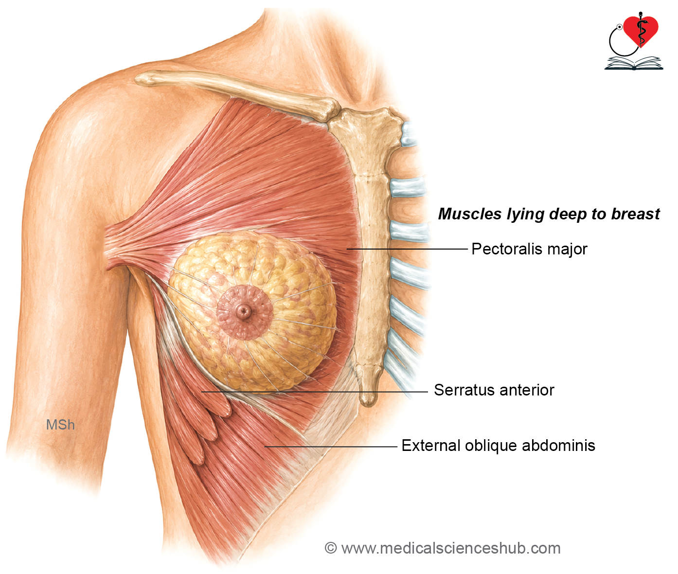
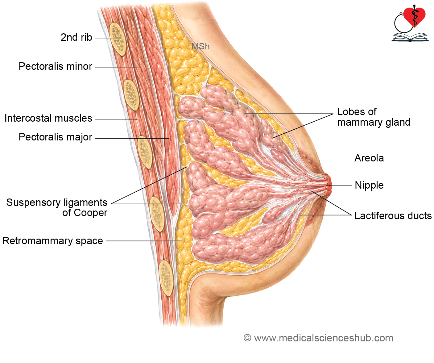
- The posterior (deep) surface of the breast is related to the pectoral fascia, underlying muscles, and the retromammary space.
- A thick pectoral fascia lies immediately deep to the breast and covers the underlying muscles.
- The breast rests mainly on the pectoralis major muscle, with smaller portions supported by the serratus anterior and external oblique muscles.
- A layer of loose areolar tissue, known as the retromammary space, separates the breast from the pectoral fascia.
- The retromammary space permits the breast to move freely over the underlying deep fascia and muscles, facilitating normal mobility.
Structure of Breast
Breast consists of the following three components:
- Skin
- Stroma
- Parenchyma: Parenchyma is the mammary gland proper.
Skin Covering the Breast
The skin covering the breast contains the nipple and the surrounding areola.
Nipple
- The nipple is a conical projection located slightly below the center of the breast. It usually lies opposite the fourth intercostal space, approximately 10 centimetres from the midline in adults.
- The nipple is pierced by 15 to 20 lactiferous ducts, which convey milk to the surface during lactation.
- It has a rich sensory nerve supply and contains smooth muscle fibers that cause it to become erect or flattened in response to stimulation.
Areola
- The areola is a circular pigmented area surrounding the nipple.
- It contains numerous modified sebaceous glands that produce oily secretions to lubricate the nipple and areola, thereby reducing drying and cracking during lactation.
- Small nodular elevations, called Montgomery tubercles, represent enlarged sebaceous glands and become more prominent during pregnancy.
- The skin of the nipple and areola is hairless, and the areola becomes darker during pregnancy.
Stroma
- The stroma forms the supporting framework of the mammary gland.
- It is composed of fibrous connective tissue and adipose (fat) tissue.
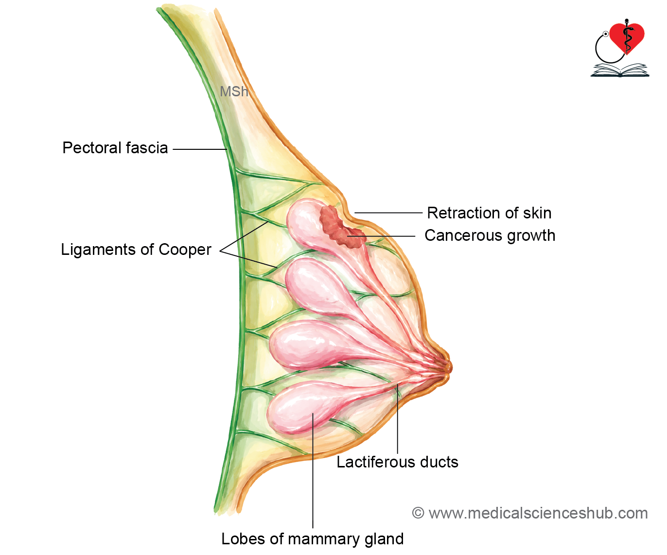
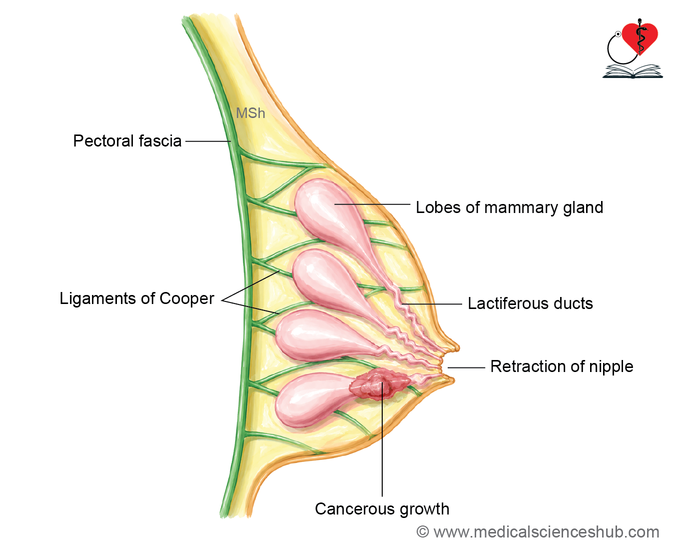
- The fibrous tissue forms fibrous septa known as the suspensory ligaments of Cooper.
- These ligaments extend from the deep (pectoral) fascia and clavicular region to the overlying skin of the breast.
- The suspensory ligaments support the breast, anchor it to the underlying structures, and help preserve its normal shape and contour.
- Adipose tissue constitutes the major bulk of the mammary gland and largely determines its size and consistency.
- Fat is distributed throughout most of the breast but is absent beneath the nipple and areola, where glandular tissue lies directly beneath the skin.
Parenchyma (mammary gland)
- The parenchyma is the glandular component of the breast and forms the functional mammary gland.
- It is a modified sweat gland specialized for milk production and secretion.
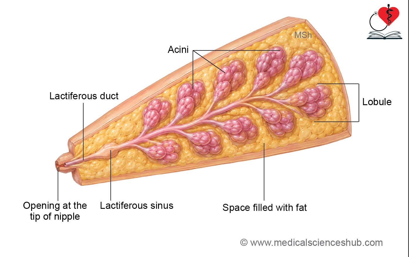
- The parenchyma contains the milk-secreting units, which consist of secretory acini (alveoli).
- The mammary gland is divided into 15–20 lobes that are arranged radially around the nipple.
- Each lobe contains a lactiferous duct surrounded by clusters of acini or alveoli.
- The lactiferous ducts converge toward the nipple and open separately at its tip.
- Near the nipple, each lactiferous duct expands to form a lactiferous sinus.
- During lactation, the lactiferous sinus serves as a temporary reservoir for milk before it is released through the nipple.
Blood Supply and Innervation
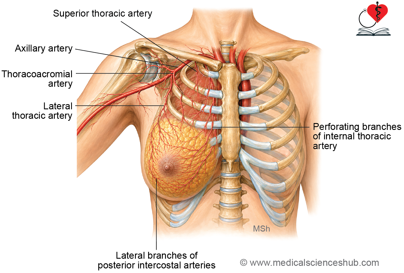
Arterial supply
- The breast receives its arterial supply from multiple sources.
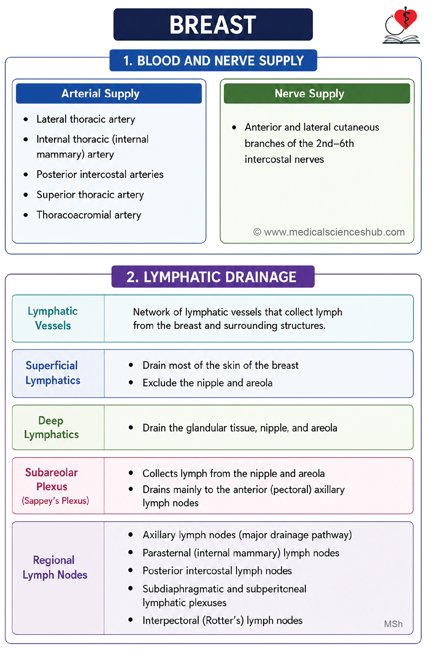
- The lateral thoracic artery is the principal artery supplying the breast.
- Additional arterial supply is provided by the internal thoracic (internal mammary) artery, lateral branches of the posterior intercostal arteries, the pectoral branch of the thoracoacromial artery, and the superior thoracic artery.
- The internal thoracic artery arises from the subclavian artery, and its perforating branches supply the medial part of the breast.
- The lateral thoracic and superior thoracic arteries are branches of the axillary artery.
Venous drainage
- Venous drainage generally follows the course of the arteries.
- Breast veins first unite beneath the nipple to form an anastomotic venous circle.
- Superficial veins drain into the internal thoracic vein, whereas deep veins drain into the axillary and posterior intercostal veins.
Innervation (nerve supply)
- The breast receives its nerve supply mainly from the 2nd to 6th intercostal nerves.
- These nerves reach the breast through their anterior and lateral cutaneous branches.
- The nerves provide sensory innervation to the skin, nipple, and areola and also supply the surrounding tissues.
- The nerves of the mammary gland do not regulate milk production.
- Milk secretion is controlled by prolactin, a hormone released from the anterior pituitary gland.
- Sensory stimulation of the nipple and areola initiates the milk ejection (let-down) reflex, which results in milk release from the breast.
Lymphatic Drainage of Breast
- Lymphatic drainage of the breast is of great clinical importance because it is the primary route for the spread of breast cancer.
- Breast cancer is the most common malignancy in women worldwide.
- Most lymph from the breast drains into the axillary lymph nodes, making them the principal lymphatic drainage pathway.
- Assessment of the axillary lymph nodes is essential for the staging, prognosis, and management of breast cancer.
Lymphatic vessels
- The lymphatic vessels of the breast are classified into superficial and deep lymphatics.
- Superficial lymphatics drain the skin covering the breast, except the nipple and areola.
- Deep lymphatics drain the breast parenchyma, nipple, and areola.
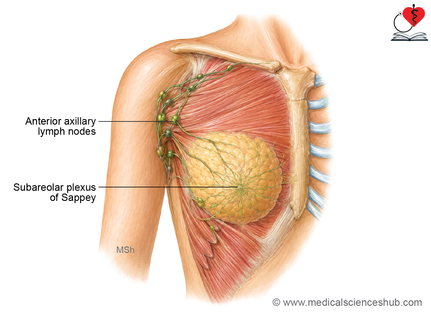
- The subareolar plexus of Sappey is a network of lymphatic vessels located deep to the areola.
- Lymph from the subareolar plexus primarily drains into the anterior (pectoral) group of axillary lymph nodes.
- Earlier, the breast was divided into four quadrants (upper lateral, upper medial, lower lateral, and lower medial) to describe lymphatic drainage. This quadrant-based concept is no longer recommended in current anatomical descriptions, including recent editions of Gray’s Anatomy.
Lymph nodes draining breast
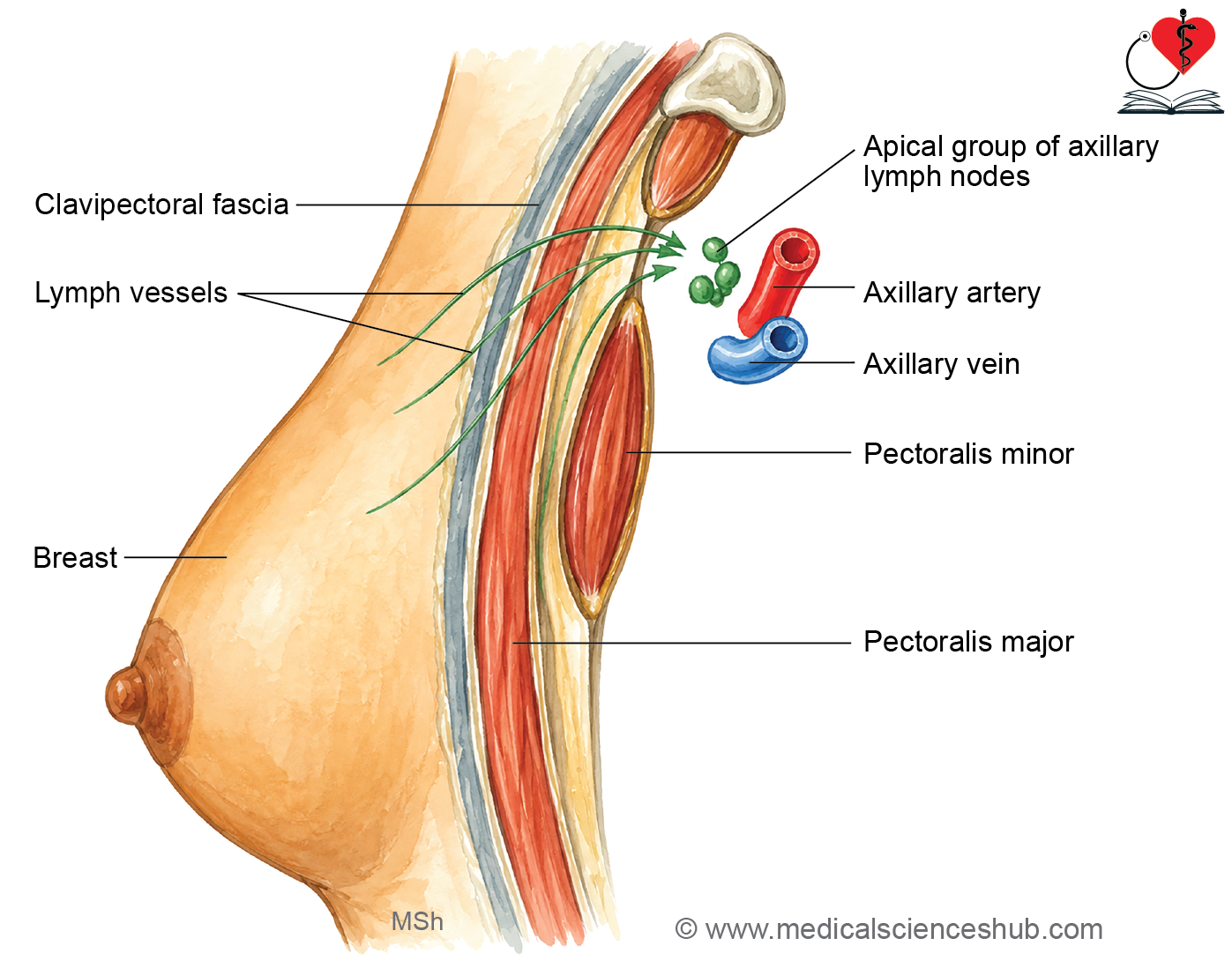
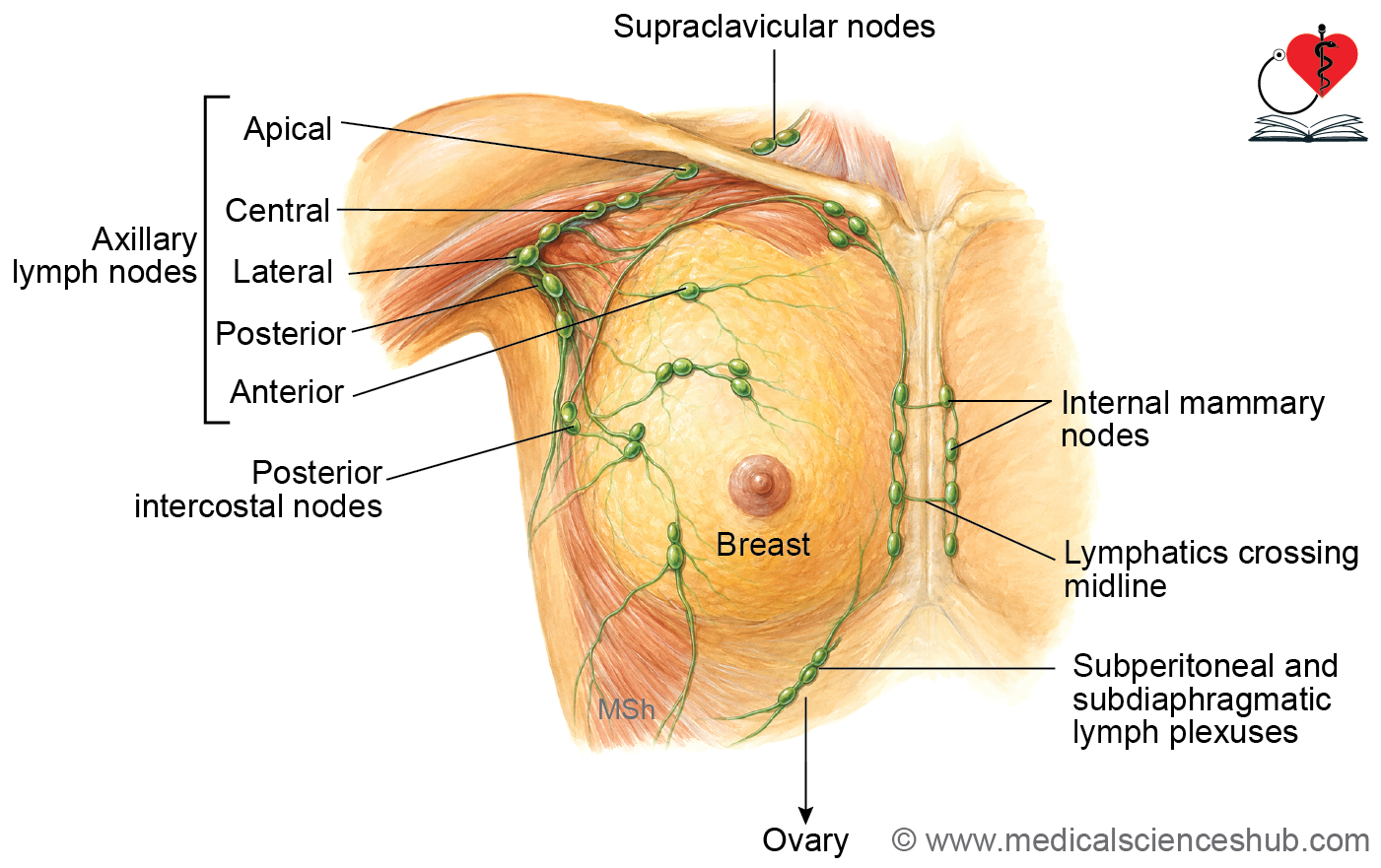
- The axillary lymph nodes receive most of the lymph from the breast, with the anterior (pectoral) group draining the largest share.
- The internal mammary (parasternal) lymph nodes, located along the internal thoracic artery, drain the medial part of the breast.
- A few lymphatic vessels from the lateral part of the breast accompany the posterior intercostal vessels and terminate in the posterior intercostal lymph nodes.
- Some lymphatics from the inferior part of the breast pierce the anterior abdominal wall and drain into the subdiaphragmatic and subperitoneal lymphatic plexuses.
- The interpectoral (Rotter’s) lymph nodes, situated between the pectoralis major and pectoralis minor muscles, also receive lymph from the breast.
- Lymphatics from the deep surface of the breast pierce the pectoralis major muscle and clavipectoral fascia to drain into the apical axillary lymph nodes.
Development of the Breast
Refer Development of Brest
CLINICAL INTEGRATION
- Breast cancer (carcinoma of the breast)
- It is the most common malignancy affecting women worldwide. It accounts for approximately 14% of cancers in women globally. It most commonly occurs between 40 and 60 years of age. Most breast cancers arise from the epithelial cells of the lactiferous ducts (ductal carcinoma).
- Major risk factors include hereditary predisposition, genetic mutations (such as BRCA1/BRCA2), alcohol consumption, obesity, advancing age, hormonal factors, and unhealthy dietary habits.
- Clinical features include:
- A painless breast lump.
- A fixed or immobile breast mass.
- Retraction or dimpling of the overlying skin.
- Retraction or inversion of the nipple.
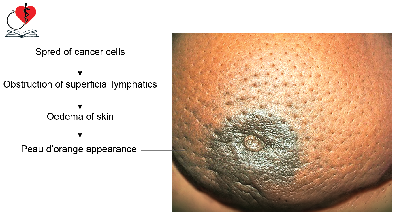
- Peau d’orange (orange-peel appearance) of the skin.
- Enlarged axillary lymph nodes.
- Abnormal nipple discharge, which may be bloody.
- Diagnostic investigations include:
- Regular breast self-examination to detect asymmetry, skin changes, nipple abnormalities, discharge, or palpable lumps.
- Mammography to identify suspicious breast lesions.
- Fine-needle aspiration cytology (FNAC) or biopsy to confirm malignant cells.
- Radical mastectomy involves removal of the entire breast, axillary lymph nodes, and the pectoralis major and minor muscles. It is now less commonly performed than modified radical mastectomy.
- Peau d’orange develops when cancer obstructs superficial lymphatic vessels, causing lymphatic edema that produces an orange-peel appearance.
- Breast cancer may spread through lymphatic channels to the peritoneum and ovaries, producing a secondary ovarian tumor known as a Krukenberg tumor.
- Congenital breast anomalies include:
- Athelia: Absence of the nipple.
- Amastia: Absence of the mammary gland.
- Polythelia: Presence of one or more accessory nipples.
- Polymastia: Presence of accessory mammary glands, occurring in about 1% of women.
- Inverted (crater) nipple: Failure of nipple eversion due to inadequate proliferation of the underlying mesenchyme.
- Gynecomastia is the abnormal enlargement of the male mammary glands.
- Mastectomy refers to partial or complete surgical removal of the breast, depending on the extent of disease.
Important Questions
- Describe the mammary gland under the following heads: Location, extent, morphology, structure, external features,relations, blood supply, and nerve supply.
- Write a short note on applied aspects of mammary gland.
- Write a short note on lymphatic drainage of breast.
- Explain: Metastasis from carcinoma of inferomedial quadrant of breast may take place in pelvic cavity.
- Write a short note on retraction and puckering of its skin, peau d’orange, and retraction of nipple.
- Write a short note on breast cancer.
📝 Test Your Knowledge – Practice MCQs
Attempt the chapter MCQ quiz and assess your understanding of key concepts.
Click to see: Medical Disclaimer and Copyright Notice