
Competencies
- AN11.5: Identify and describe boundaries and contents of cubital fossa.
- AN11.3: Describe the anatomical basis of venipuncture of cubital veins.
- AN11.6: Describe the anastomosis around the elbow joint.
Introduction
- The cubital fossa is a triangular depression located on the front of the elbow. It is a potential space that contains a variable amount of fat.
- The base of the triangular fossa is directed upward.
- The apex is directed downward.
- The cubital fossa is the upper limb equivalent (homologous structure) of the popliteal fossa, which is located behind the knee in the lower limb.
Boundaries, Roof, and Floor
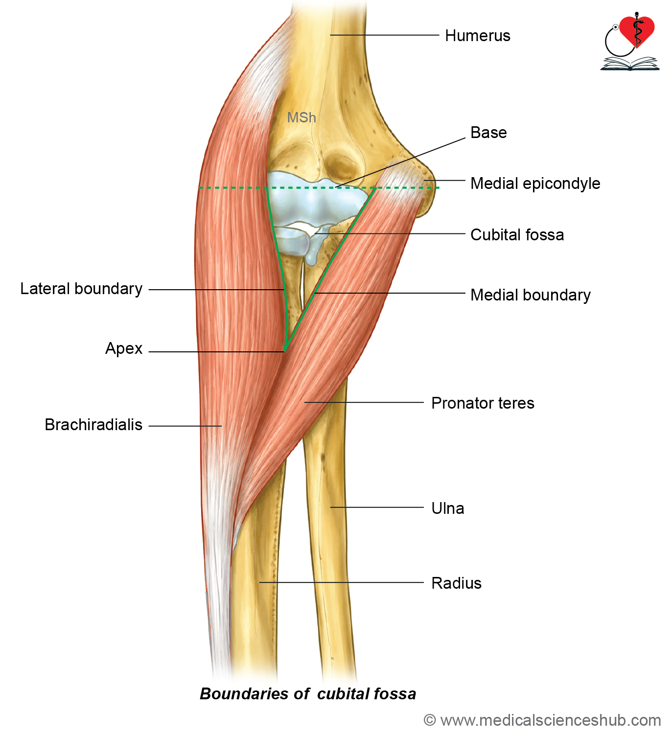
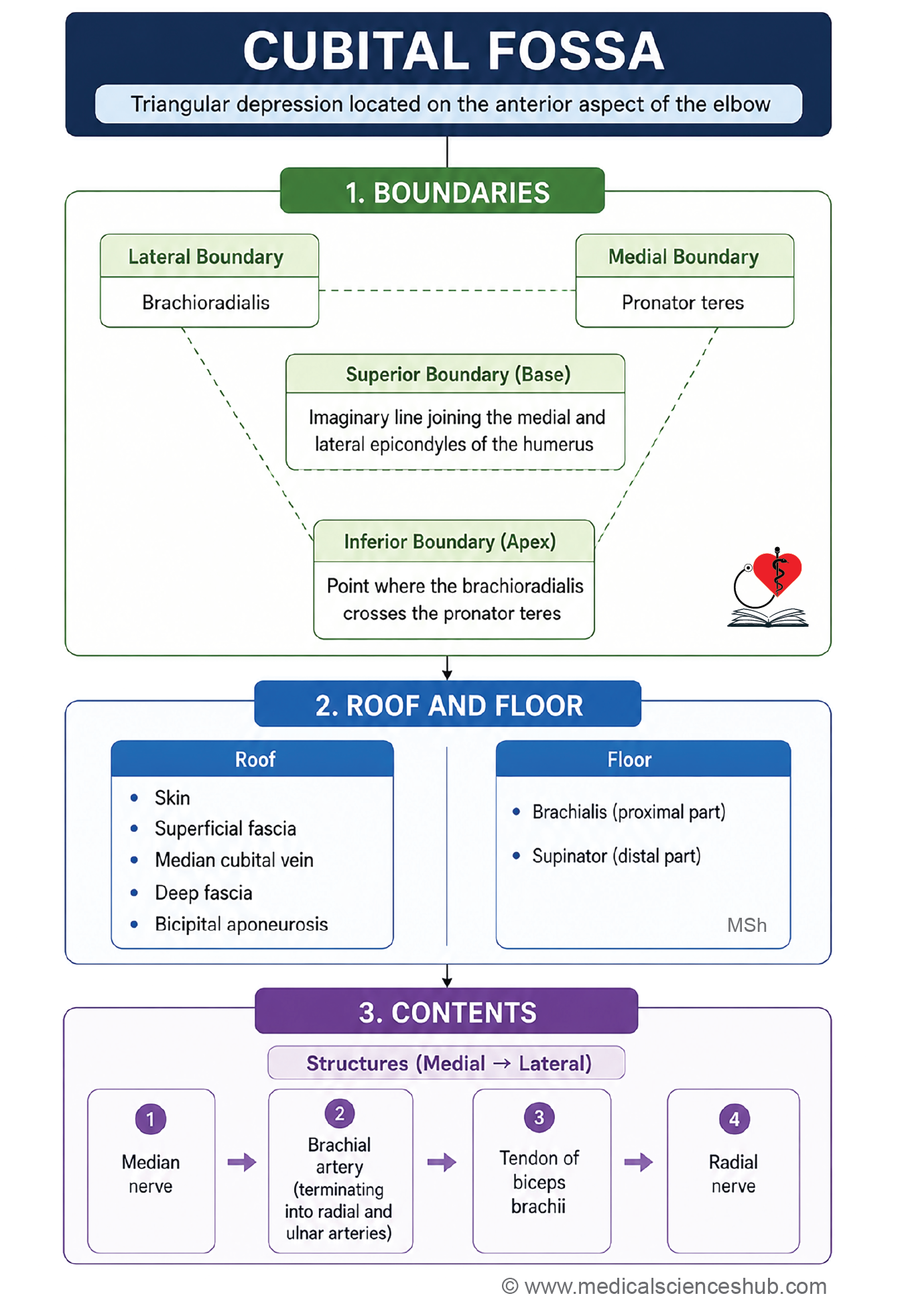
- Cubital fossa is a triangular hollow in front of the elbow. It has two boundaries, roof and floor.
Boundaries
- Medial boundary: Formed by the lateral border of the pronator teres muscle.
- Lateral boundary: Formed by the medial border of the brachioradialis muscle.
- Base (superior boundary): Formed by an imaginary line joining the medial and lateral epicondyles of the humerus.
- Apex: Formed at the point where the brachioradialis crosses the pronator teres muscle.
Roof
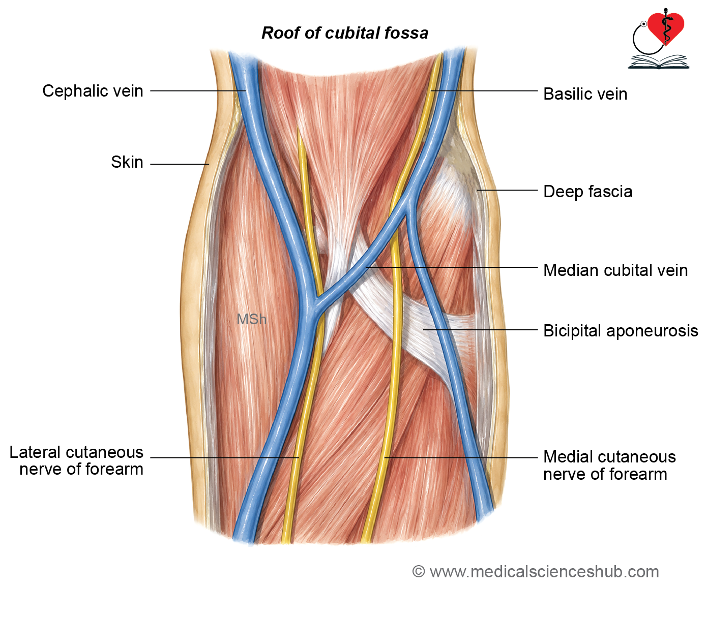
The roof of the cubital fossa is formed by:
- Skin.
- Superficial fascia.
- Median cubital vein.
- Medial cutaneous nerve of the forearm.
- Lateral cutaneous nerve of the forearm.
- Deep fascia reinforced by the bicipital aponeurosis.
Floor
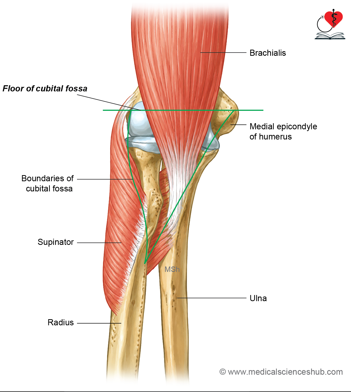
The floor of the cubital fossa is formed by:
- Brachialis muscle.
- Supinator muscle.
Contents of Cubital Fossa
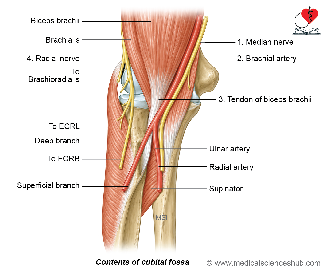
- The major contents of the cubital fossa are arranged from medial to lateral in the following order:
- Median nerve.
- Brachial artery.
- Tendon of the biceps brachii.
- Radial nerve.
- A simple mnemonic to remember this arrangement is MBBR:
- M – Median nerve
- B – Brachial artery
- B – Biceps brachii tendon
- R – Radial nerve
- This arrangement is clinically important during procedures such as arterial cannulation, venipuncture, and surgical exploration of the cubital fossa.
Key Description
Bicipital Aponeurosis (Lacertus Fibrosus)
- The bicipital aponeurosis is a flat fibrous expansion that arises from the medial side of the lower part of the biceps brachii tendon.
- It forms an important part of the roof of the cubital fossa.
- It passes downward and medially to blend with the deep fascia covering the pronator teres muscle.
- Through the deep fascia, it is indirectly attached to the posterior border of the ulna.
- The upper border of the bicipital aponeurosis is sharp and concave, with the concavity directed upward.
- It separates the major contents of the cubital fossa from the superficial structures, including the skin and superficial veins.
Median Nerve
- In the cubital fossa, the median nerve gives muscular branches to:
- Flexor carpi radialis.
- Palmaris longus.
- Flexor digitorum superficialis.
- The branch to the pronator teres muscle arises above the elbow, in the lower part of the arm.
- The median nerve is the most medial structure in the cubital fossa.
- It leaves the cubital fossa by passing between the humeral and ulnar heads of the pronator teres muscle.
Brachial Artery
- The brachial artery ends in the cubital fossa at the level of the neck of the radius.
- It divides into:
- Ulnar artery, the larger medial terminal branch.
- Radial artery, the smaller lateral terminal branch.
Ulnar Artery
- The ulnar artery passes deep into the forearm.
- It gives rise to the common interosseous artery, which further divides into the anterior and posterior interosseous arteries.
Radial Nerve
- In the cubital fossa, the radial nerve is largely covered by the brachioradialis muscle.
- It divides into two terminal branches:
- Superficial branch, which continues deep to the brachioradialis and is mainly sensory.
- Deep branch (posterior interosseous nerve), which supplies the extensor carpi radialis brevis and supinator muscles before piercing the supinator to enter the posterior compartment of the forearm.
CLINICAL INTEGRATION
Intravenous Injections
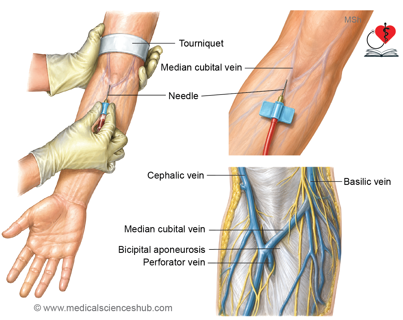
- The median cubital vein is the preferred vein for intravenous injections and venipuncture.
- It is stabilized by perforating veins that pass through the bicipital aponeurosis and connect it to the deep veins.
- This fixation prevents the vein from moving during needle insertion.
Recording Blood Pressure
- The brachial artery is auscultated in the cubital fossa while measuring blood pressure.
- A stethoscope is placed over the artery to hear the Korotkoff sounds during cuff deflation.
Protective Role of the Bicipital Aponeurosis
- The bicipital aponeurosis protects the brachial artery and median nerve from accidental injury during procedures such as venipuncture.
Biceps Jerk
- The biceps tendon reflex (biceps jerk) is elicited by tapping the tendon of the biceps brachii in the cubital fossa with a tendon hammer.
- This reflex is used to assess the integrity of the musculocutaneous nerve and the C5–C6 spinal cord segments.
Surgical Safety in the Cubital Fossa
- The median nerve gives its branches from the medial side, whereas the radial nerve gives its branches from the lateral side.
- Therefore, the lateral side of the median nerve and the medial side of the radial nerve are considered the safe surgical zones in the cubital fossa, as they minimize the risk of damaging the branches of these nerves.
Anastomosis around elbow joint
- The arterial anastomosis around the elbow provides an alternative pathway for blood flow when the main arteries are compressed or blocked.
- It maintains continuous blood supply between the brachial artery and the radial and ulnar arteries during movements of the elbow.
Sites of Anastomosis
In Front of the Medial Epicondyle of the Humerus
- Inferior ulnar collateral artery (branch of the brachial artery).
- Anterior ulnar recurrent artery (branch of the ulnar artery).
Behind the Medial Epicondyle of the Humerus
- Superior ulnar collateral artery (branch of the brachial artery).
- Posterior ulnar recurrent artery (branch of the ulnar artery).
In Front of the Lateral Epicondyle of the Humerus
- Radial collateral artery (branch of the profunda brachii artery).
- Radial recurrent artery (branch of the radial artery).
Behind the Lateral Epicondyle of the Humerus
- Middle collateral (posterior descending) artery, a branch of the profunda brachii artery.
- Interosseous recurrent artery, a branch of the posterior interosseous artery.
Above the Olecranon Fossa
- Middle collateral artery (branch of the profunda brachii artery).
- Transverse branch of the posterior division of the inferior ulnar collateral artery.
Note
- The ulnar artery and ulna are located on the medial side of the forearm.
- The radial artery and radius are located on the lateral (thumb) side of the forearm.
Important Questions
- Describe the cubital fossa under following heads: Boundaries, contents, and applied aspects.
- List the contents of cubital fossa.
- Write a short note on median cubital vein or venipuncture in the cubital fossa.
- Write a short note on anastomosis around the elbow joint.
📝 Test Your Knowledge – Practice MCQs
Attempt the chapter MCQ quiz and assess your understanding of key concepts.
Click to see: Medical Disclaimer and Copyright Notice