
Competencies
- AN12.11: Identify, describe, and demonstrate important muscle groups of dorsal forearm with attachments, nerve supply, and actions.
- AN12.12: Identify and describe origin, course, relations, branches (or tributaries), termination of important nerves and vessels of back of forearm.
- AN12.13: Describe the anatomical basis of wrist drop.
- AN12.14: Identify and describe compartments deep to extensor retinaculum.
- AN12.15: Identify and describe extensor expansion formation.
- AN13.7: ….., testing of muscles: brachioradialis,….. .
Introduction
The back of the forearm and hand contains the following important structures:
- Muscles
- Superficial extensor group
- Deep extensor group
- Extensor retinaculum
- Dorsal digital expansion (extensor expansion)
- Posterior interosseous nerve
- Posterior interosseous artery
Note: The back of the hand is also known as the dorsum of the hand.
Surface Landmarks
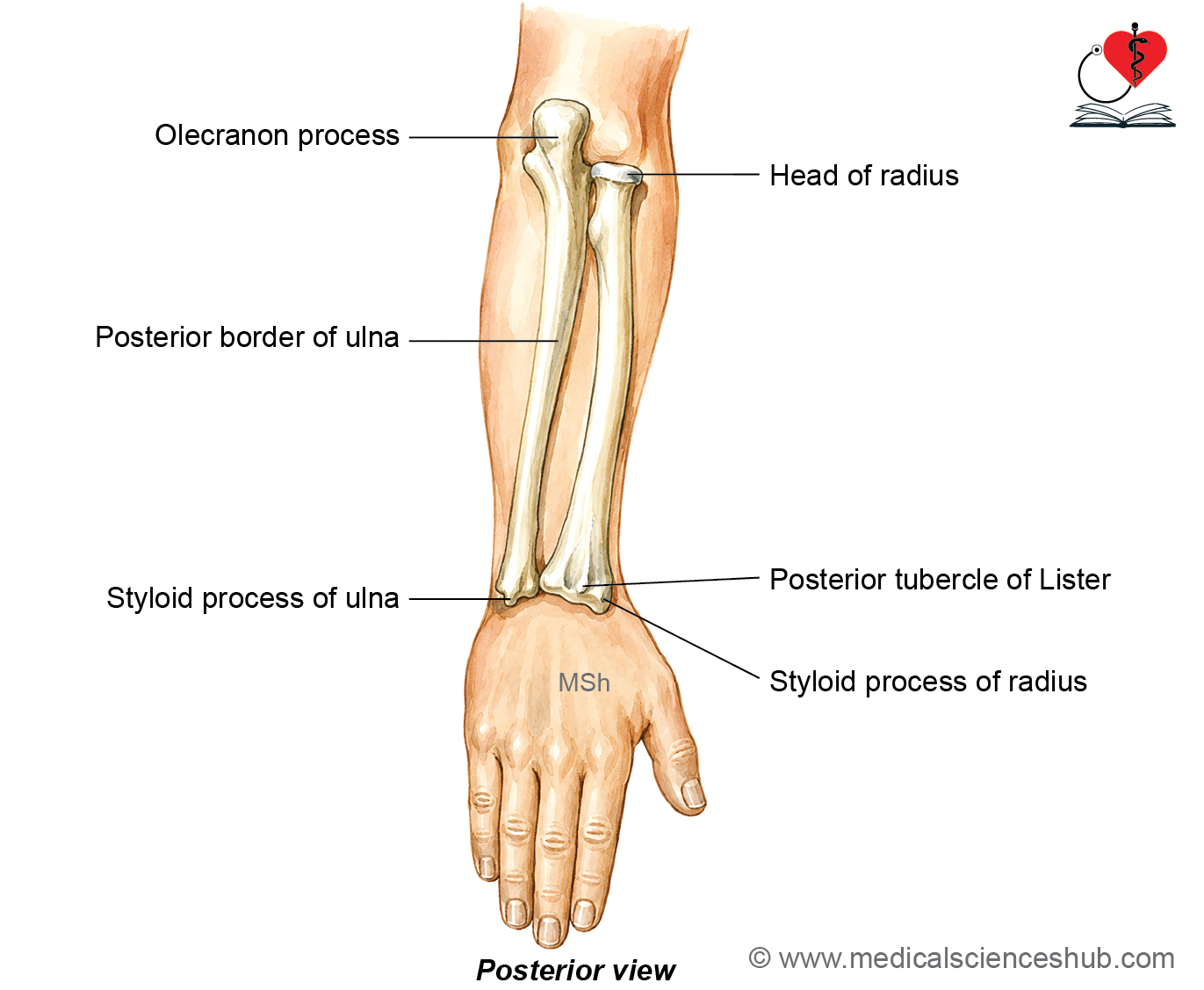
- Olecranon process: The olecranon forms the most prominent bony landmark on the posterior aspect of the elbow and is easily palpable.
- Posterior border of the ulna: This sharp subcutaneous border can be felt throughout its entire length. It extends from the apex of the triangular area over the olecranon to the base of the ulnar styloid process.
- Styloid process of the ulna: This bony projection is palpable on the medial side of the wrist.
- Head of the radius: With the elbow extended, the radial head can be palpated in the depression on the posterolateral aspect of the elbow. It becomes more distinct during pronation and supination.
- Styloid process of the radius: It is palpable on the lateral aspect of the wrist. The radial styloid projects approximately 1.25 cm distal to the ulnar styloid process.
- Lister’s dorsal tubercle: This palpable prominence is located on the posterior surface of the distal radius. It lies along a vertical line passing through the cleft between the index and middle fingers (second interdigital cleft).
Muscles of Posterior Compartment of Forearm
The muscles on the posterior aspect of the forearm are arranged into superficial and deep groups.
Superficial Group
The superficial muscles are further divided into lateral and posterior groups.
Lateral group
- Brachioradialis
- Extensor carpi radialis longus (ECRL)
- Extensor carpi radialis brevis (ECRB)
Posterior group
- Extensor digitorum (ED)
- Extensor digiti minimi (EDM)
- Extensor carpi ulnaris (ECU)
- Anconeus
Deep Group
The deep extensor muscles include:
- Supinator
- Abductor pollicis longus (APL)
- Extensor pollicis brevis (EPB)
- Extensor pollicis longus (EPL)
- Extensor indicis (EI)
Superficial Muscles
- The superficial extensor muscles of the forearm are arranged into lateral and posterior groups.
- Most of the superficial extensor muscles, namely extensor carpi radialis brevis (ECRB), extensor digitorum (ED), extensor digiti minimi (EDM), and extensor carpi ulnaris (ECU), originate from the lateral epicondyle of the humerus through a common extensor tendon (common extensor origin).
Anconeus
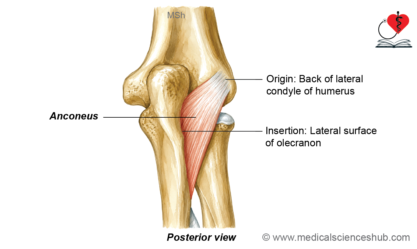
The anconeus is a small, triangular muscle situated on the posterolateral aspect of the elbow, adjacent to the olecranon process. The term anconeus is derived from the Greek word meaning “bend of the elbow.”
Identification
- The muscle is covered by a thick layer of deep fascia.
- Reflection of this fascia is necessary to expose the anconeus during dissection.
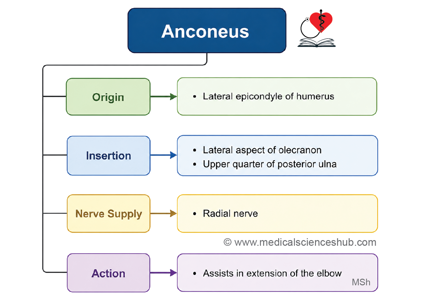
Origin
- Lateral epicondyle of the humerus
Direction of Fibres
- The fibres pass downward and medially.
Insertion
- Lateral surface of the olecranon and the adjoining upper part of the posterior surface of the ulna
Innervation
- Radial nerve (nerve to anconeus), arising from the radial nerve within the spiral groove of the humerus
Actions
- Assists in extension of the elbow joint
- Helps stabilize the elbow during extension
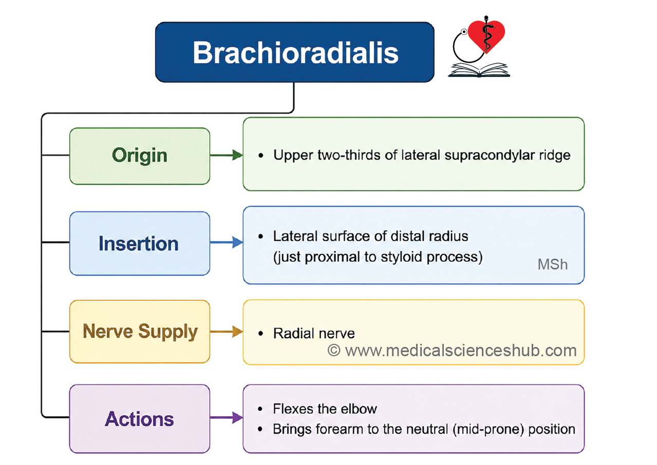
Brachioradialis
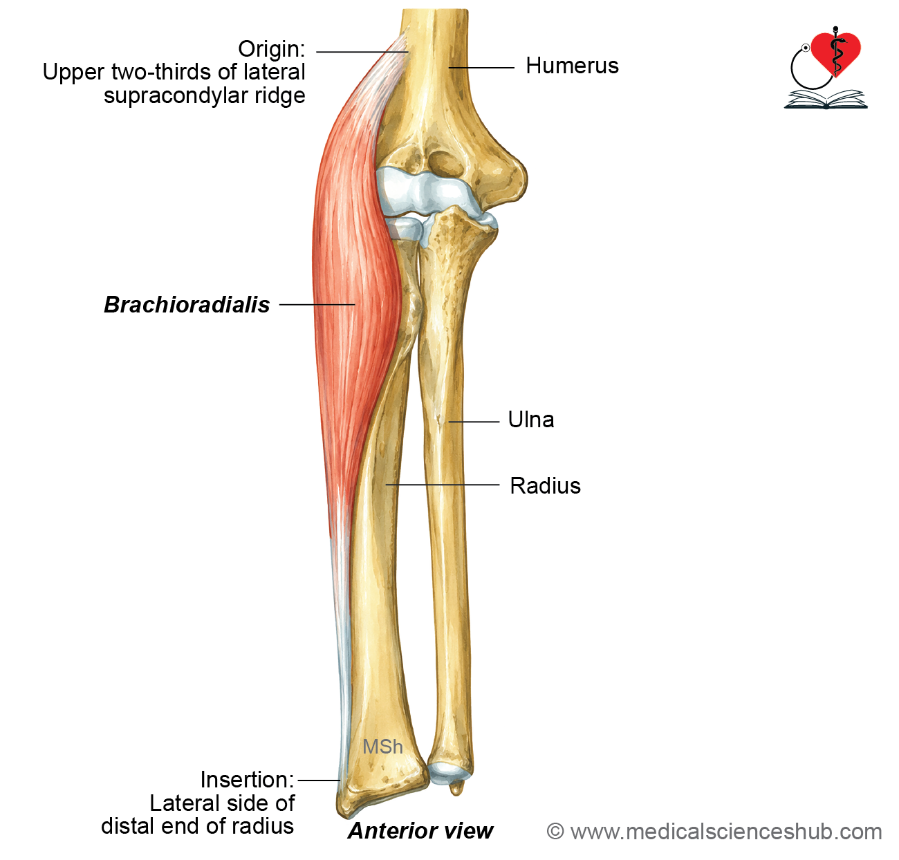
The brachioradialis is a long, fusiform muscle located on the anterolateral aspect of the forearm. Although it belongs to the posterior (extensor) compartment, it acts primarily as a flexor of the elbow. In the cubital fossa, it forms the lateral boundary.
Identification
- The muscle forms a prominent muscular ridge on the anterolateral side of the proximal forearm.
- Its muscle belly overlies the radial nerve.
Origin
- Upper two-thirds of the lateral supracondylar ridge of the humerus
Direction of Fibres
- The fibres descend vertically to form a thick muscle belly before continuing as a tendon toward the distal radius.
Insertion
- Lateral surface of the distal radius, just proximal to the styloid process
Innervation
- Radial nerve
Actions
- Flexes the forearm at the elbow, especially when the forearm is in the mid-prone (neutral) position.
- Assists in returning the forearm to the mid-prone position from either full pronation or full supination.
CLINICAL ANATOMY
Mobile Wad of Henry
- The Mobile Wad of Henry consists of three muscles on the lateral side of the posterior compartment of the forearm:
- Brachioradialis
- Extensor carpi radialis longus (ECRL)
- Extensor carpi radialis brevis (ECRB)
- These muscles form a mobile muscular mass along the lateral forearm. The brachioradialis acts as an elbow flexor, while ECRL and ECRB also assist in elbow flexion in addition to extending and abducting the wrist.
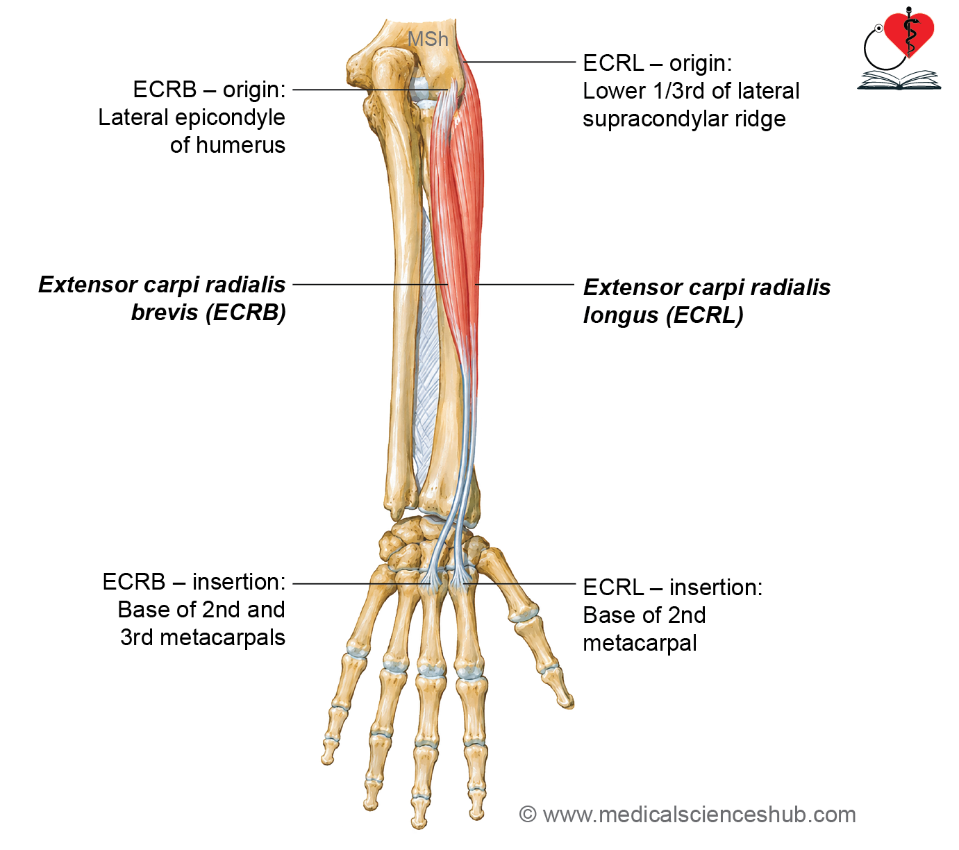
Extensor Carpi Radialis Longus (ECRL)
The extensor carpi radialis longus (ECRL) is a long, fusiform muscle situated on the lateral side of the posterior compartment of the forearm. In the proximal forearm, it lies posterolateral to the brachioradialis and is partially covered by it.
Identification
- The muscle is partly concealed by the brachioradialis in the upper forearm.
- It gives rise to a long, slender tendon that becomes prominent in the distal forearm.
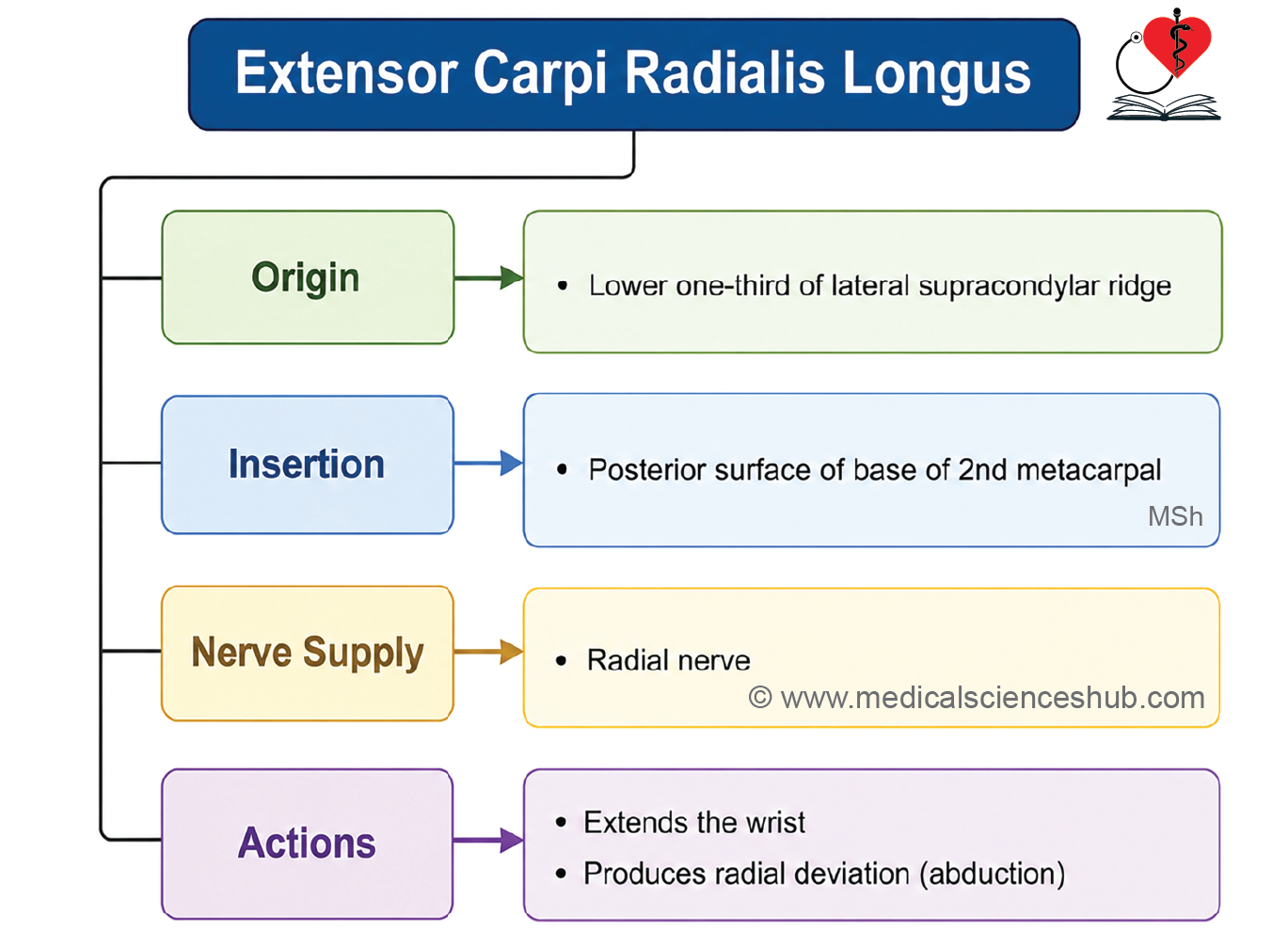
Origin
- Lower one-third of the lateral supracondylar ridge of the humerus
Direction of Fibres
- The fibres descend to form a muscle belly in the proximal forearm, which continues as a narrow tendon in the distal forearm.
Insertion
- Posterior surface of the base of the second metacarpal
Innervation
- Radial nerve
Actions
- Extends the wrist, acting together with the extensor carpi radialis brevis (ECRB) and extensor carpi ulnaris (ECU).
- Abducts (radially deviates) the hand at the wrist, in association with the flexor carpi radialis (FCR) and extensor carpi radialis brevis (ECRB).
Extensor Carpi Radialis Brevis (ECRB)
The extensor carpi radialis brevis (ECRB) is a short extensor muscle of the posterior compartment of the forearm. It lies deep and slightly medial to the extensor carpi radialis longus (ECRL) and has a smaller muscle belly.
Identification
- The ECRB is partially covered by the ECRL.
- It has a relatively short muscle belly that continues as a long tendon running medial to the tendon of the ECRL.
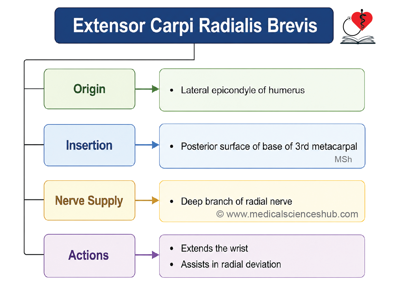
Origin
- Lateral epicondyle of the humerus through the common extensor tendon
Direction of Fibres
- The fibres form a short muscular belly that narrows into a long tendon in the distal forearm.
Insertion
- Posterior surface of the base of the third metacarpal
Innervation
- Posterior interosseous nerve (continuation of the deep branch of the radial nerve)
Actions
- Extends the wrist, acting with the extensor carpi radialis longus (ECRL) and extensor carpi ulnaris (ECU).
- Abducts (radially deviates) the hand at the wrist, together with the flexor carpi radialis (FCR) and extensor carpi radialis longus (ECRL).
Extensor Digitorum (Extensor Digitorum Communis)
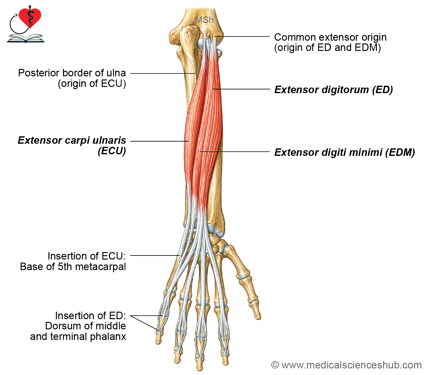
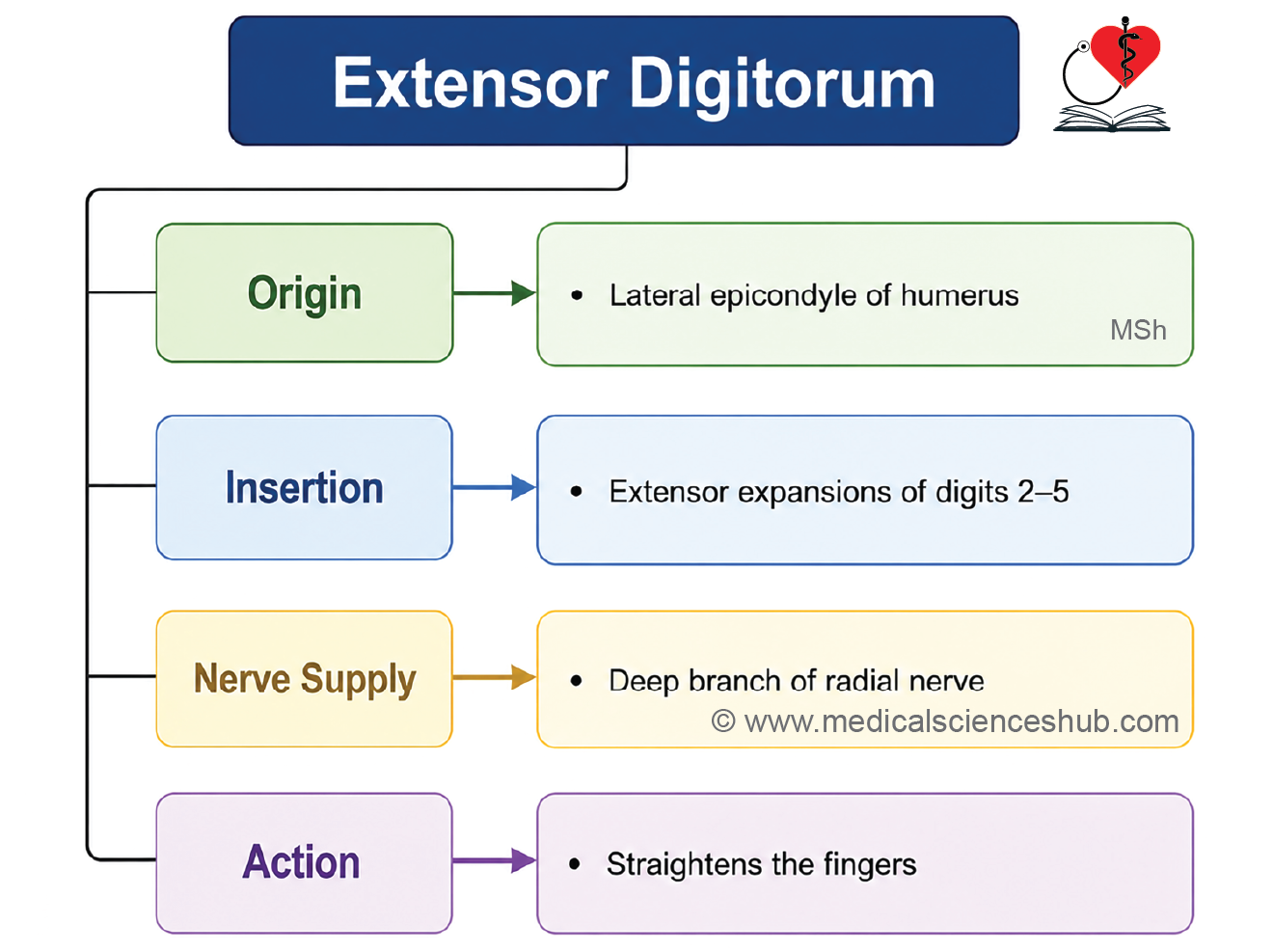
The extensor digitorum is the largest and most prominent muscle of the superficial extensor group of the forearm. It gives rise to four tendons that extend to the medial four digits.
Identification
- The muscle has a broad, bulky belly in the proximal forearm.
- In the distal forearm, it divides into four tendons, one for each of the medial four fingers.
Origin
- Lateral epicondyle of the humerus through the common extensor tendon
Direction of Fibres
- The muscle fibres converge distally to form four tendons that pass beneath the extensor retinaculum before spreading across the dorsum of the hand.
Insertion
- One tendon is distributed to each of the index, middle, ring, and little fingers.
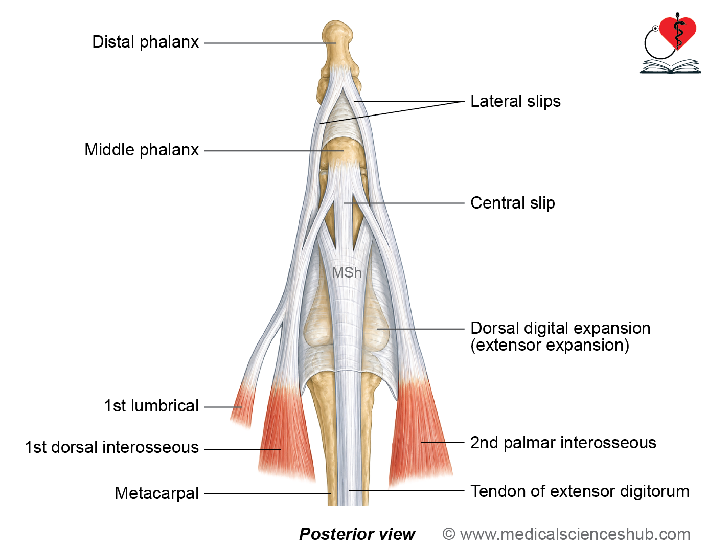
- Each tendon expands to form the dorsal digital (extensor) expansion, through which it is attached to the base of the middle phalanx and the base of the distal phalanx.
Innervation
- Posterior interosseous nerve (continuation of the deep branch of the radial nerve)
Actions
- Extends the medial four fingers at the metacarpophalangeal joints.
- Assists in extension of the interphalangeal joints through the dorsal digital expansion.
- Helps extend the wrist.
Extensor Digiti Minimi (EDM)
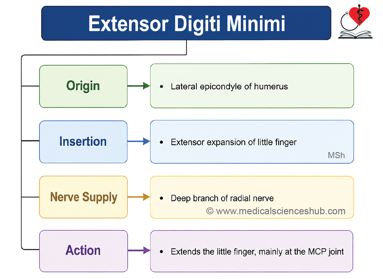
The extensor digiti minimi (EDM), also known as the extensor digiti quinti proprius, is a slender muscle of the superficial extensor compartment of the forearm. It lies medial to the extensor digitorum and is often regarded as a specialized muscle that has separated from it.
Identification
- A thin, narrow muscle situated on the medial side of the extensor digitorum.
- Its tendon is dedicated exclusively to the little finger.
Origin
- Lateral epicondyle of the humerus through the common extensor tendon
Insertion
- Dorsal digital (extensor) expansion of the little finger
Innervation
- Posterior interosseous nerve (continuation of the deep branch of the radial nerve)
Actions
- Extends the little finger, primarily at the metacarpophalangeal joint.
- Assists in extension of the interphalangeal joints through the dorsal digital expansion.
- Contributes to wrist extension along with the other extensor muscles.
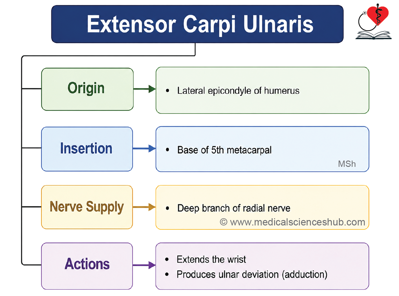
Extensor Carpi Ulnaris
The extensor carpi ulnaris (ECU) is the most medial long muscle of the superficial extensor compartment of the forearm. It extends along the ulnar side of the forearm and terminates as a tendon that passes behind the ulnar styloid process.
Identification
- The ECU occupies the medial border of the posterior forearm.
- Its tendon becomes prominent in the distal forearm and passes posterior to the ulnar styloid process beneath the extensor retinaculum.
Origin
- Lateral epicondyle of the humerus through the common extensor tendon
Direction of Fibres
- The muscle fibres descend distally and converge into a tendon that courses behind the ulnar styloid process before reaching the hand.
Insertion
- Medial aspect of the base of the fifth metacarpal
Innervation
- Posterior interosseous nerve (continuation of the deep branch of the radial nerve)
Actions
- Extends the wrist, acting together with the extensor carpi radialis longus (ECRL) and extensor carpi radialis brevis (ECRB).
- Adducts (ulnar deviates) the hand at the wrist, in conjunction with the flexor carpi ulnaris (FCU).
Deep Muscles
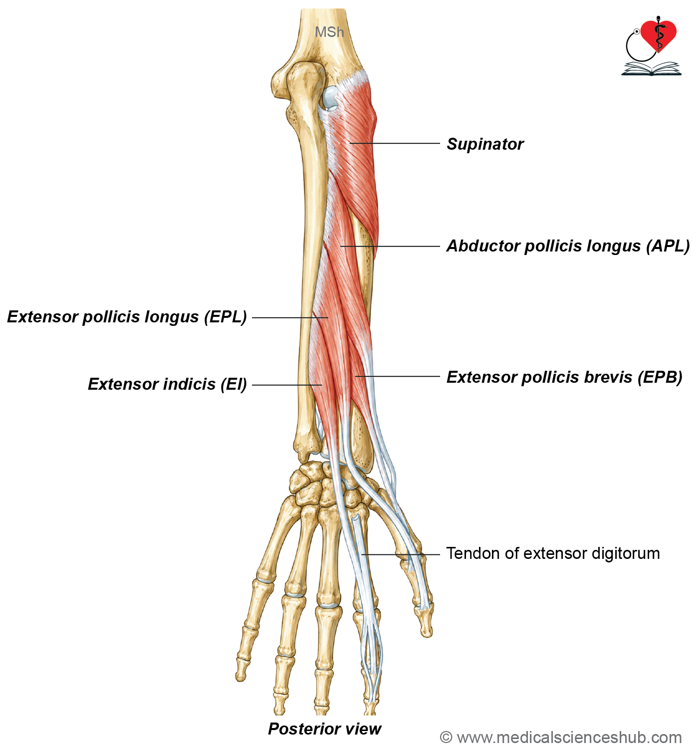
- There are five deep muscles on the back of forearm:
- Supinator
- Abductor pollicis longus (APL)
- Extensor pollicis brevis (EPB)
- Extensor pollicis longus (EPL)
- Extensor indicis (EI)
Abductor Pollicis Longus (APL)
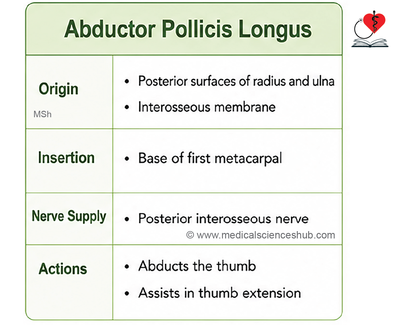
The abductor pollicis longus (APL) is a fusiform muscle of the deep extensor compartment of the forearm. Its muscle belly lies beneath the superficial extensor muscles, while its tendon emerges in the distal forearm to form the lateral boundary of the anatomical snuffbox.
Origin
- Posterior surface of the ulna
- Posterior surface of the interosseous membrane
- Posterior surface of the radius
Direction of Fibres
- The fibres run obliquely downward and laterally, continuing as a tendon that passes beneath the extensor retinaculum on the lateral side of the distal radius.
Insertion
- Lateral aspect of the base of the first metacarpal
Innervation
- Posterior interosseous nerve (continuation of the deep branch of the radial nerve)
Actions
- Abducts the thumb at the carpometacarpal joint.
- Assists in extension of the thumb at the carpometacarpal joint.
Clinical Testing
- Ask the patient to abduct the thumb against resistance.
- A normally functioning APL tendon becomes prominent along the lateral boundary of the anatomical snuffbox.
Extensor Pollicis Brevis (EPB)
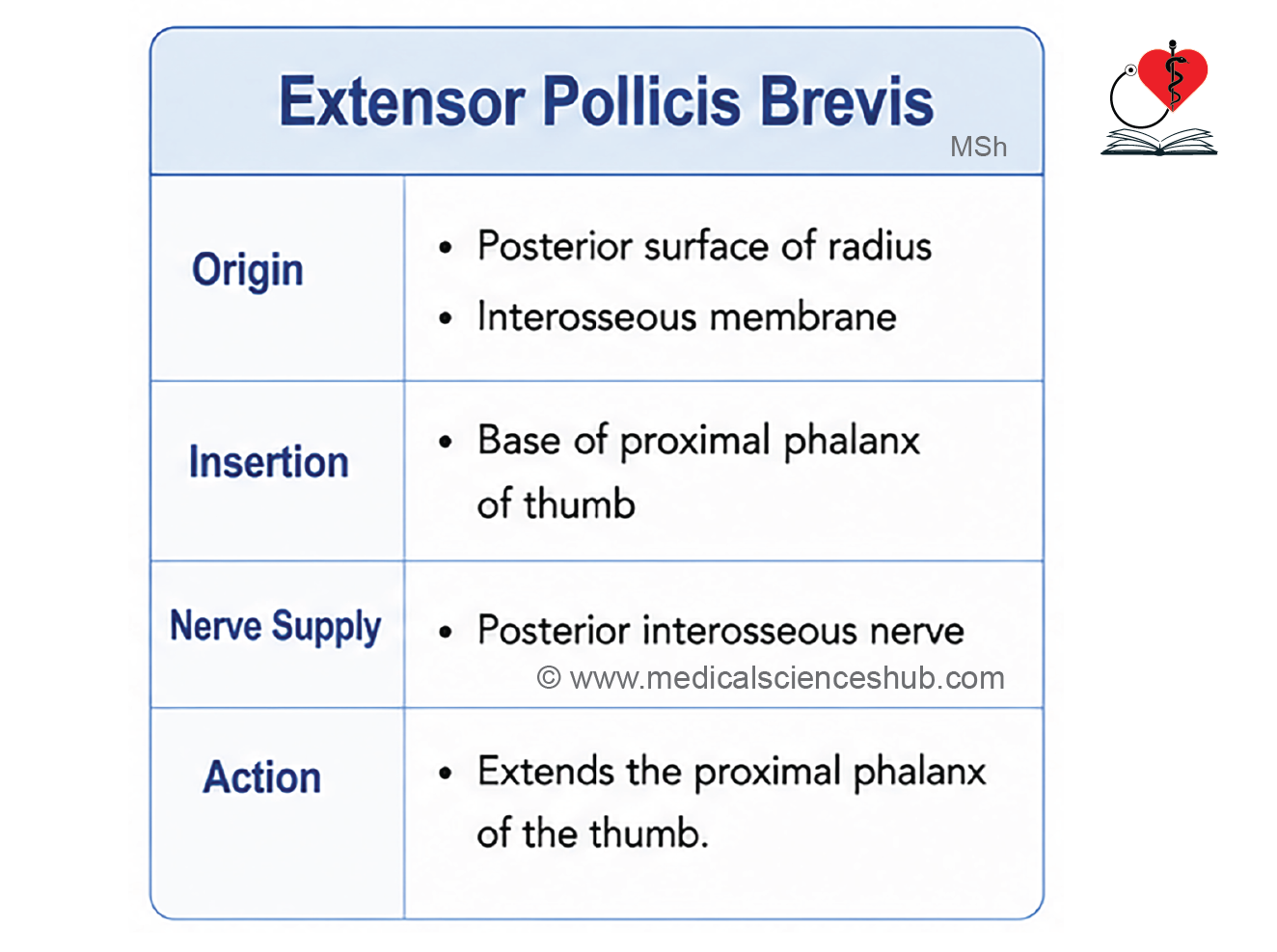
The extensor pollicis brevis (EPB) is a small fusiform muscle of the deep extensor compartment of the forearm. It lies deep to the superficial extensor muscles and accompanies the abductor pollicis longus (APL) in the distal forearm.
Origin
- Posterior surface of the radius, inferior to the origin of the abductor pollicis longus
- Adjacent posterior surface of the interosseous membrane
Direction of Fibres
- The fibres run obliquely downward and laterally, following a course similar to that of the abductor pollicis longus before continuing as a tendon.
Insertion
- Dorsal surface of the base of the proximal phalanx of the thumb
Innervation
- Posterior interosseous nerve (continuation of the deep branch of the radial nerve)
Actions
- Extends the proximal phalanx of the thumb at the metacarpophalangeal joint.
- Assists in extension of the first metacarpal at the carpometacarpal joint.
Clinical Testing
- Ask the patient to extend the metacarpophalangeal joint of the thumb against resistance.
- The EPB tendon becomes prominent along the lateral (radial) boundary of the anatomical snuffbox.
Dorsal digital expansion
The dorsal digital expansion is a specialized triangular aponeurotic structure on the dorsum of each finger. It is also known as the extensor hood, extensor expansion, or dorsal aponeurosis. It serves as the insertion site for the extensor tendons and provides attachment to the intrinsic muscles of the hand, thereby coordinating finger movements.
Location
- Situated on the dorsal aspect of the proximal phalanx of each finger.
- Extends over the metacarpophalangeal (MCP) joint and continues distally toward the middle and distal phalanges.
Shape
- Thin, triangular aponeurosis.
Parts of the Dorsal Digital Expansion
Base
- Broad proximal part directed upwards over the metacarpophalangeal joint.
- Receives the tendon of the extensor digitorum in its central portion.
Posterolateral Margins
- Provide attachment for the lumbrical and interosseous muscles.
Apex
- Narrow distal end directed toward the fingertip.
- Distally, the expansion divides into:
- Central (middle) slip, which inserts into the base of the middle phalanx.
- Two lateral bands, which reunite distally and insert into the base of the distal phalanx.
Muscular Attachments
| Finger | Lateral (Radial) Attachment | Medial (Ulnar) Attachment |
| Index | First lumbrical, first dorsal interosseous | First palmar interosseous |
| Middle | Second lumbrical, second dorsal interosseous | Third dorsal interosseous |
| Ring | Third lumbrical, fourth dorsal interosseous | Second palmar interosseous |
| Little | Fourth lumbrical | Extensor digiti minimi and third palmar interosseous |
Functions
The dorsal digital expansion acts as a common insertion for both the extrinsic extensor muscles and the intrinsic muscles of the hand, allowing coordinated finger movements.
Its functions include:
- Integrates the actions of the extensor digitorum, lumbricals, and interossei.
- Redistributes the pull of these muscles to produce smooth and coordinated finger movements.
- Alters the direction of muscle force, improving the efficiency of finger extension.
Through the dorsal digital expansion:
- Lumbricals flex the metacarpophalangeal joints while extending the proximal and distal interphalangeal joints.
- Interossei assist in the same combined movement.
- Extensor digitorum extends the metacarpophalangeal joints and, through the expansion, contributes to extension of the interphalangeal joints.
CLINICAL CORRELATION
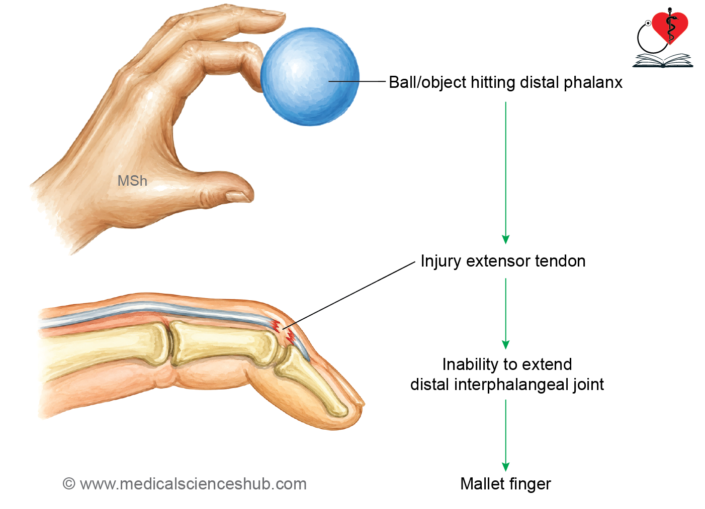
Mallet Finger
- Also known as baseball finger, cricketer’s finger, or hammer finger.
- Usually results from a forceful blow to the tip of an extended finger, causing sudden flexion of the distal phalanx.
- The injury avulses or ruptures the terminal tendon of the extensor digitorum from its insertion on the distal phalanx.
- The patient is unable to actively extend the distal interphalangeal (DIP) joint, resulting in a flexed fingertip.
Busch Fracture
- An avulsion fracture involving the base of the distal phalanx, where the terminal extensor tendon remains attached to the avulsed bony fragment.
- It represents the bony variant of a mallet finger injury.
Extensor Pollicis Longus
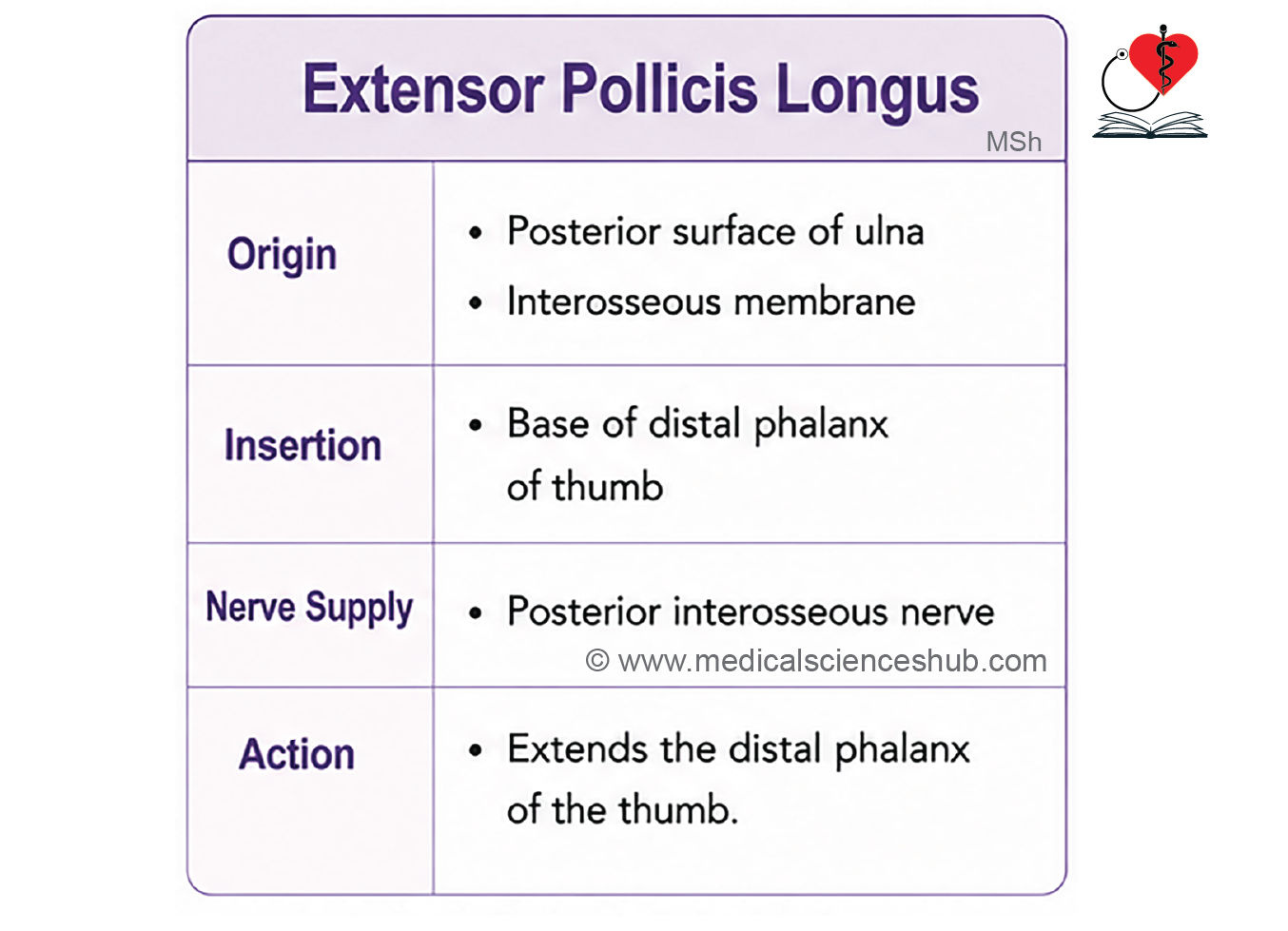
The extensor pollicis longus (EPL) is a long, fusiform muscle of the deep extensor compartment of the forearm. It is larger and longer than the extensor pollicis brevis (EPB) and is the principal extensor of the distal phalanx of the thumb.
Origin
- Posterior surface of the ulna, inferior to the origin of the abductor pollicis longus
- Adjacent posterior surface of the interosseous membrane
Direction of Fibres
- The fibres descend to form a fusiform muscle belly that continues as a long tendon.
- The tendon curves around Lister’s tubercle, changing the direction of pull before reaching the thumb.
Insertion
- Posterior surface of the base of the distal phalanx of the thumb
Innervation
- Posterior interosseous nerve (continuation of the deep branch of the radial nerve)
Actions
- Extends the distal phalanx of the thumb at the interphalangeal joint.
- Assists in extension of the proximal phalanx at the metacarpophalangeal joint.
- Contributes to extension of the first metacarpal at the carpometacarpal joint.
CLINICAL ANATOMY
Clinical Testing
- Ask the patient to extend the interphalangeal joint of the thumb against resistance.
- A functioning EPL tendon becomes prominent along the medial boundary of the anatomical snuffbox.
Extensor Indicis
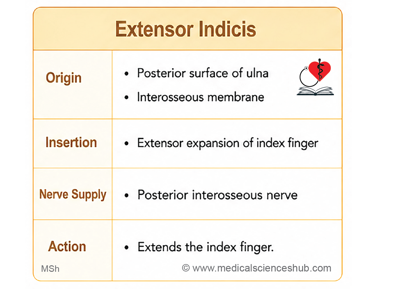
The extensor indicis (EI) is a slender muscle of the deep extensor compartment of the forearm. It provides an additional extensor tendon to the index finger, allowing it to move independently of the other fingers.
Identification
- The extensor indicis is a thin, fusiform muscle located deep to the superficial extensor muscles.
- Its tendon passes beneath the extensor retinaculum through the fourth dorsal compartment, deep to the tendons of the extensor digitorum.
- On the dorsum of the hand, the tendon lies medial (ulnar) to the extensor digitorum tendon destined for the index finger.
Origin
- Posterior surface of the shaft of the ulna, inferior to the origin of the extensor pollicis longus
- Adjacent posterior surface of the interosseous membrane
Direction of Fibres
- The fibres form a small fusiform muscle belly that continues as a long tendon passing beneath the extensor retinaculum before reaching the index finger.
Insertion
- Dorsal digital (extensor) expansion of the index finger
Innervation
- Posterior interosseous nerve (continuation of the deep branch of the radial nerve)
Actions
- Produces independent extension of the index finger, particularly at the metacarpophalangeal joint.
- Assists in extension of the interphalangeal joints through the dorsal digital expansion.
- Works with the extensor digitorum during extension of the index finger.
CLINICAL ANATOMY
Clinical Testing
- Ask the patient to extend the index finger while keeping the remaining fingers flexed.
- Isolated extension of the index finger demonstrates the integrity of the extensor indicis and the posterior interosseous nerve.
Supinator
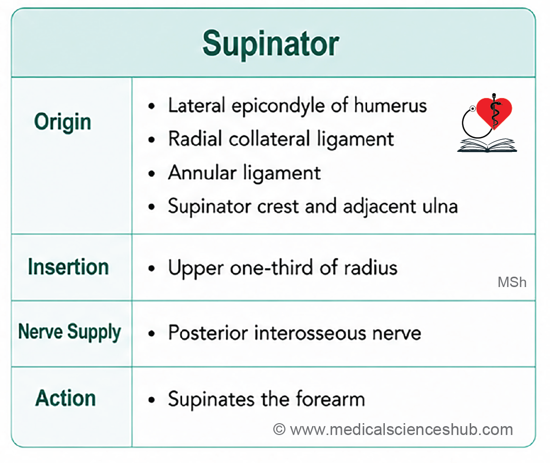
The supinator is a broad, sheet-like muscle of the deep extensor compartment of the forearm. It surrounds the upper part of the radius and is the principal muscle responsible for supination of the forearm, particularly when the elbow is extended.
Origin
From the humerus
- Lateral epicondyle of the humerus
From the elbow joint
- Annular ligament of the superior radioulnar joint
From the ulna
- Supinator crest
- Triangular area (supinator fossa) anterior to the supinator crest
Direction of Fibres
- The fibres wind spirally around the upper part of the radius from medial to lateral before inserting onto the bone.
Insertion
- Lateral, anterior, and posterior surfaces of the upper one-third of the radius
Innervation
- Deep branch of the radial nerve (before it continues as the posterior interosseous nerve)
Note: Although many textbooks describe the nerve supply as the deep branch of the radial nerve, the terminal part of this nerve continues as the posterior interosseous nerve after emerging from the supinator.
Actions
- Produces supination of the forearm, especially during slow or unresisted movements.
- Acts regardless of the position of the elbow joint.
Functional Note
- The supinator can supinate the forearm whether the elbow is flexed or extended.
- The biceps brachii is a powerful supinator only when the elbow is partially flexed.
CLINICAL CORRELATION
- The deep branch of the radial nerve may become compressed as it passes through the supinator (Arcade of Frohse), producing posterior interosseous nerve syndrome, characterized by weakness of finger and thumb extension without sensory loss.
Extensor Retinaculum of Hand
The extensor retinaculum is a thickened transverse band of deep fascia located across the posterior aspect of the distal forearm. It secures the extensor tendons as they pass from the forearm to the dorsum of the hand.
Features
- Width: Approximately 2.5 cm
- Direction: Runs obliquely downward and medially across the back of the wrist.
Osteofibrous Compartments
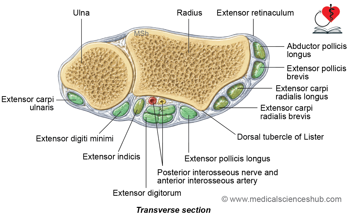
Fibrous septa extend from the deep surface of the extensor retinaculum to the radius and ulna, dividing the space beneath it into six osteofibrous compartments. These compartments are numbered from lateral (radial) to medial (ulnar).
| Compartment | Structures Passing Through |
| First | Abductor pollicis longus (APL), Extensor pollicis brevis (EPB) |
| Second | Extensor carpi radialis longus (ECRL), Extensor carpi radialis brevis (ECRB) |
| Third | Extensor pollicis longus (EPL) |
| Fourth | Extensor digitorum (ED), Extensor indicis (EI), Posterior interosseous nerve, Posterior interosseous artery* |
| Fifth | Extensor digiti minimi (EDM) |
| Sixth | Extensor carpi ulnaris (ECU) |
*The posterior interosseous nerve and artery reach the dorsum of the wrist in association with the fourth compartment.
Synovial Sheaths
- The extensor tendons are enclosed within synovial tendon sheaths, which reduce friction and facilitate smooth tendon movement beneath the extensor retinaculum.
Functions
- Prevents bowstringing of the extensor tendons during wrist and finger extension.
- Maintains the tendons close to the underlying bones, thereby improving the efficiency of muscle action.
- Provides a smooth fibro-osseous tunnel for the passage of the extensor tendons.
CLINICAL CORRELATION
- De Quervain’s tenosynovitis results from inflammation of the tendons within the first dorsal compartment (abductor pollicis longus and extensor pollicis brevis), causing pain over the radial side of the wrist.
Nerve and Vessels of Posterior Compartment of Forearm
Posterior Interosseous Nerve
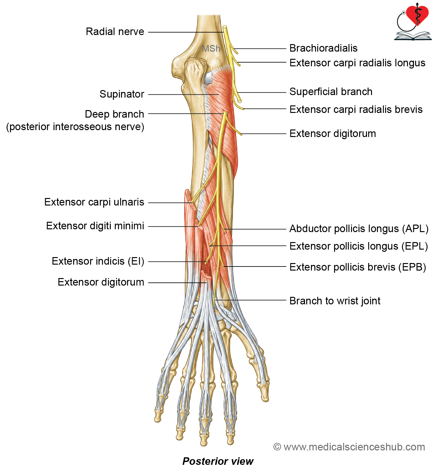
The posterior interosseous nerve (PIN) is the principal motor nerve of the posterior compartment of the forearm. It is the continuation of the deep branch of the radial nerve after it emerges from the supinator muscle.
Beginning
- The posterior interosseous nerve begins as the deep branch of the radial nerve in the cubital fossa, beneath the cover of the brachioradialis.
Course and Relations
- From the cubital fossa, the deep branch of the radial nerve passes posteriorly by entering the supinator muscle.
- Within the supinator, it traverses between the superficial and deep layers of the muscle.
- On emerging from the distal border of the supinator, it is known as the posterior interosseous nerve.
- It descends on the posterior surface of the interosseous membrane, deep to the superficial extensor muscles and in close relation to the posterior interosseous artery.
- Distally, the nerve reaches the dorsum of the wrist by passing beneath the extensor retinaculum, in association with the fourth dorsal compartment.
- Note: The nerve does not enter the palm.
Termination
- Near the wrist, the nerve expands into a pseudoganglion (a fusiform enlargement without nerve cell bodies).
- It terminates by supplying the wrist joint, intercarpal joints, and adjacent ligaments.
Branches
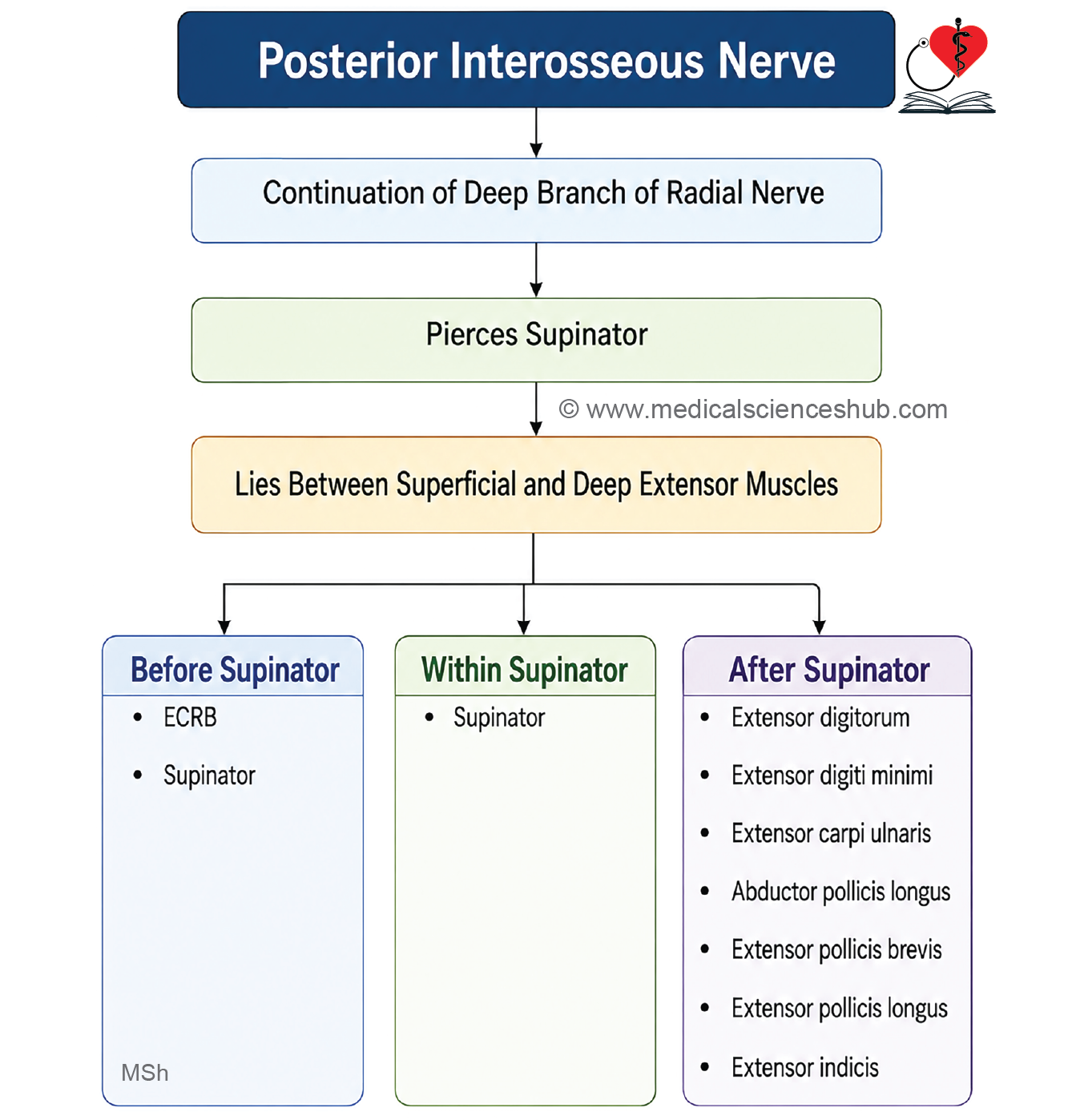
Before entering the supinator
- Extensor carpi radialis brevis (ECRB)
- Supinator
Within the supinator
- Muscular branches to the supinator
After emerging from the supinator
- Extensor digitorum
- Extensor digiti minimi
- Extensor carpi ulnaris
- Abductor pollicis longus
- Extensor pollicis brevis
- Extensor pollicis longus
- Extensor indicis
Distribution
- Supplies all muscles of the posterior compartment of the forearm except:
- Brachioradialis
- Extensor carpi radialis longus
- Anconeus
- These three muscles receive direct branches from the radial nerve.
CLINICAL CORRELATION
Posterior Interosseous Nerve Syndrome
- Compression of the posterior interosseous nerve, commonly within the supinator (especially at the Arcade of Frohse), produces weakness or paralysis of the finger and thumb extensors.
- Wrist extension is usually preserved but may show radial deviation because the extensor carpi radialis longus remains functional.
- Since the posterior interosseous nerve is a pure motor nerve, sensory loss is absent.
Posterior Interosseous Artery
The posterior interosseous artery is the principal artery supplying the posterior compartment of the forearm. It is a branch of the common interosseous artery, which arises from the ulnar artery.
Beginning
- Arises as the smaller terminal branch of the common interosseous artery in the proximal forearm.
Course
- Shortly after its origin, the artery enters the posterior compartment by passing above the upper border of the interosseous membrane through the interval between the oblique cord and the proximal margin of the interosseous membrane.
- It descends between the supinator and abductor pollicis longus muscles.
- Throughout most of its course, it accompanies the posterior interosseous nerve on the posterior surface of the interosseous membrane.
- Distally, it continues toward the dorsum of the wrist beneath the extensor muscles.
Termination
- Ends in the distal forearm by anastomosing with the anterior interosseous artery, contributing to the arterial network around the wrist.
Branches
- Muscular branches supplying the extensor muscles of the posterior compartment of the forearm.
- Interosseous recurrent artery, which ascends to participate in the periarticular arterial anastomosis around the elbow.
- Small branches to the wrist and adjacent structures.
CLINICAL CORRELATION
- The posterior interosseous artery accompanies the posterior interosseous nerve; therefore, injuries or surgical procedures in the posterior forearm may affect both structures.
- Its anastomosis with the anterior interosseous artery provides an important collateral pathway for maintaining blood supply to the distal forearm and wrist.
Anatomical Snuffbox
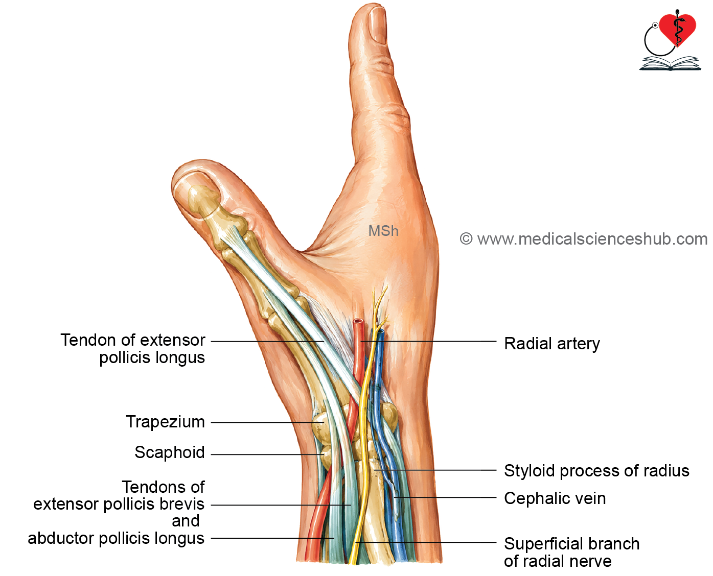
The anatomical snuffbox is a triangular depression located on the posterolateral aspect of the wrist. It becomes prominent when the thumb is fully extended. Historically, this depression was used to hold powdered tobacco (snuff) before it was inhaled, giving rise to its name.
Boundaries
Medial (Ulnar) Border
- Tendon of extensor pollicis longus (EPL)
Lateral (Radial) Border
- Tendons of:
- Abductor pollicis longus (APL)
- Extensor pollicis brevis (EPB)
Proximal Boundary
- Styloid process of the radius
Distal End (Apex)
- Formed where the medial and lateral tendon boundaries converge.
Floor
The floor is formed by:
- Scaphoid
- Trapezium
Roof
The roof consists of:
- Skin
- Superficial fascia
- Cephalic vein
- Superficial branch of the radial nerve
Contents
The principal content of the anatomical snuffbox is the:
- Radial artery, which crosses the floor of the snuffbox before entering the palm.
Branches of the Radial Artery in the Snuffbox
Within the anatomical snuffbox, the radial artery gives articular branches that contribute to the blood supply of:
- Wrist joint
- Distal radioulnar joint
- Intercarpal joints
- Intermetacarpal joints
CLINICAL IMPORTANCE
- The radial pulse can be palpated in the anatomical snuffbox.
- Tenderness in this region following a fall on an outstretched hand is highly suggestive of a scaphoid fracture.
- The radial artery can be compressed against the scaphoid to control bleeding from its distal branches.
Important Questions
- Write a short note on brachioradialis muscle.
- Write a short note on dorsal digital expansion.
- Write a short note on supinator.
- Write a short note on extensor retinaculum of hand.
- List the structures passing deep to extensor retinaculum of hand.
- Write a short note on posterior interosseous nerve.
- White a short note on anatomical snuffbox.
📝 Test Your Knowledge – Practice MCQs
Attempt the chapter MCQ quiz and assess your understanding of key concepts.
Click to see: Medical Disclaimer and Copyright Notice