
- The upper limb is supplied by five major peripheral nerves, all of which arise from the brachial plexus.
- These nerves provide motor innervation to the muscles and sensory innervation to the skin and joints of the upper limb.
- The five major nerves of the upper limb are:
- Axillary nerve
- Musculocutaneous nerve
- Radial nerve
- Median nerve
- Ulnar nerve
Axillary Nerve
- The axillary nerve is one of the terminal branches of the posterior cord of the brachial plexus.
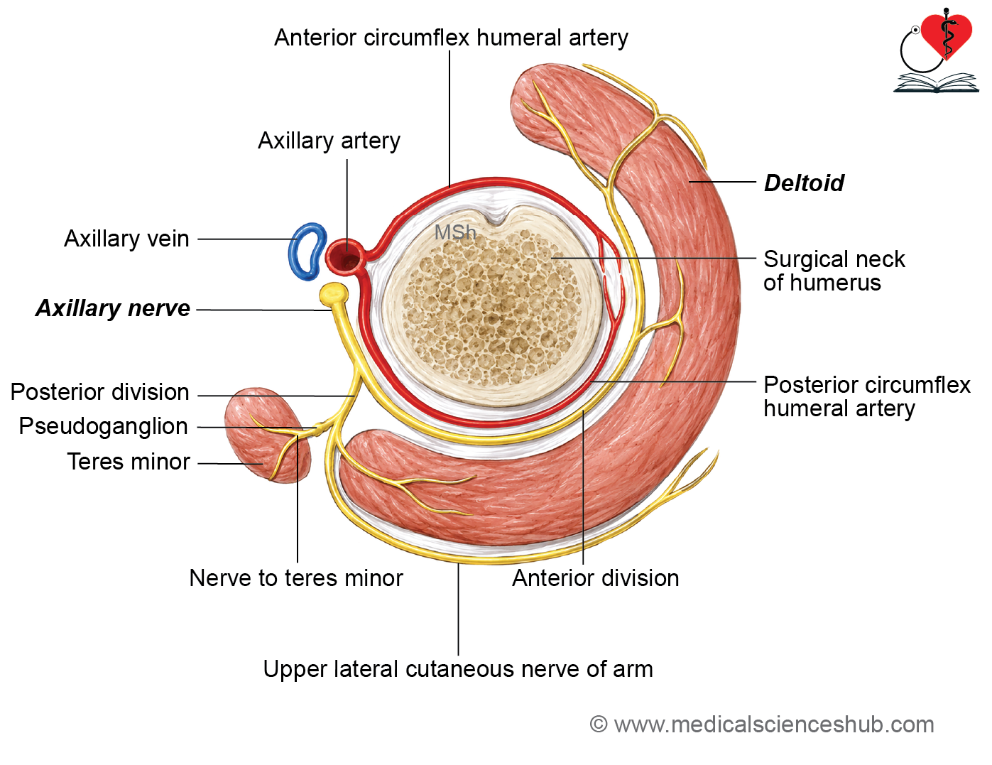
- It is also known as the circumflex nerve because it winds around the surgical neck of the humerus.
- The root value of the axillary nerve is C5 and C6..
Beginning
The axillary nerve arises from the posterior cord of the brachial plexus.
Course and relations
In the Axilla
- The axillary nerve originates from the posterior cord of the brachial plexus.
- It passes posterolateral to the third part of the axillary artery.
- It lies on the anterior surface of the subscapularis muscle.
In the Quadrangular Space
- The axillary nerve leaves the axilla by passing through the quadrangular space.
- It is accompanied by the posterior circumflex humeral artery and vein.
Relations of the Quadrangular Space
- Superior: Subscapularis muscle and the capsule of the shoulder joint
- Inferior: Teres major muscle
- Lateral: Surgical neck of the humerus
- Medial: Long head of the triceps brachii muscle
Termination
- In quadrangular space, axillary nerve divides into anterior and posterior branches Within the quadrangular space, the axillary nerve divides into:
- Anterior branch
- Posterior branch
Branches
Articular Branch
- The axillary nerve gives an articular branch to the shoulder joint.
Anterior Branch
- The anterior branch accompanies the posterior circumflex humeral vessels.
- It winds around the surgical neck of the humerus deep to the deltoid muscle.
- It supplies:
- Deltoid muscle
- Skin over the anteroinferior part of the deltoid region
Posterior Branch
- The posterior branch winds around the posterior border of the deltoid muscle.
- It supplies:
- Teres minor muscle
- Posterior part of the deltoid muscle
- The nerve to the teres minor contains a pseudoganglion, which has no functional significance.
- The posterior branch continues as the upper lateral cutaneous nerve of the arm.
- The upper lateral cutaneous nerve supplies the skin over the lower half of the deltoid region.
CLINICAL INTEGRATION
Axillary Nerve Palsy
Causes
- Fracture of the surgical neck of the humerus
- Dislocation of the shoulder joint
- Improper intramuscular injection into the upper part of the deltoid muscle
Signs and Symptoms
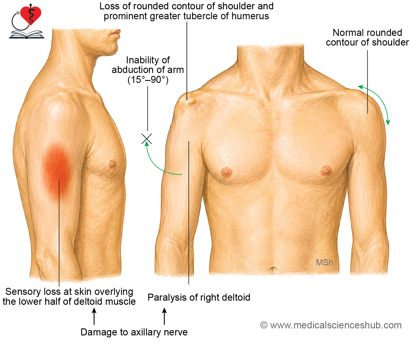
- Inability to abduct the arm from 15° to 90° due to paralysis of the deltoid muscle.
- Loss of the normal rounded contour of the shoulder as a result of deltoid muscle wasting.
- Regimental badge anesthesia, characterized by loss of sensation over the skin covering the lower half of the deltoid region, supplied by the upper lateral cutaneous nerve of the arm.
Musculocutaneous Nerve
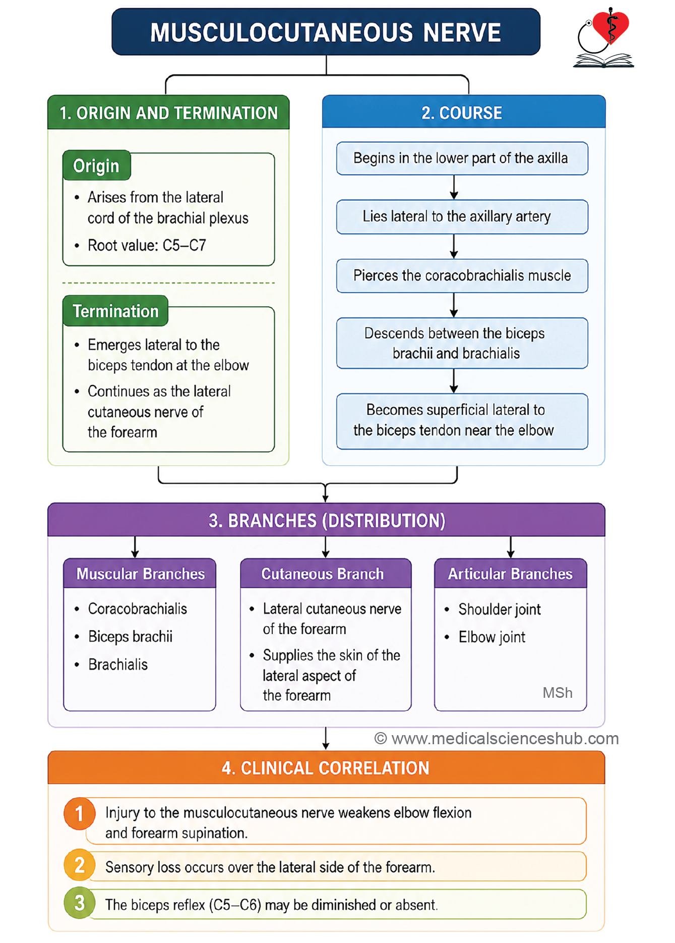
- The musculocutaneous nerve is the main motor nerve of the anterior compartment of the arm.
- It supplies all the muscles in the front of the arm.
Beginning
- The musculocutaneous nerve arises from the lateral cord of the brachial plexus.
- The root value of the musculocutaneous nerve is C5–C7.
- It contains fibers from the ventral primary rami of the fifth, sixth, and seventh cervical spinal nerves.
Termination
- The musculocutaneous nerve terminates by continuing as the lateral cutaneous nerve of the forearm, approximately 2 cm above the elbow.
Course
- The musculocutaneous nerve arises from the lateral cord of the brachial plexus in the lower part of the axilla.
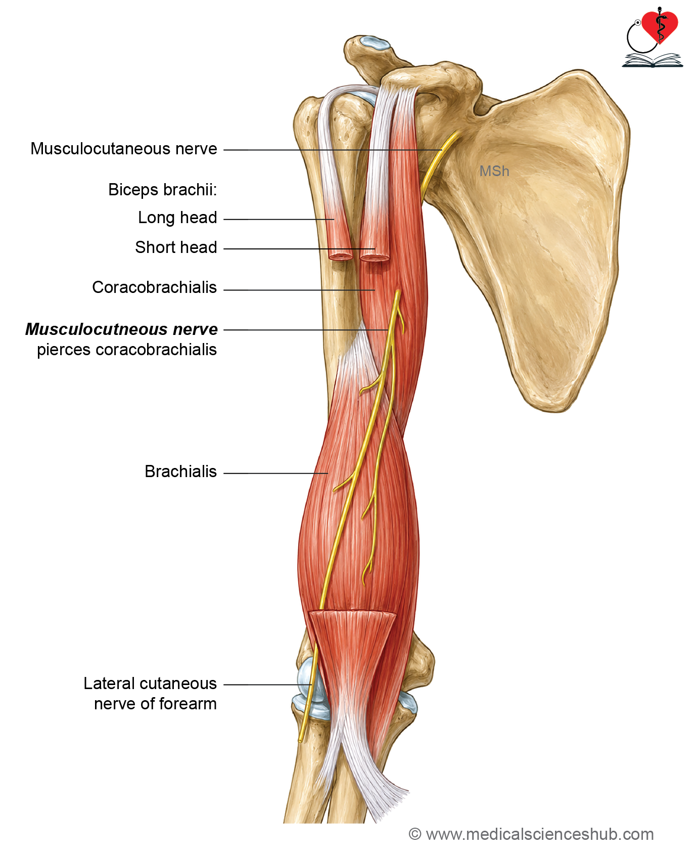
- It pierces the coracobrachialis muscle, which is an important identifying feature.
- It then descends downward and laterally between the biceps brachii and brachialis muscles.
- About 2 cm above the elbow, it emerges lateral to the tendon of the biceps brachii, pierces the deep fascia, and continues as the lateral cutaneous nerve of the forearm.
Relations
In the Lower Part of the Axilla
- Anterior: Pectoralis major muscle
- Posterior: Subscapularis muscle
- Medial: Axillary artery
- Lateral: Coracobrachialis muscle
In the Upper Part of the Arm
- The musculocutaneous nerve lies within the substance of the coracobrachialis muscle after piercing it.
In the Middle Part of the Arm
- The nerve lies between the biceps brachii and brachialis muscles.
- It pierces the deep fascia approximately 2 cm above the elbow joint.
Branches and distribution
Muscular Branches
- The musculocutaneous nerve supplies the following muscles:
- Coracobrachialis
- Biceps brachii
- Brachialis (mainly its large medial part)
Cutaneous Branch
- The terminal lateral cutaneous nerve of the forearm supplies the skin over the lateral aspect of the forearm.
Articular Branches
- The musculocutaneous nerve gives articular branches to:
- Shoulder joint
- Elbow joint
CLINICAL INTEGRATION
Injury to the Musculocutaneous Nerve
Effects
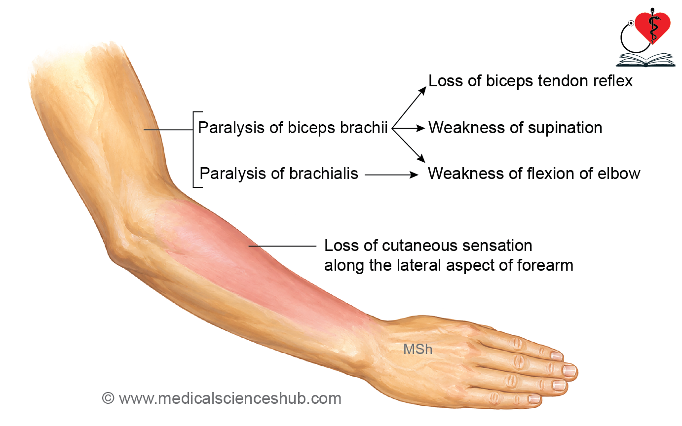
- Paralysis of the biceps brachii results in:
- Weakness of forearm supination
- Loss or reduction of the biceps tendon reflex
- Paralysis of the brachialis muscle causes weakness of elbow flexion.
- Loss of cutaneous sensation occurs over the lateral aspect of the forearm due to involvement of the lateral cutaneous nerve of the forearm.
Radial Nerve
- The radial nerve, also known as the musculospiral nerve, is the main nerve of the posterior compartment of the arm and forearm.
- It also provides sensory innervation to the dorsum of the hand.
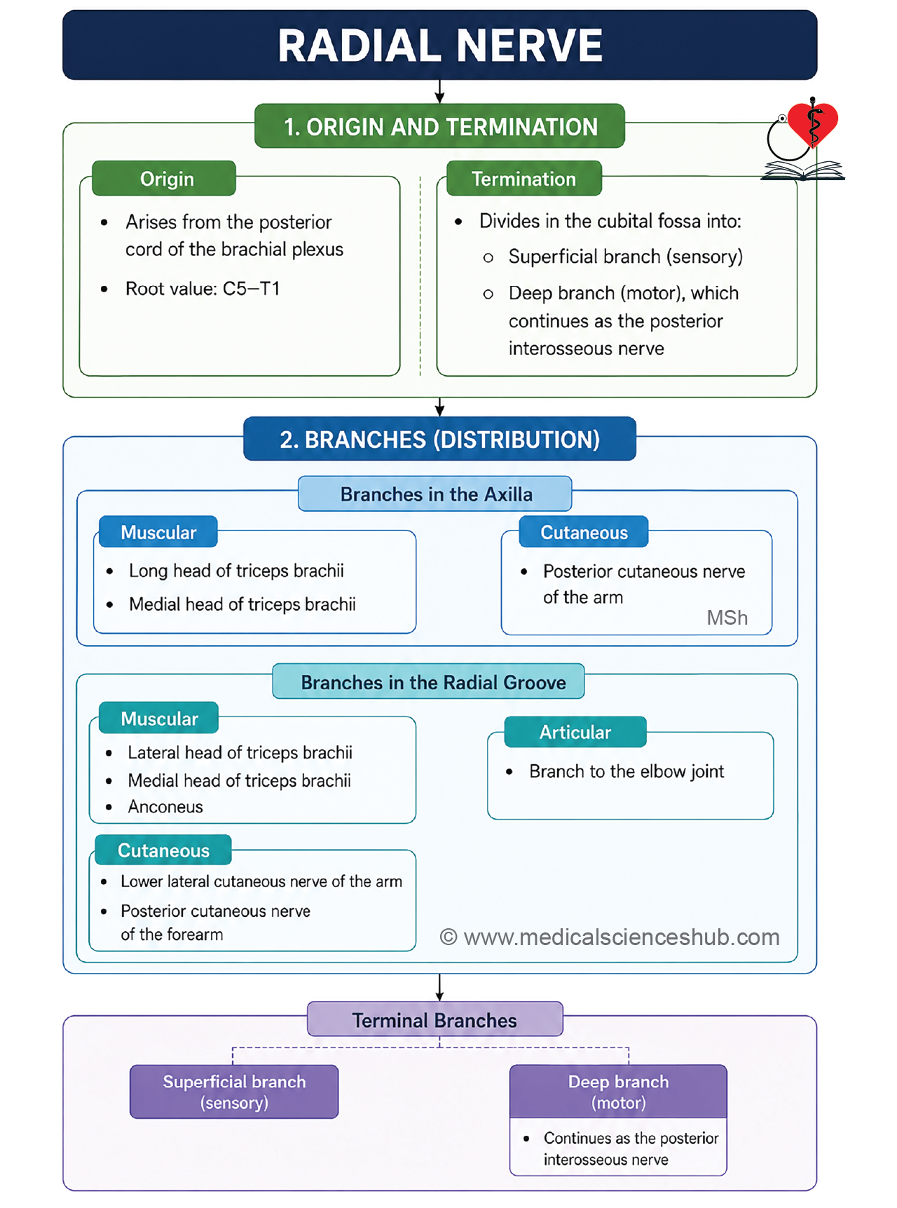
- The radial nerve is the continuation of the posterior cord of the brachial plexus.
- It is the largest terminal branch of the brachial plexus.
- The radial nerve has a root value of C5–T1.
- It contains fibers from the ventral primary rami of C5, C6, C7, C8, and T1 spinal nerves.
Beginning
- The radial nerve arises as the continuation of the posterior cord of the brachial plexus.
- It originates behind the third part of the axillary artery in the lower part of the axilla.
Course and relations
In the Axilla
- The radial nerve passes posterior to the axillary artery.
- It continues for a short distance behind the upper part of the brachial artery.
In the Arm
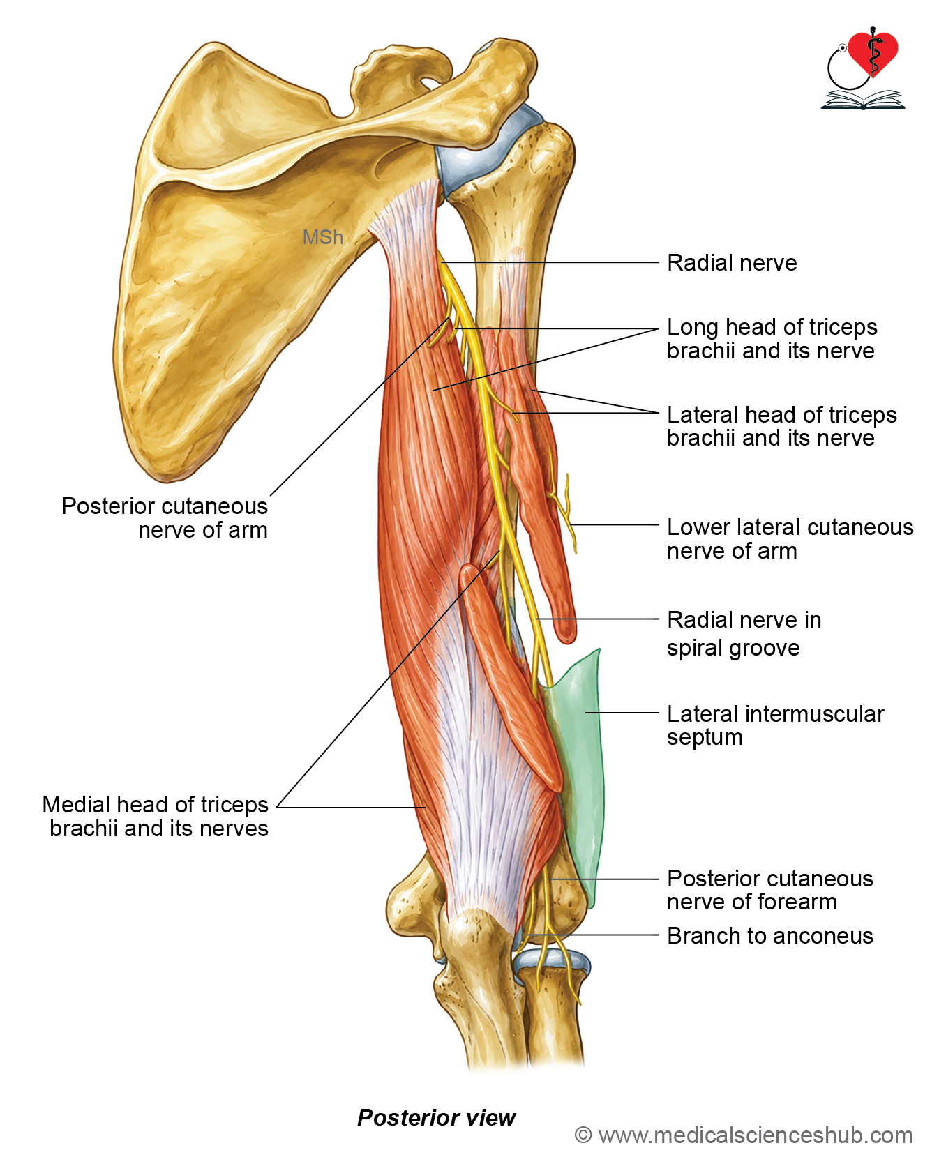
- The radial nerve enters the posterior compartment of the arm through the lower triangular space (triangular interval) together with the profunda brachii artery.
- It descends downward and laterally within the radial groove of the humerus.
- It pierces the lateral intermuscular septum to enter the anterior compartment of the arm.
- In the anterior compartment, it lies deep to the brachioradialis and extensor carpi radialis longus muscles.
In the Forearm
- Just below the lateral epicondyle of the humerus, the radial nerve divides into:
- Superficial branch
- Deep branch (posterior interosseous nerve)
Branches
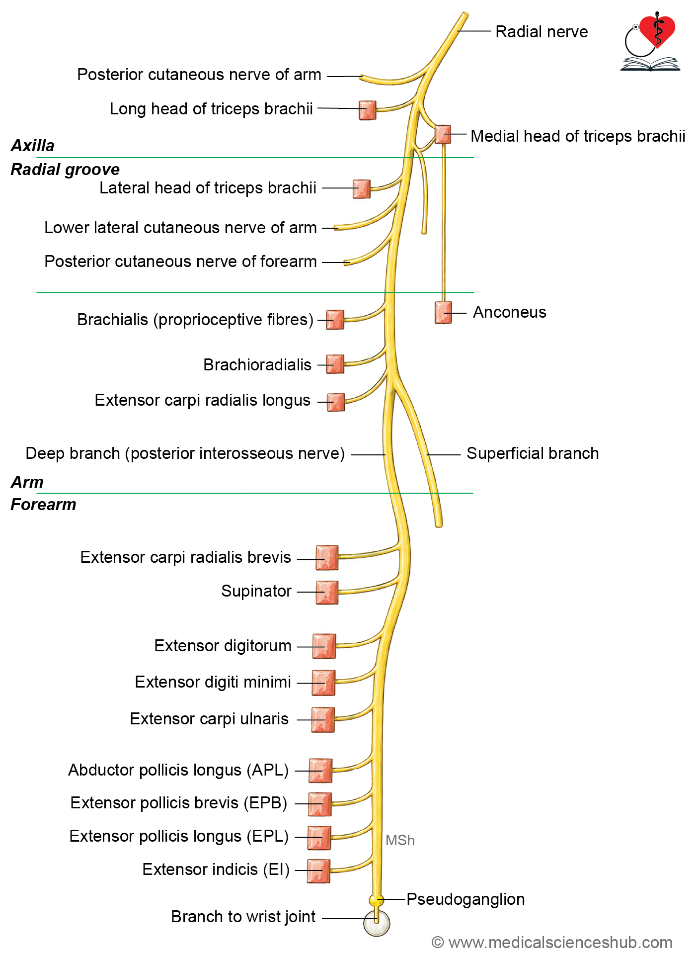
Branches in the Axilla
- Muscular branches to:
- Long head of triceps brachii
- Medial head of triceps brachii
- Posterior cutaneous nerve of the arm, which supplies the skin over the posterior aspect of the arm.
Branches in the Radial Groove
- Muscular branches to:
- Lateral head of triceps brachii
- Medial head of triceps brachii
- The branch to the medial head is also called the ulnar collateral nerve because it lies close to the ulnar nerve.
- Anconeus muscle (through the branch to the medial head of the triceps)
- Cutaneous branches:
- Lower lateral cutaneous nerve of the arm
- Posterior cutaneous nerve of the forearm
Branches in the Lower Part of the Arm
- Muscular branches to:
- Brachialis (lateral part)
- Brachioradialis
- Extensor carpi radialis longus
- Articular branch to the elbow joint
Terminal Branches
Superficial Branch
- The superficial branch is purely sensory.
- It supplies:
- The lateral half of the dorsum of the hand
- The dorsal surface of the lateral 3½ digits (except the distal phalanges)
Deep Branch (Posterior Interosseous Nerve)
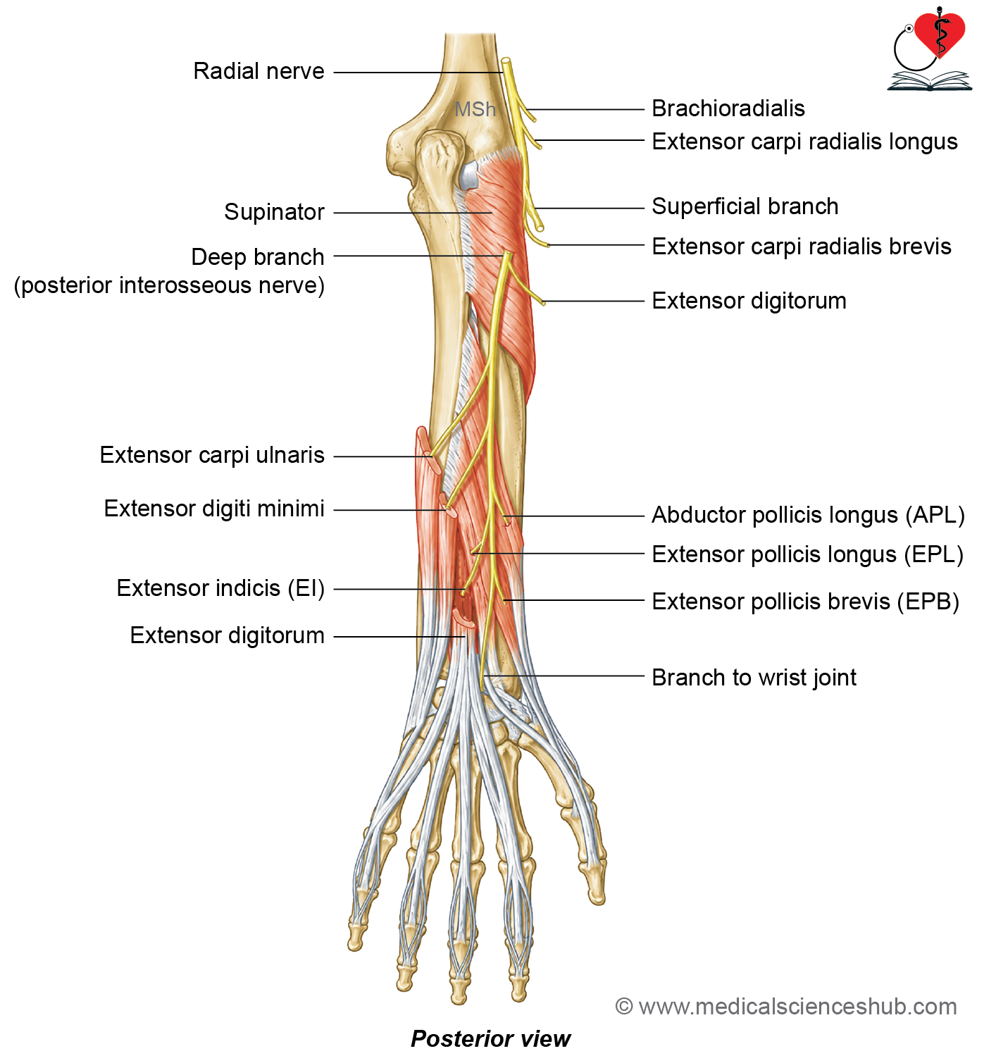
- The deep branch pierces the supinator muscle.
- It continues as the posterior interosseous nerve.
- It supplies all the remaining muscles of the posterior compartment of the forearm.
CLINICAL INTEGRATION
Radial Nerve Injury
- The clinical features of radial nerve injury depend on the site of the lesion.
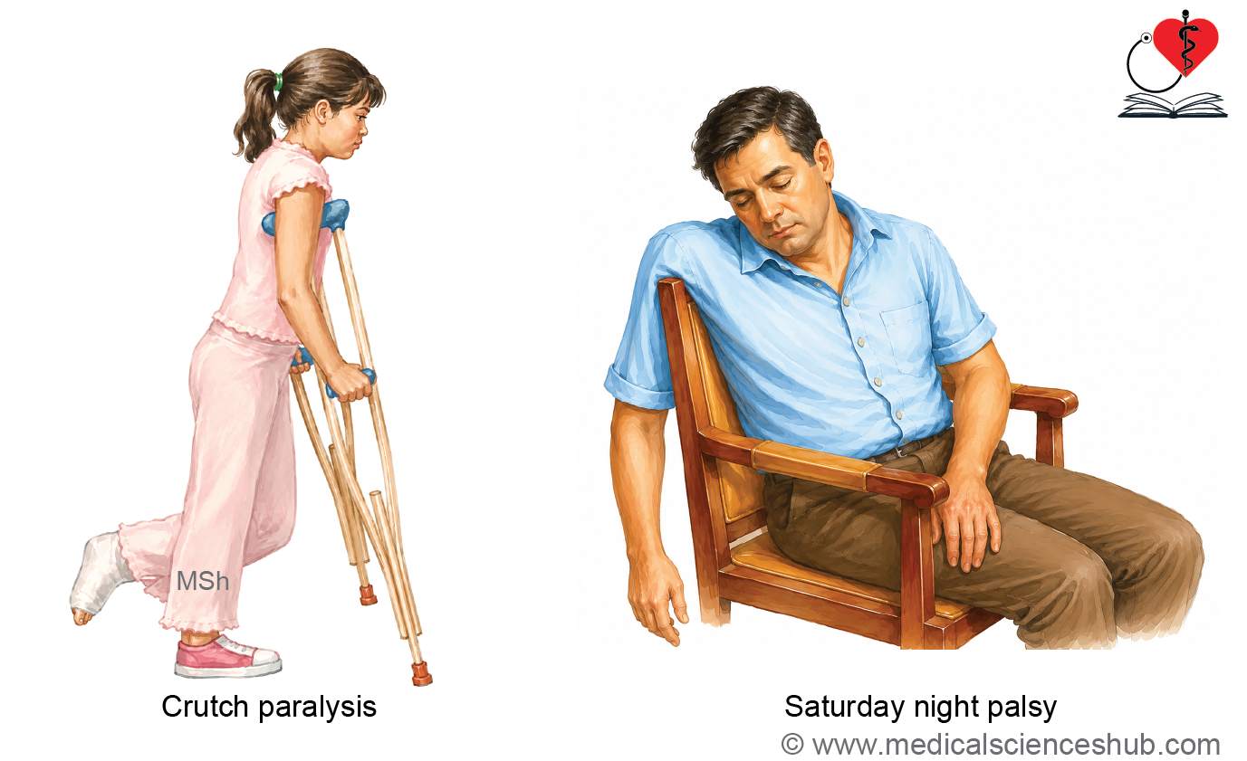
Injury in the Axilla (Saturday Night Palsy)
Causes
- Compression of the radial nerve due to:
- Sleeping with the arm hanging over the back of a chair (Saturday night palsy).
- Improper use of crutches (crutch palsy).
Clinical Features
- Loss of extension of the forearm due to paralysis of the triceps brachii.
- Weakness of forearm supination.
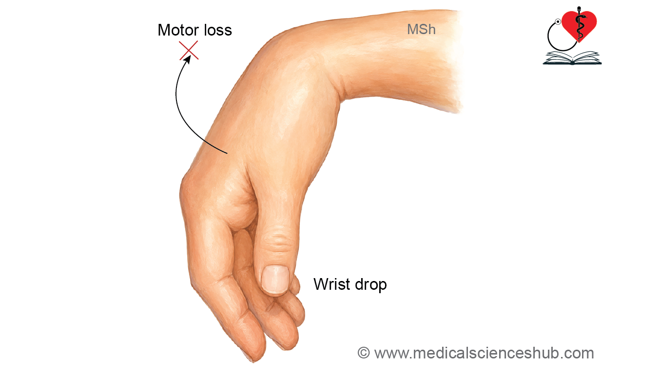
- Loss of wrist extension, producing wrist drop.
- Loss of extension of the fingers.
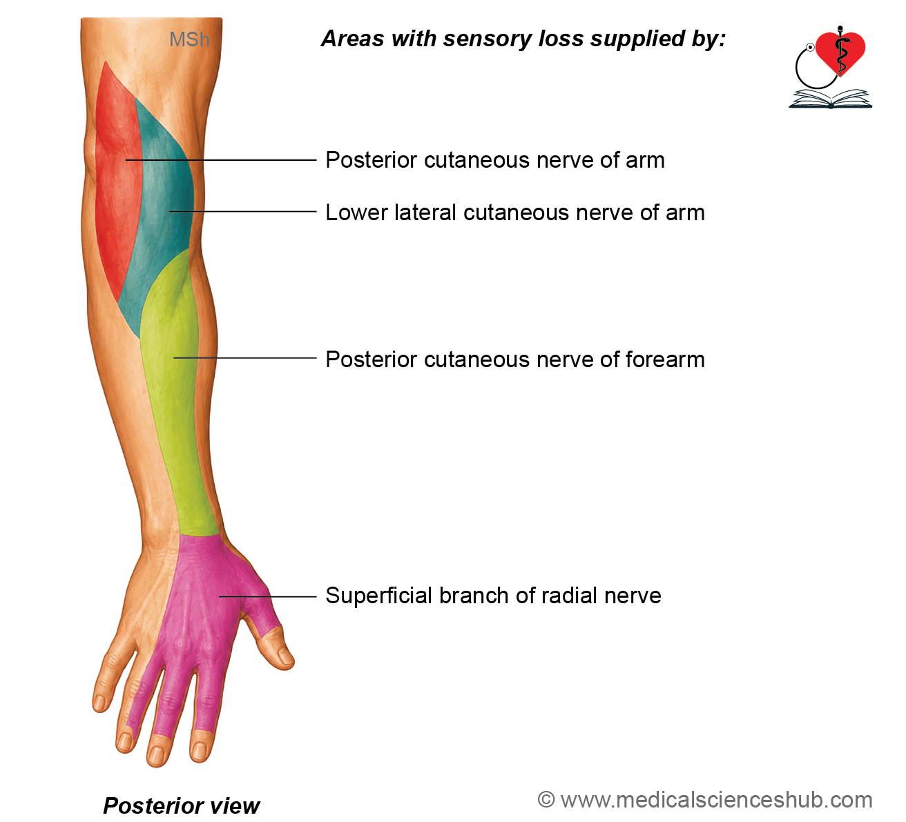
- Loss of sensation over:
- The lateral and posterior aspects of the arm
- The posterior aspect of the forearm
- The dorsum of the hand
- The lateral 3½ digits (except the fingertips)
Injury in the Radial Groove
- The clinical features are similar to those of axillary injury.
- The long and medial heads of the triceps brachii are usually spared, because their branches arise proximal to the radial groove.
- As a result, elbow extension is largely preserved, although it may be slightly weak.
- Wrist drop and finger drop are present due to paralysis of the extensor muscles distal to the lesion.
Injury at the Elbow (Radial Tunnel Syndrome)
- Injury at the elbow produces radial tunnel syndrome, an entrapment neuropathy of the deep branch of the radial nerve.
Causes
- Compression by a fibrous band or by the proximal fibrous edge of the supinator muscle (arcade of Frohse).
Clinical Features
- Loss of extension of the fingers and thumb.
- Weakness of wrist extension, but complete wrist drop is usually absent because the extensor carpi radialis longus remains functional.
- Pain and tenderness over the extensor aspect of the proximal forearm.
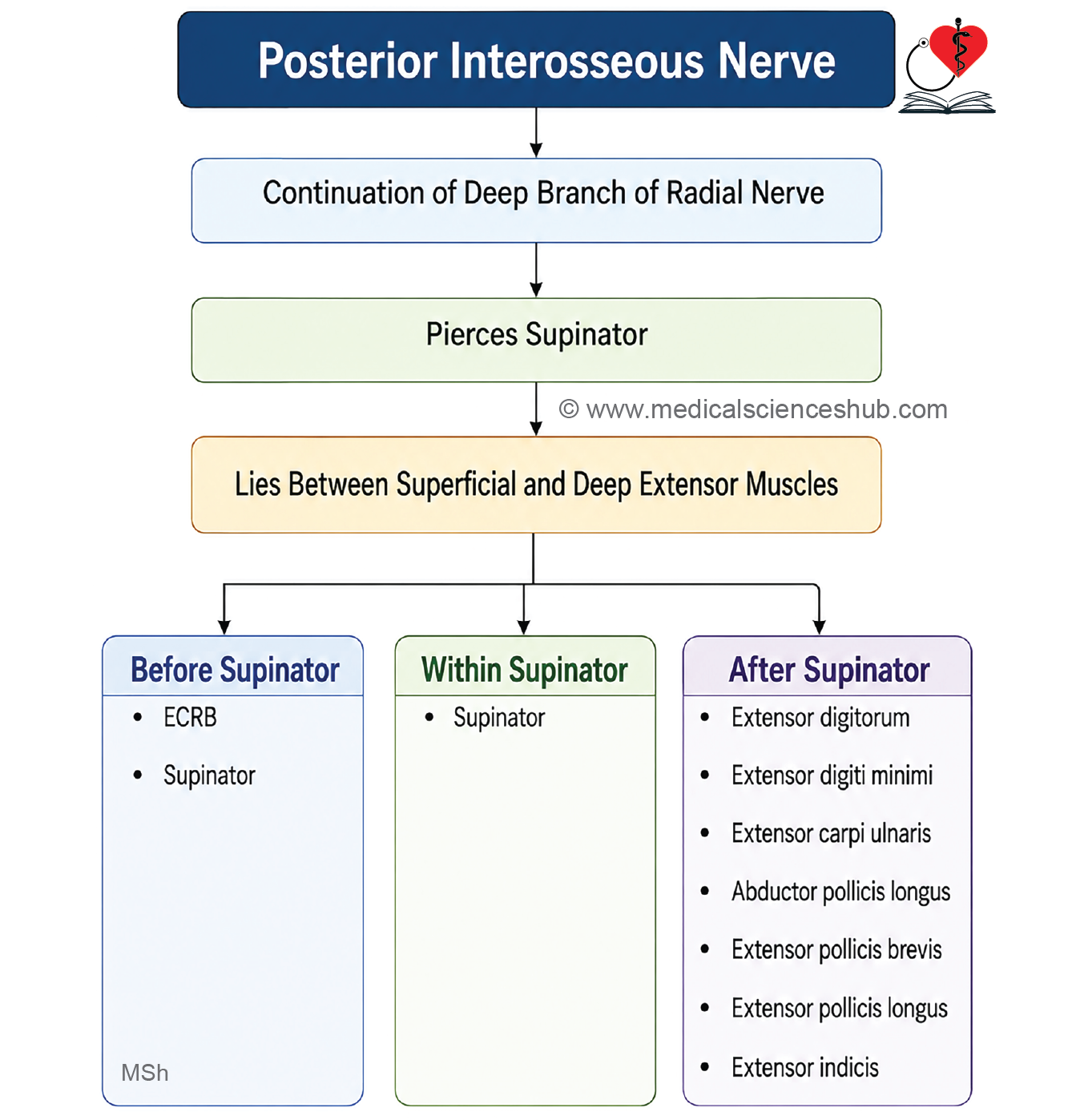
Posterior Interosseous Nerve
- The posterior interosseous nerve is the continuation of the deep branch of the radial nerve.
- It is the principal motor nerve of the posterior compartment of the forearm.
Beginning
- The posterior interosseous nerve begins as the deep branch of the radial nerve in the cubital fossa.
- It arises deep to the brachioradialis muscle.
Course and Relations
- The nerve leaves the cubital fossa by entering the substance of the supinator muscle.
- It passes through the supinator to enter the posterior compartment of the forearm.
- In the posterior compartment, it descends deep to the superficial extensor muscles.
- It reaches the dorsum of the wrist by passing deep to the extensor retinaculum through the fourth extensor compartment.
Termination
- Near the wrist, the posterior interosseous nerve exhibits a pseudoganglion.
- It terminates by supplying the:
- Wrist joint
- Intercarpal joints
Branches
Muscular Branches
Before Piercing the Supinator
- Extensor carpi radialis brevis
- Supinator
Within the Supinator
- Supinator
After Emerging from the Supinator
- Extensor digitorum
- Extensor digiti minimi
- Extensor carpi ulnaris
- Abductor pollicis longus
- Extensor pollicis longus
- Extensor pollicis brevis
- Extensor indicis
Articular Branches
- The posterior interosseous nerve supplies the:
- Wrist joint
- Distal radioulnar joint
- Intercarpal joints
- Intermetacarpal joints
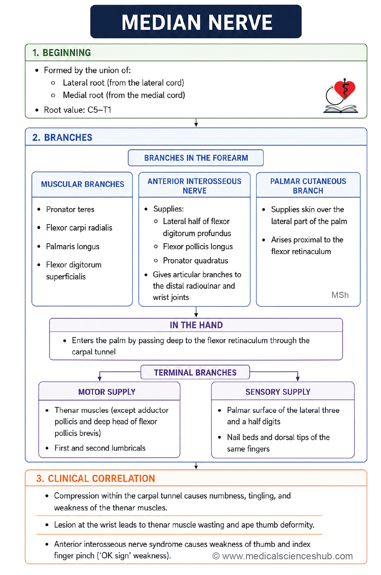
Median Nerve
- The median nerve, also known as the laborer’s nerve, is the main nerve of the forearm.
- It is called the laborer’s nerve because it supplies most of the muscles responsible for the powerful and precise movements of the forearm and thumb.
- The median nerve is formed by the union of the lateral root (from the lateral cord) and the medial root (from the medial cord) of the brachial plexus in the lower part of the axilla.
- The median nerve, also known as the laborer’s nerve, is the main nerve of the forearm.
- It is called the laborer’s nerve because it supplies most of the muscles responsible for the powerful and precise movements of the forearm and thumb.
- The median nerve is formed by the union of the lateral root (from the lateral cord) and the medial root (from the medial cord) of the brachial plexus in the lower part of the axilla.
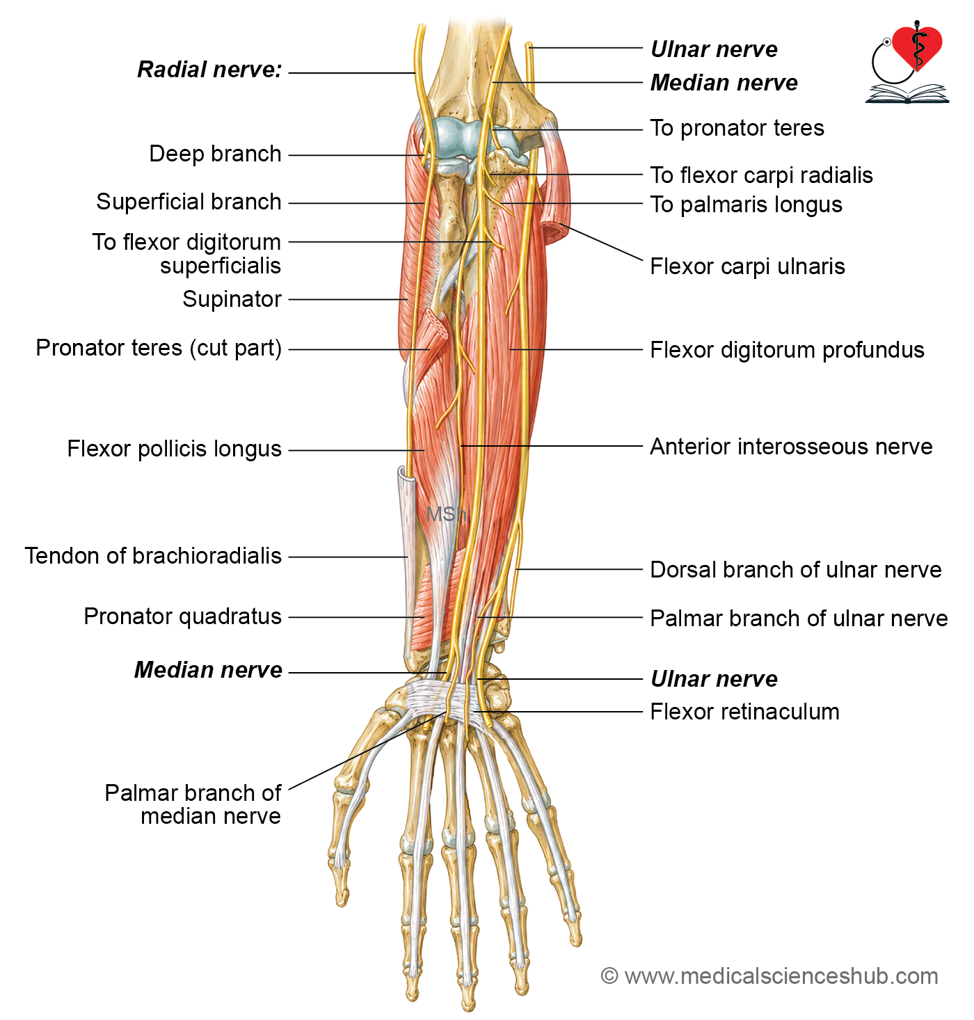
Course and main relations
In the Axilla
- The median nerve is formed anterior to the third part of the axillary artery.
- It descends lateral to the artery and crosses anterior to the brachial artery from lateral to medial.
In the Arm
- The median nerve descends alongside the brachial artery without giving any major muscular branches.
- In the lower part of the arm, it lies medial to the brachial artery.
In the Cubital Fossa
- The median nerve lies medial to the brachial artery.
- It enters the forearm by passing between the humeral and ulnar heads of the pronator teres muscle.
In the Forearm
- The median nerve passes between the two heads of the pronator teres.
- At this level, it is separated from the ulnar artery by the deep head of the pronator teres.
- It then passes deep to the fibrous arch of the flexor digitorum superficialis.
- Approximately 5 cm proximal to the flexor retinaculum, the nerve becomes superficial and lies lateral to the tendons of the flexor digitorum superficialis.
At the Wrist
- The median nerve enters the palm by passing deep to the flexor retinaculum through the carpal tunnel.
In the Palm
- In the palm, the median nerve lies medial to the thenar muscles.
- It divides into medial and lateral terminal branches, which supply the thenar muscles, the first two lumbricals, and the lateral digits.
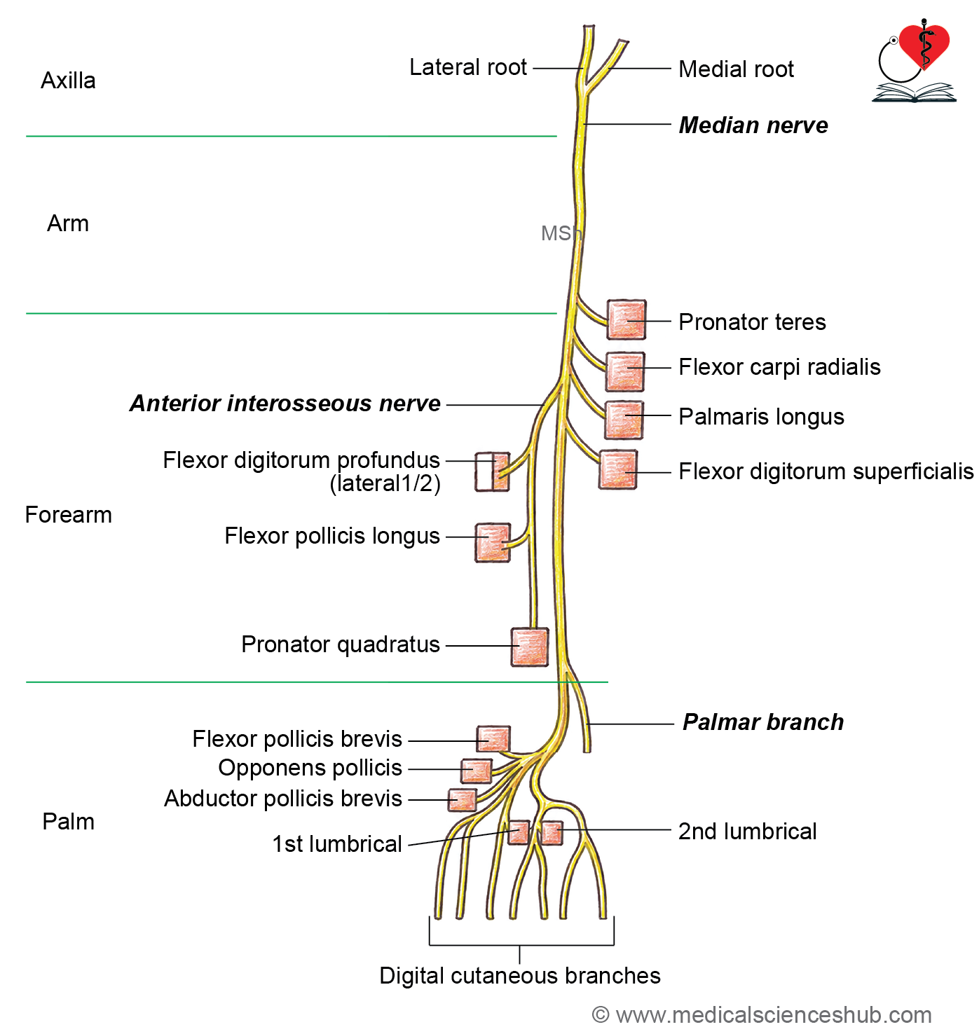
Branches
In the Axilla
- The median nerve gives no branches in the axilla.
In the Arm
- The median nerve normally gives no muscular branches in the arm.
In the Cubital Fossa
Muscular Branches
- Pronator teres
- Flexor carpi radialis
- Palmaris longus
- Flexor digitorum superficialis
In the Forearm
Anterior Interosseous Nerve
- The anterior interosseous nerve is the largest branch of the median nerve in the forearm.
- It supplies:
- Flexor pollicis longus
- Lateral (radial) half of the flexor digitorum profundus
- Pronator quadratus
- It also provides articular branches to the:
- Distal radioulnar joint
- Wrist joint
Palmar Cutaneous Branch
- The palmar cutaneous branch arises proximal to the flexor retinaculum.
- It passes superficial to the flexor retinaculum.
- It supplies the skin over the thenar eminence and the lateral part of the palm.
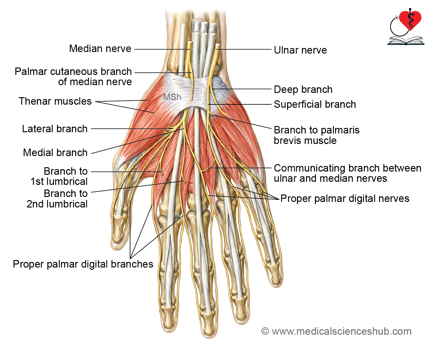
Terminal Branches in the Palm
- The median nerve divides into medial and lateral branches.
Medial Branch
- The medial branch gives two common palmar digital nerves.
Lateral Common Digital Nerve
- Supplies the adjacent sides of the index and middle fingers.
- Supplies the second lumbrical muscle.
Medial Common Digital Nerve
- Supplies the adjacent sides of the middle and ring fingers.
Lateral Branch
Muscular Branch: Supplies the thenar muscles:
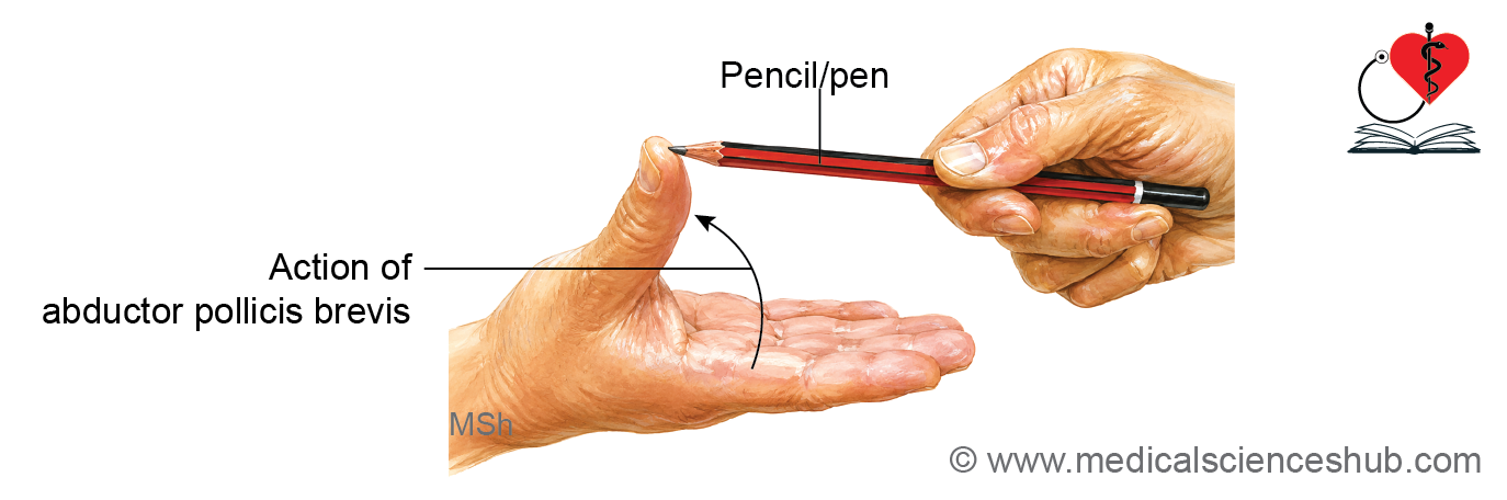
- Abductor pollicis brevis
- Opponens pollicis
- Superficial head of flexor pollicis brevis
Proper Digital Branches
- Gives three proper palmar digital nerves to:
- Lateral side of the thumb
- Medial side of the thumb
- Lateral side of the index finger
- The branch to the lateral side of the index finger also supplies the first lumbrical muscle.
CLINICAL INTEGRATION
Injury to the Median Nerve at the Elbow
Causes
- Supracondylar fracture of the humerus
- Entrapment of the nerve between the two heads of the pronator teres muscle (pronator syndrome)
Clinical Features
- Paralysis of the pronator teres and pronator quadratus causes loss of forearm pronation, leaving the forearm in a supinated position.
- Paralysis of all wrist flexors except the flexor carpi ulnaris and the medial half of the flexor digitorum profundus results in weakness of wrist flexion.
- Paralysis of the flexor carpi radialis produces adduction of the wrist during flexion because of the unopposed action of the flexor carpi ulnaris.
- Paralysis of the flexor digitorum superficialis and the lateral half of the flexor digitorum profundus causes loss of flexion at the interphalangeal joints of the index and middle fingers.
- Paralysis of the flexor pollicis longus results in loss of flexion of the terminal phalanx of the thumb.
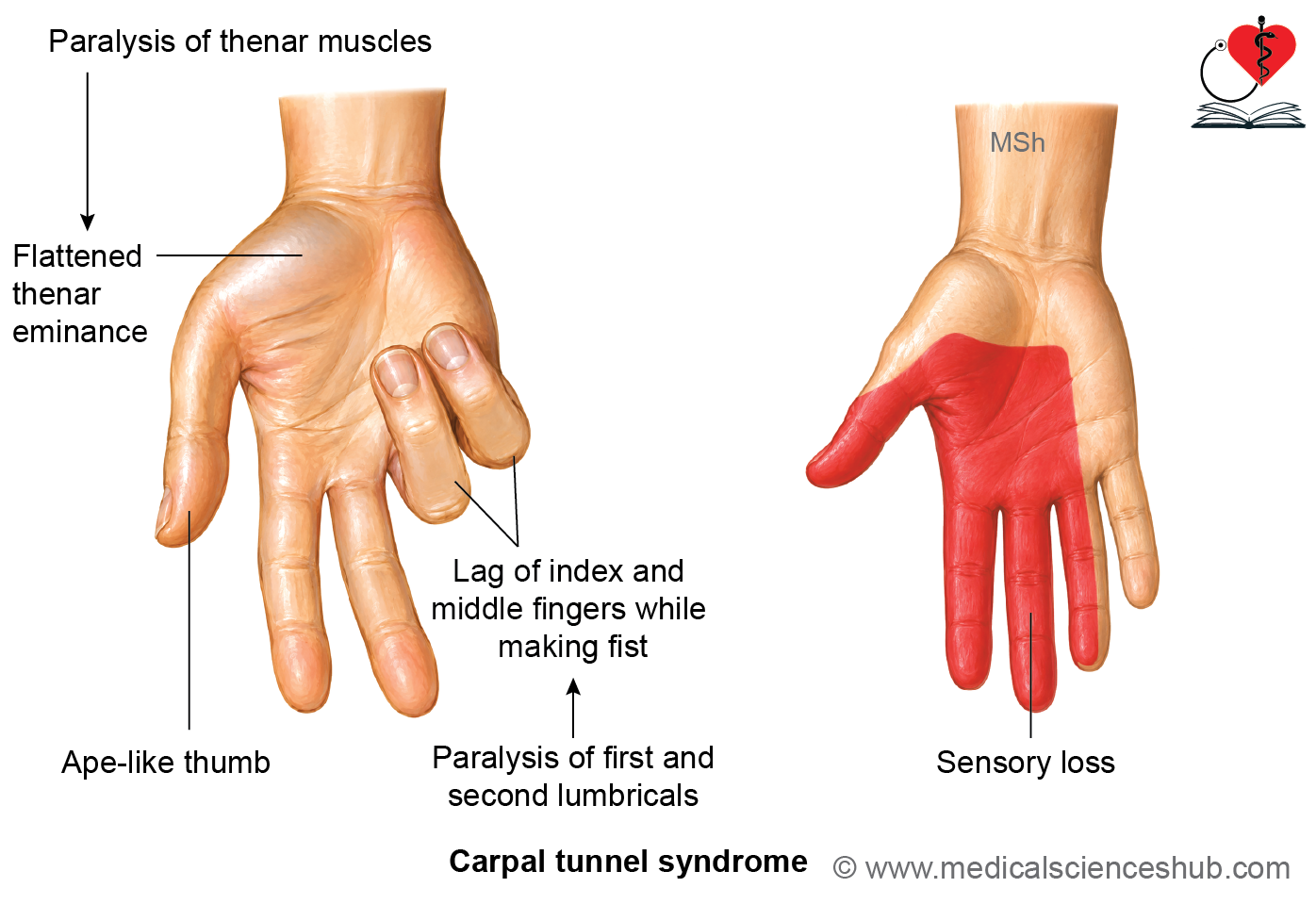
- Paralysis of the thenar muscles leads to thenar wasting and an ape-like thumb deformity, characterized by loss of thumb opposition and abduction.
- Loss of sensation occurs over the lateral 3½ digits, including their palmar surfaces, nail beds, and dorsal distal phalanges.
- Paralysis of the first and second lumbricals produces difficulty flexing the metacarpophalangeal joints of the index and middle fingers, resulting in a characteristic lag of these fingers while making a fist, also known as the hand of benediction when attempting to close the hand.
Carpal Tunnel Syndrome
- Carpal tunnel syndrome is the most common entrapment neuropathy of the median nerve.
- The carpal tunnel is an osseofibrous canal formed by the carpal bones posteriorly and the flexor retinaculum anteriorly.
- The carpal tunnel transmits:
- The median nerve
- The flexor tendons of the forearm
- Compression of the median nerve within the carpal tunnel produces carpal tunnel syndrome.
Clinical Features
Motor Deficits
- Paralysis of the thenar muscles results in an ape-like thumb deformity.
- The thumb becomes extended and adducted due to the inability to abduct, oppose, and flex the thumb effectively.
- Paralysis of the first and second lumbricals causes difficulty flexing the metacarpophalangeal joints of the index and middle fingers, producing a lag of these fingers while making a fist.
Sensory Deficits
- There is loss of sensation over the lateral 3½ digits, including their palmar surfaces, nail beds, and dorsal distal phalanges.
- Patients commonly complain of pain, tingling, numbness, and paresthesia in the thumb, index finger, middle finger, and lateral half of the ring finger.
Autonomic Changes
- Compression of sympathetic fibers may produce vasomotor disturbances.
- The skin over the lateral 3½ digits may become dry because of reduced sweating.
Clinical Tests
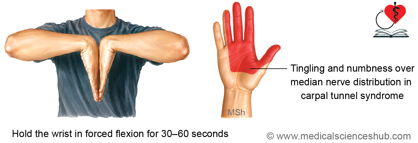
Phalen’s Maneuver
- The patient is asked to maintain maximum wrist flexion for about 60 seconds.
- The test is positive if it reproduces pain, tingling, or numbness in the lateral 3½ digits.
- Wrist flexion increases pressure within the carpal tunnel, compressing the median nerve.
Durkan’s Test (Carpal Compression Test)
- Direct pressure is applied over the carpal tunnel for approximately 30 seconds.
- The test is positive if it reproduces pain or paresthesia in the distribution of the median nerve.
Causes
- The cause is idiopathic (unknown) in most patients.
- Other common causes include:
- Repetitive occupational activities
- Obesity
- Pregnancy
- Rheumatoid arthritis
- Hypothyroidism
- Inflammatory disorders
Treatment
- Physiotherapy and activity modification.
- Wrist splinting, particularly in a neutral position, to reduce pressure within the carpal tunnel.
- Surgical decompression by division of the flexor retinaculum in patients with persistent or severe symptoms.
Ulnar Nerve
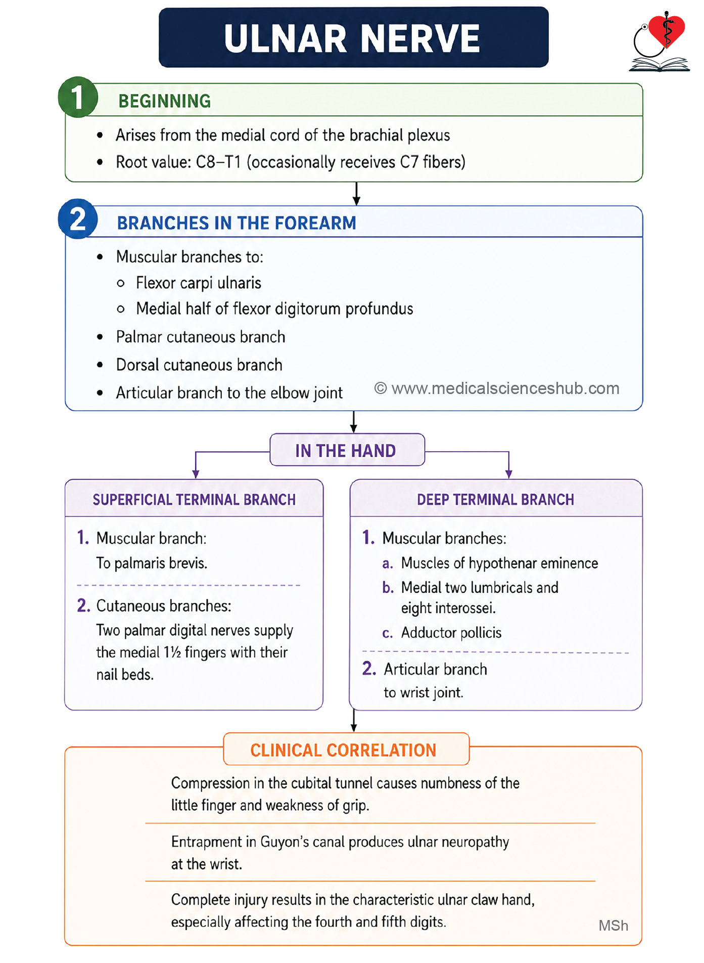
- The ulnar nerve is a terminal branch of the medial cord of the brachial plexus.
- It has a root value of C7, C8, and T1.
- The ulnar nerve is commonly known as the “musician’s nerve” because it supplies most of the intrinsic muscles of the hand responsible for the fine movements of the fingers.
Course and Main Relations
In the Axilla
- The ulnar nerve arises in the lower part of the axilla.
- It descends between the axillary artery and the axillary vein.
In the Arm
- The ulnar nerve runs along the medial side of the brachial artery as far as the insertion of the coracobrachialis muscle.
- It pierces the medial intermuscular septum together with the superior ulnar collateral artery to enter the posterior compartment of the arm.
- It descends behind the medial epicondyle of the humerus, where it lies superficially and is vulnerable to injury.
In the Forearm
- The ulnar nerve enters the forearm by passing through the cubital tunnel, a gap between the humeral and ulnar heads of the flexor carpi ulnaris.
- It then descends vertically along the forearm.
- At the wrist, it passes superficial to the flexor retinaculum, lateral to the pisiform bone, and medial to the ulnar artery.
In the Palm
- At the distal border of the flexor retinaculum, the ulnar nerve divides into:
- Superficial branch
- Deep branch
Branches and Distribution
In the Axilla and Arm
- The ulnar nerve gives no branches in the axilla or arm.
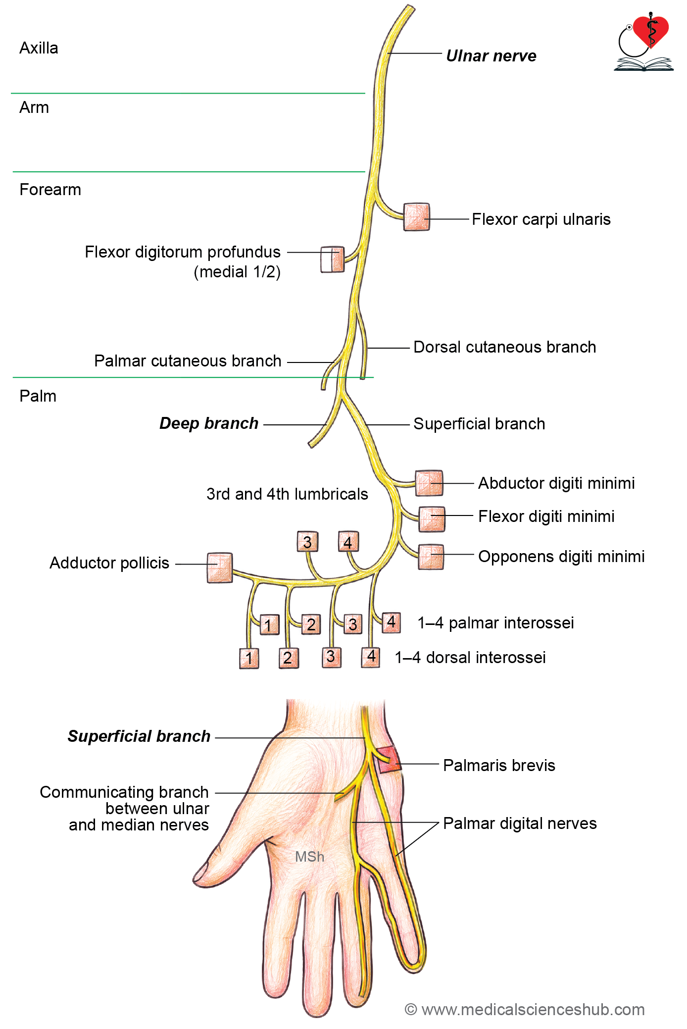
In the Forearm
Muscular Branches
- Flexor carpi ulnaris
- Medial (ulnar) half of the flexor digitorum profundus
Articular Branch
- Elbow joint
Palmar Cutaneous Branch
- The palmar cutaneous branch arises just proximal to the wrist.
- It passes superficial to the flexor retinaculum.
- It supplies the skin over the hypothenar eminence.
Dorsal Cutaneous Branch
- The dorsal cutaneous branch arises approximately 7.5 cm proximal to the wrist beneath the flexor carpi ulnaris.
- It winds around the ulna to reach the dorsum of the hand.
- It supplies:
- The proximal dorsal surface of the medial 2½ digits
- The adjacent medial part of the dorsum of the hand
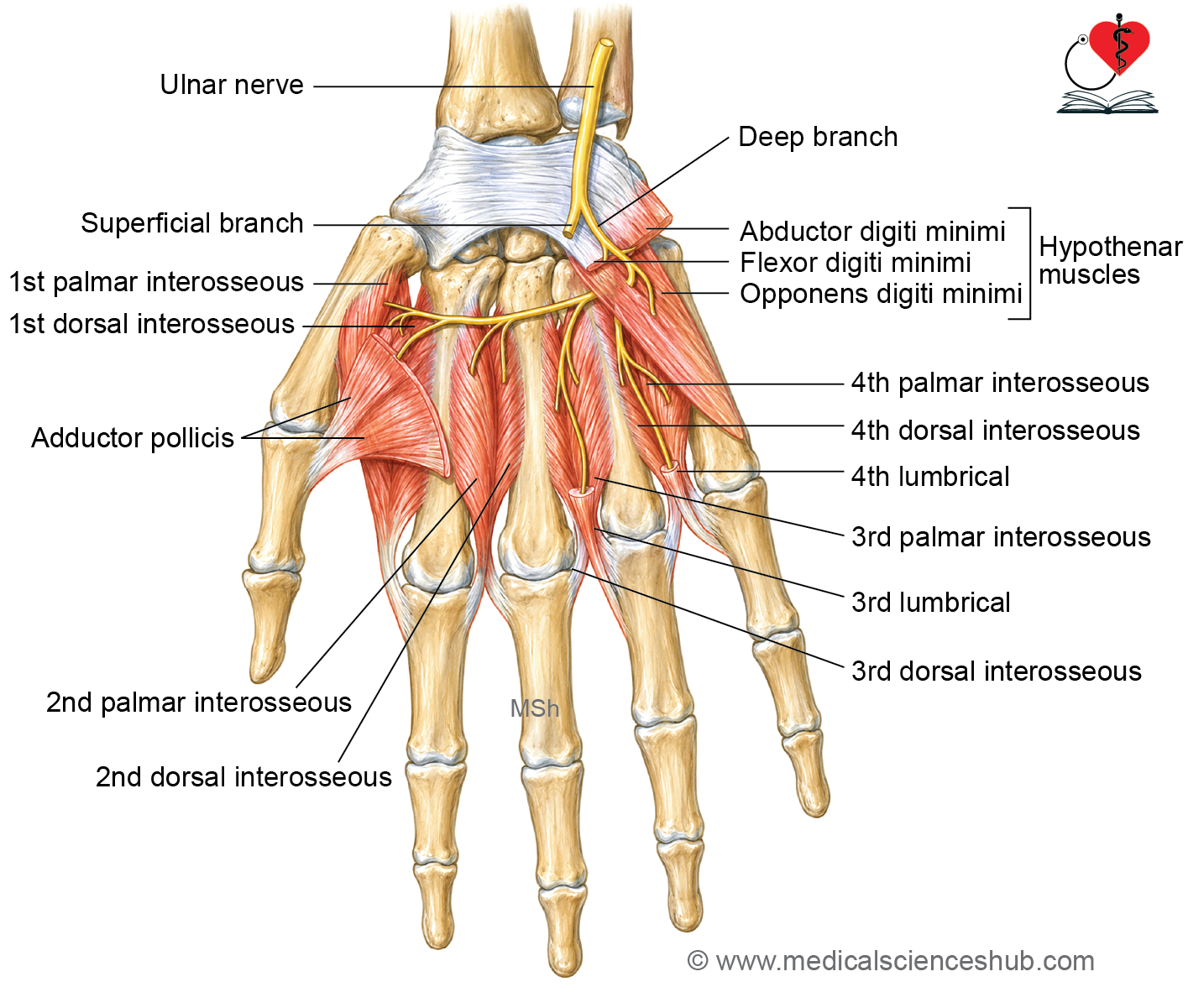
In the Palm
Superficial Branch
Muscular branch for palmaris brevis
Cutaneous Branches: The superficial branch gives two palmar digital nerves.
- Medial Palmar Digital Nerve: Supplies the medial side of the little finger.
- Lateral Palmar Digital Nerve: Divides into two branches supplying the adjacent sides of the ring and little fingers.
Deep Branch
Muscular Branches: The deep branch supplies:
- Hypothenar muscles
- Abductor digiti minimi
- Flexor digiti minimi brevis
- Opponens digiti minimi
- Four palmar interossei
- Four dorsal interossei
- Third and fourth lumbricals
- Adductor pollicis
Articular Branches
- Intercarpal joints
- Carpometacarpal joints
- Intermetacarpal joints
Some Interesting Facts
- Palmar cutaneous branch arises just above the wrist and enters in the palm along with ulnar nerve by passing superficial to flexor retinaculum.
- Dorsal cutaneous branch: It arises 7.5 cm above the wrist under the cover of flexor carpi ulnaris. It turns backward around ulna to enter on posterior side of forearm.
- In lower part of forearm, the ulnar nerve lies superficially, medial to the tendon of FCU and lateral to the ulnar artery.
- Ulnar paradox
- The lesion of ulnar or median nerve at more higher levels induces less prominent deformity.
CLINICAL INTEGRATION
Common Sites of Ulnar Nerve Injury
- Behind the medial epicondyle of the humerus
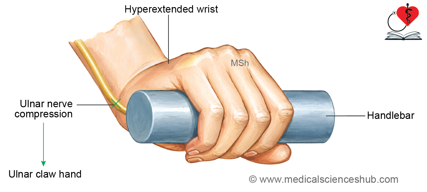
- At the wrist within Guyon’s canal
Lesion at the Wrist (Guyon’s Canal Syndrome)
Clinical Features
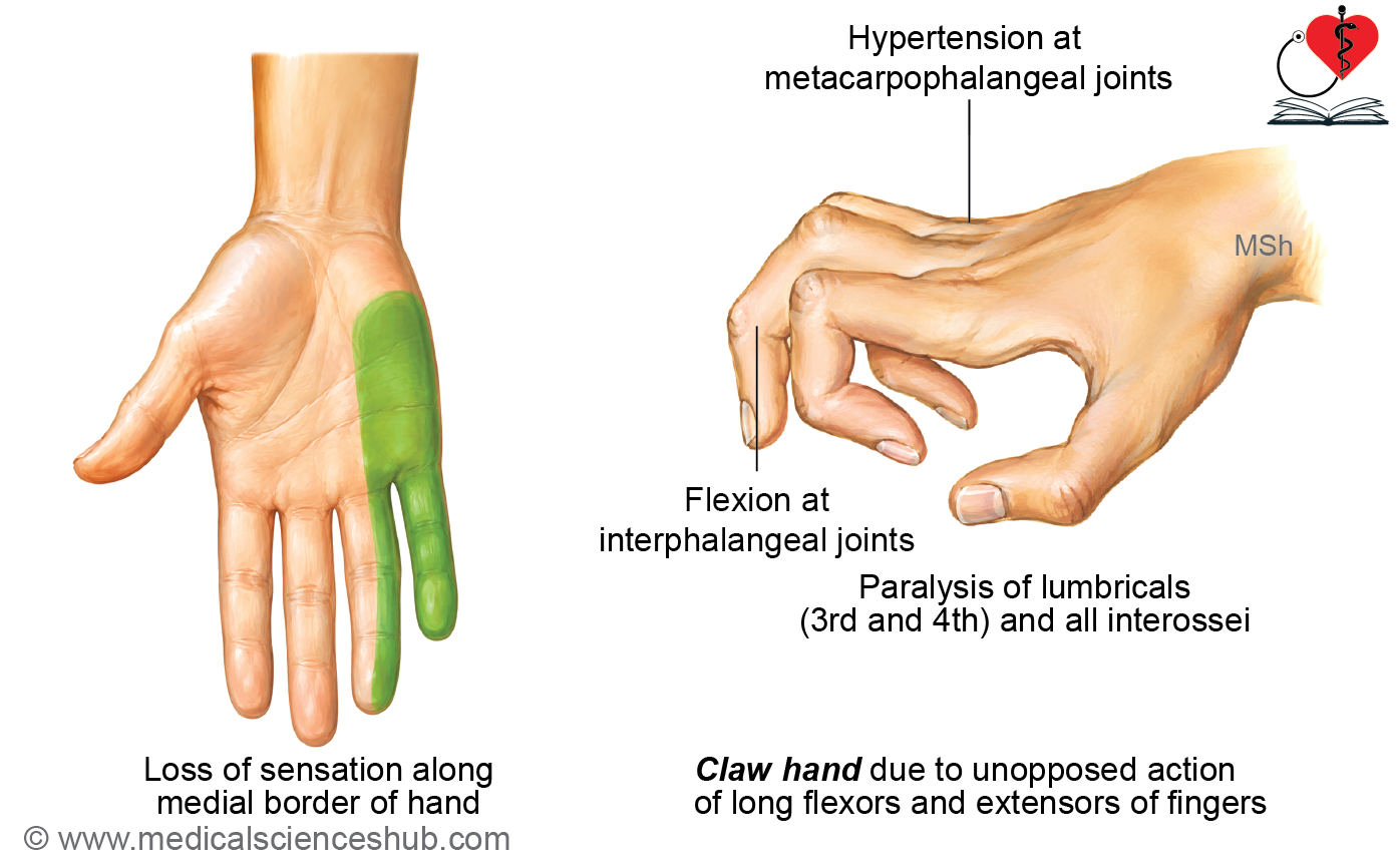
- Produces ulnar claw hand (spinster’s claw deformity).
Effects
- Paralysis of the third and fourth lumbricals causes:
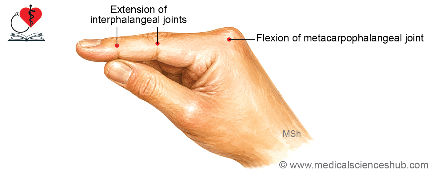
- Hyperextension at the metacarpophalangeal joints
- Flexion at the interphalangeal joints
- Clawing of the ring and little fingers
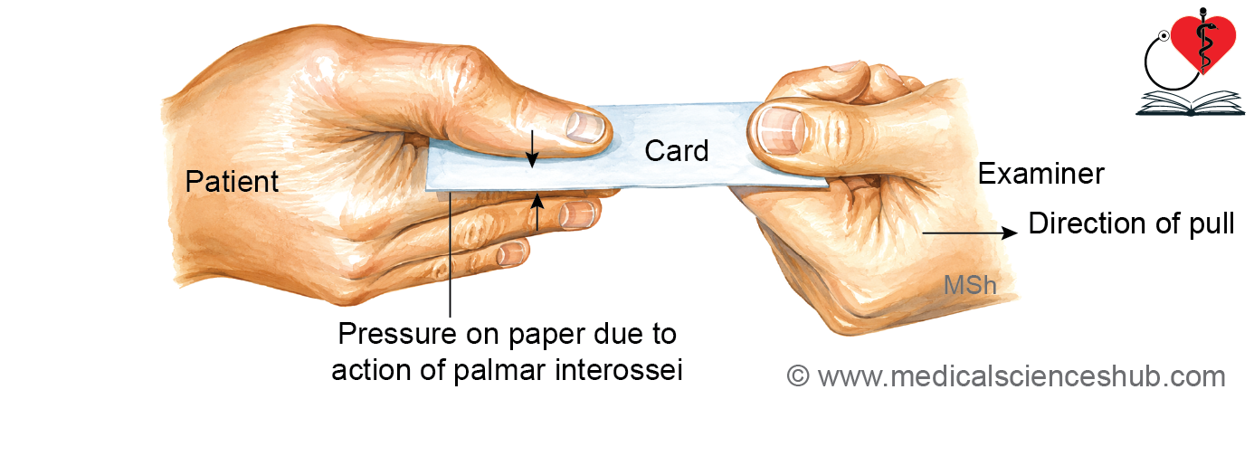
- Paralysis of the palmar interossei results in loss of finger adduction.
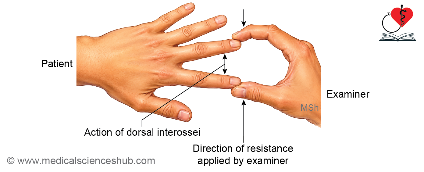
- Paralysis of the dorsal interossei results in loss of finger abduction.
- Loss of sensation occurs over the medial 1½ digits.
- Loss of sympathetic fibers produces vasomotor changes, including dry skin over the medial 1½ digits.
Lesion at the Elbow (Behind the Medial Epicondyle)
Clinical Features
- Produces ulnar claw hand, although the deformity is less pronounced than in a wrist lesion because of the ulnar paradox.
Effects
- Paralysis of the flexor carpi ulnaris causes:
- Weakness of wrist flexion
- Loss of wrist adduction
- Paralysis of the medial half of the flexor digitorum profundus causes:
- Loss of flexion at the distal interphalangeal joints of the ring and little fingers
- Sensory loss involves:
- The medial 1½ digits
- The hypothenar eminence, because the lesion is proximal to the origin of the palmar cutaneous branch.
Ulnar Paradox
- The ulnar paradox refers to the phenomenon in which a higher lesion of the ulnar nerve produces a less obvious claw hand deformity than a lower lesion.
- This occurs because paralysis of the medial half of the flexor digitorum profundus in high lesions reduces flexion of the distal interphalangeal joints of the ring and little fingers, thereby decreasing the severity of clawing.
Clinical Anatomy
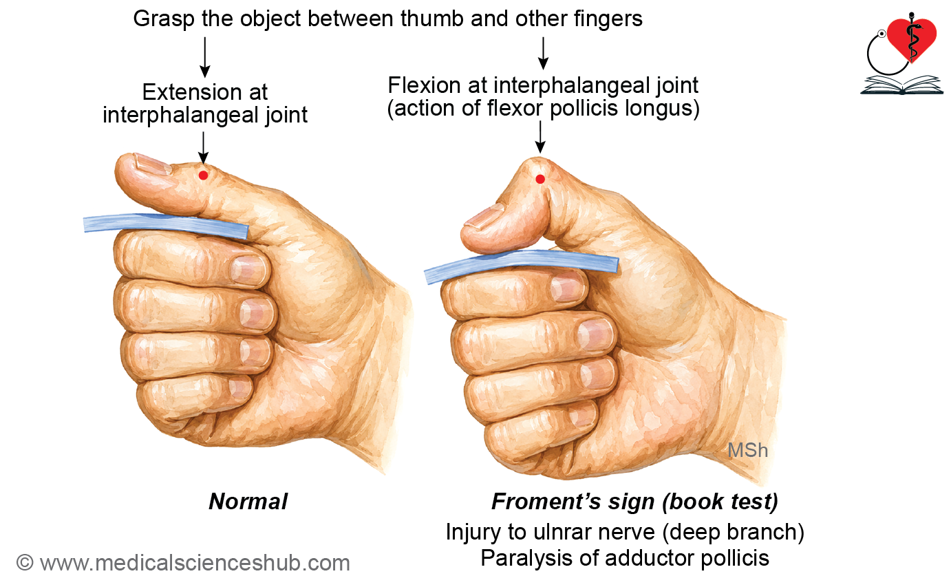
Froment’s Sign
Procedure
- Ask the patient to hold a sheet of paper or a book between the thumb and the lateral side of the index finger while the examiner attempts to pull it away.
Interpretation
- Normal: The thumb remains straight at the interphalangeal (IP) joint, indicating a functioning adductor pollicis.
Positive Froment’s sign: The IP joint of the thumb flexes while gripping the paper due to compensatory action of the flexor pollicis longus, indicating paralysis or weakness of the adductor pollicis from ulnar nerve injury.
Important Questions
- Write a short note on axillary nerve.
- Write a short note on musculocutaneous nerve.
- Write a short note on radial (musculospiral) nerve in radial groove.
- Write a short note on wrist drop.
- Write a short note on posterior interosseous nerve.
- Write a short note on carpal tunnel syndrome.
- Write a short note on ape thumb deformity.
- Write a short note on claw hand (ulnar claw hand).
📝 Test Your Knowledge – Practice MCQs
Attempt the chapter MCQ quiz and assess your understanding of key concepts.
Click to see: Medical Disclaimer and Copyright Notice