
Competencies
- AN8.1: Features of individual bones (upper limb): Identify the given bone, its side, important features, and keep it in anatomical position.
- AN8.2: Features of individual bones (upper limb): Identify and describe joints formed by the given bone.
- AN8.3: Enumerate peculiarities of clavicle.
- AN8.4: Features of individual bones (upper limb): Demonstrate important muscle attachment on the given bone. This chapter includes description of scapula and clavicle.
Introduction
- The upper limb contains 32 bones: scapula and clavicle (pectoral girdle), humerus (arm), radius and ulna (forearm), 8 carpals, 5 metacarpals, and 14 phalanges.
- The pectoral girdle connects the upper limb to the axial skeleton and comprises the scapula and clavicle.
- In certain lower vertebrates, the pectoral girdle additionally includes a third bone called the coracoid.
Clavicle
- The clavicle, or collar bone, is a modified long bone located at the front of the neck root.
- It connects the upper limb to the trunk.
- Due to its curvature, the clavicle resembles the letter “f,” being convex anteriorly along its medial two-thirds and concave anteriorly along its lateral one-third.
- It has a flattened lateral end and a thickened medial end.
- Its inferior surface bears a depression called the subclavian groove.
Anatomical Position and Side Determination
- To determine the anatomical position of the clavicle, hold it horizontally with the medial two-thirds of the shaft convex anteriorly and the lateral one-third concave anteriorly.
- The inferior surface should display a visible depression, the subclavian groove.
- The thicker end points medially, while the flattened end points laterally, with the flat end placed on the corresponding side of the body, for instance, the right clavicle’s flat end faces right.
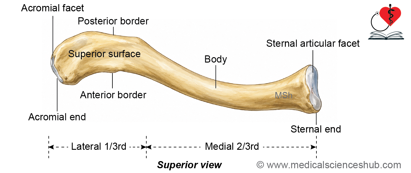
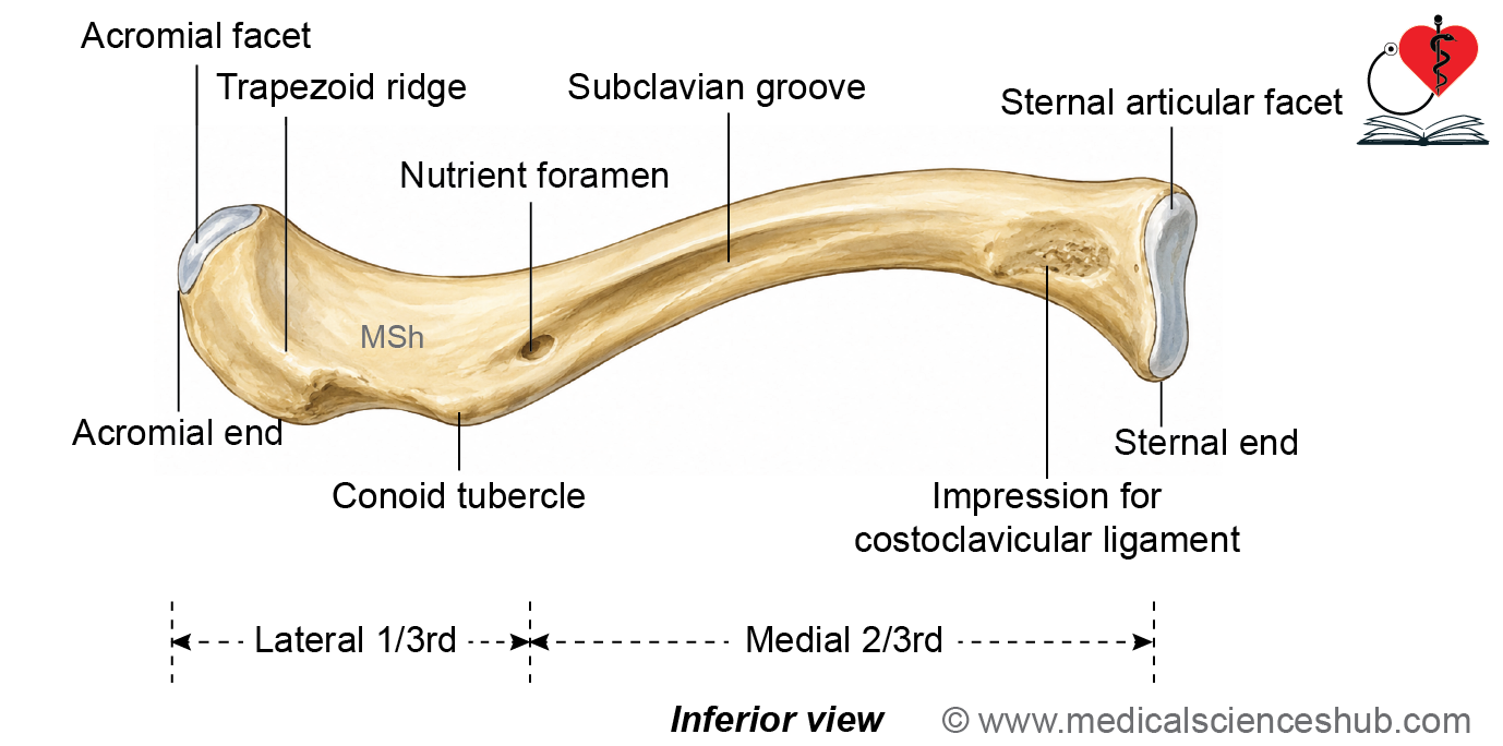
Figure H1.1.1: Hand-drawn diagram showing features of clavicle (Click to see figure)
Peculiarities of Clavicle
The clavicle shows several distinctive features among long bones.
- It is the only long bone positioned horizontally and remains subcutaneous along its entire length.
- It is the first bone in the body to begin ossification, yet it completes ossification relatively late, around 21 to 25 years of age.
- It is the only long bone that ossifies through the intramembranous method and possesses two primary ossification centers.
- A medullary cavity is present within its medial two-thirds.
Functions of Clavicle
- The clavicle transmits the weight of the upper limb to the axial skeleton through the coracoclavicular ligament.
- It functions as a strut, holding the upper limb away from the trunk.
- This positioning allows the upper limb a wide and free range of movement at the shoulder joint.
Features of Clavicle
- The clavicle consists of a middle shaft, also called the body, positioned between two ends.
- The medial end, or sternal end, articulates with the manubrium of the sternum.
- The lateral end, or acromial end, articulates with the acromion process of the scapula.
Shaft
- The middle portion of the clavicle forms an elongated shaft, curved in the shape of the letter “f.”
- Based on this curvature, the shaft is divided into two regions.
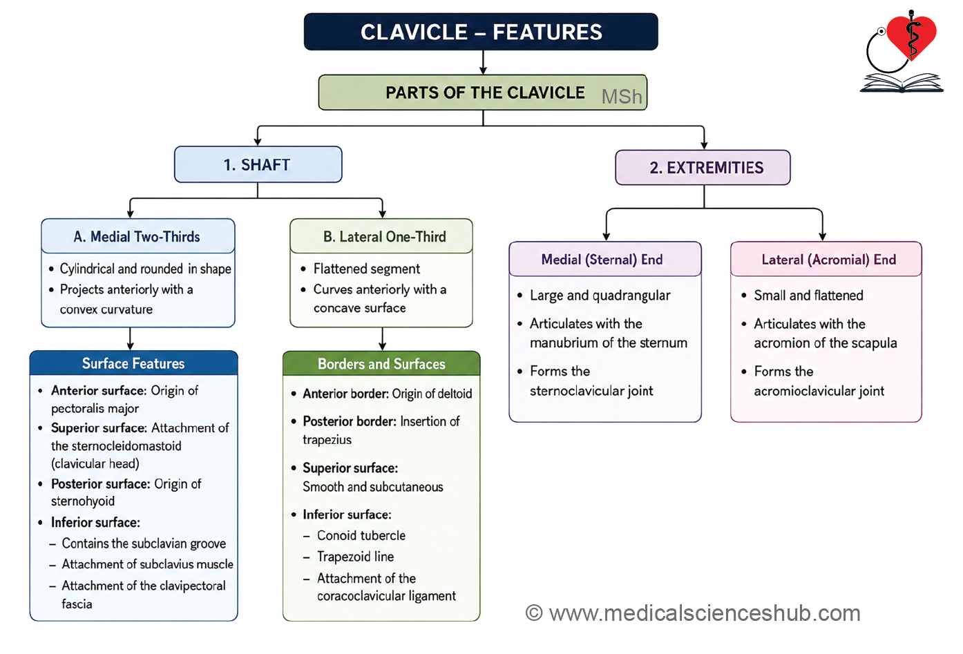
- The medial two-thirds is rounded in cross-section and convex anteriorly.
- The lateral one-third is flattened from above downward and concave anteriorly.
Medial two-thirds of clavicle
- The medial two-thirds of the clavicle is cylindrical in shape and forms the stronger medial portion of the bone.
- It has four surfaces: anterior, posterior, superior, and inferior.
- Anterior surface
- The anterior surface is smooth and convex.
- It provides the origin for the clavicular head of the pectoralis major muscle.
- Posterior surface
- The posterior surface is smooth and concave posteriorly.
- Near the medial end, it gives origin to the sternohyoid muscle.
- Posterior to the medial end, the internal jugular vein joins the subclavian vein to form the brachiocephalic vein.
- The posterior aspect of the medial two-thirds is also related to the trunks of the brachial plexus and the first part of the subclavian artery.
- Superior surface
- The medial part of the superior surface is rough, whereas the lateral part is smooth.
- It gives origin to the clavicular head of the sternocleidomastoid muscle.
- Inferior surface
- The inferior surface shows a rough oval impression near the medial end for ligament attachment.
- A longitudinal subclavian groove occupies the lateral part of the inferior surface.
- A nutrient foramen is usually present near the lateral end of the subclavian groove.
- The nutrient artery commonly arises from the thoracoacromial or suprascapular artery.
- The nutrient foramen is directed laterally, away from the growing end of the clavicle.
- Attachments on the inferior surface
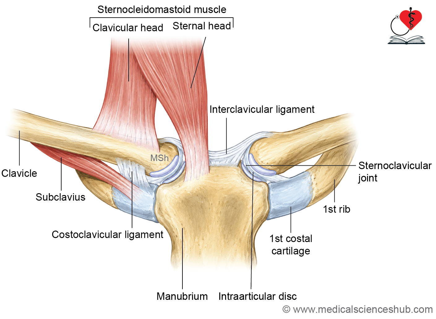
- The costoclavicular ligament attaches to the rough oval impression and extends to the first rib and its costal cartilage.
- The subclavius muscle is inserted into the subclavian groove.
- The clavipectoral fascia is attached along the margins of the subclavian groove.
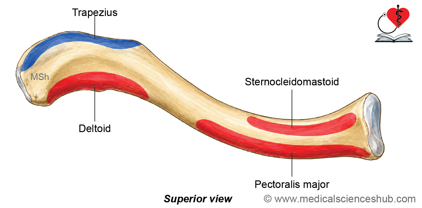
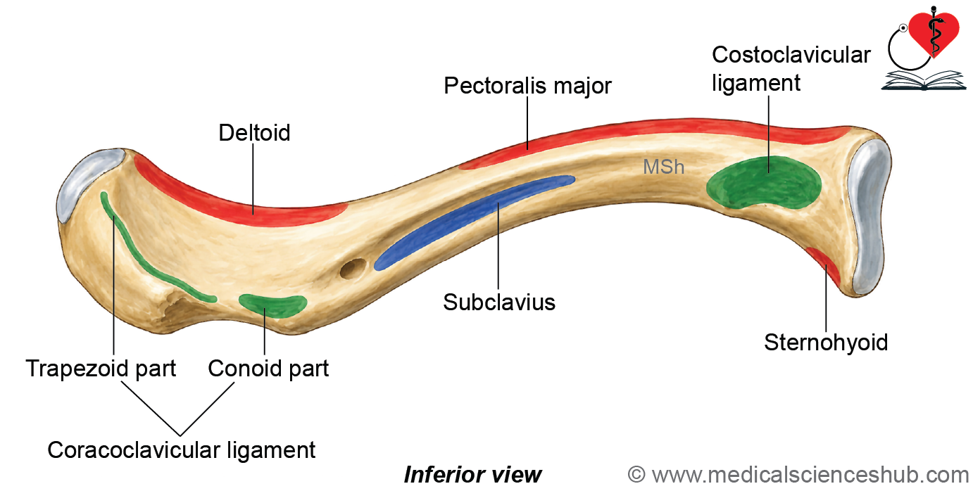
Lateral One-third of Clavicle
- The lateral one-third of the clavicle is flat and has two surfaces and two borders.
- It consists of superior and inferior surfaces, and anterior and posterior borders.
- The anterior border is rough and concave anteriorly and may show a small deltoid tubercle. The anterior border gives origin to the deltoid muscle.
- The posterior border is thick and convex posteriorly. The posterior border provides insertion to the trapezius muscle.
- The superior surface is rough at its anterior and posterior parts, while the middle part is smooth and subcutaneous.
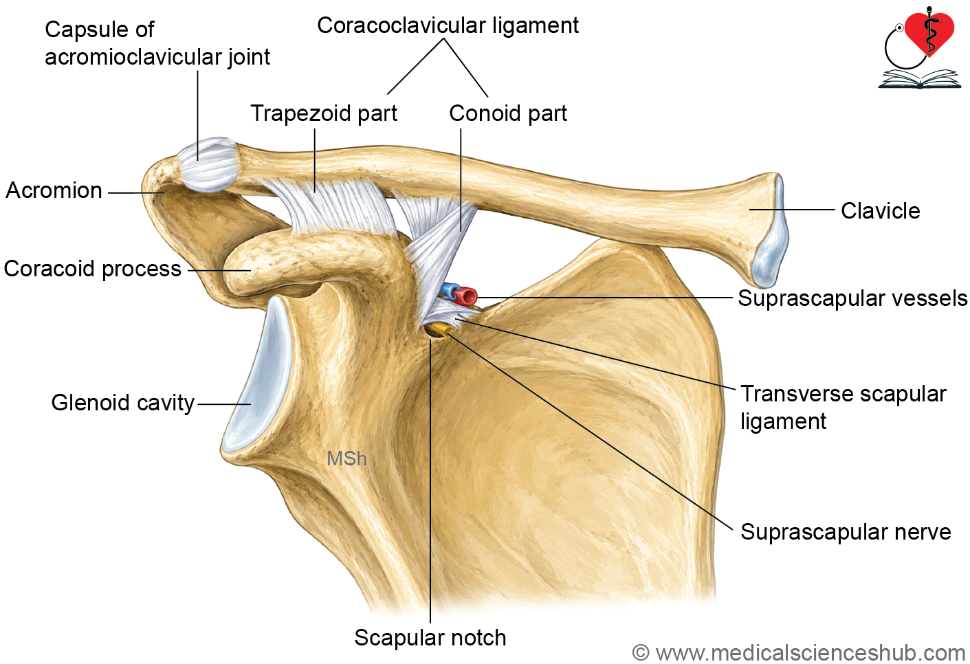
- The inferior surface bears the conoid tubercle and trapezoid line. The coracoclavicular ligament attaches to the inferior surface. Its conoid part attaches to the conoid tubercle, whereas its trapezoid part attaches to the trapezoid line.
Ends of Clavicle
- Clavicle has two ends: Medial and lateral.
Medial (sternal) end
- The medial (sternal) end of the clavicle is quadrangular in shape.
- It has a saddle-shaped articular surface for articulation.
- It articulates with the clavicular notch of the manubrium to form the sternoclavicular joint.
- It provides attachment to the fibrous capsule of the sternoclavicular joint, the articular disc, and the interclavicular ligament.
Lateral (acromial) end
- The lateral (acromial) end of the clavicle is flattened from above downward.
- It has a small oval articular facet. It articulates with the medial margin of the acromion to form the acromioclavicular joint.
- The margin of the articular facet provides attachment to the fibrous capsule of the acromioclavicular joint.
Ossification
- The clavicle is the first bone in the body to begin ossification, although it completes the process relatively late.
- It undergoes membranocartilaginous ossification, where most of the bone forms in membrane, except the medial end, which forms in cartilage.
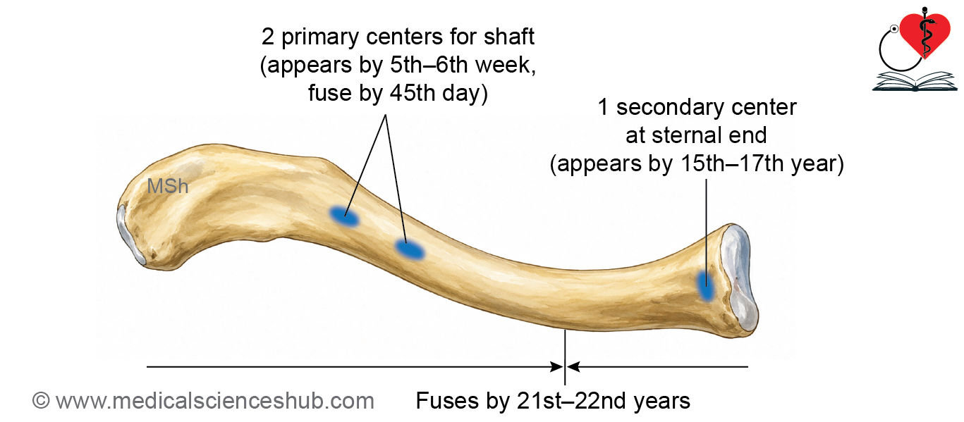
- Two primary ossification centers appear in the shaft around the fifth to sixth week of intrauterine life and fuse by the forty-fifth day.
- A secondary center appears at the medial end during fifteen to seventeen years of age, occurring earlier in females, and fuses with the shaft by twenty-one to twenty-two years.
CLINICAL INTEGRATION
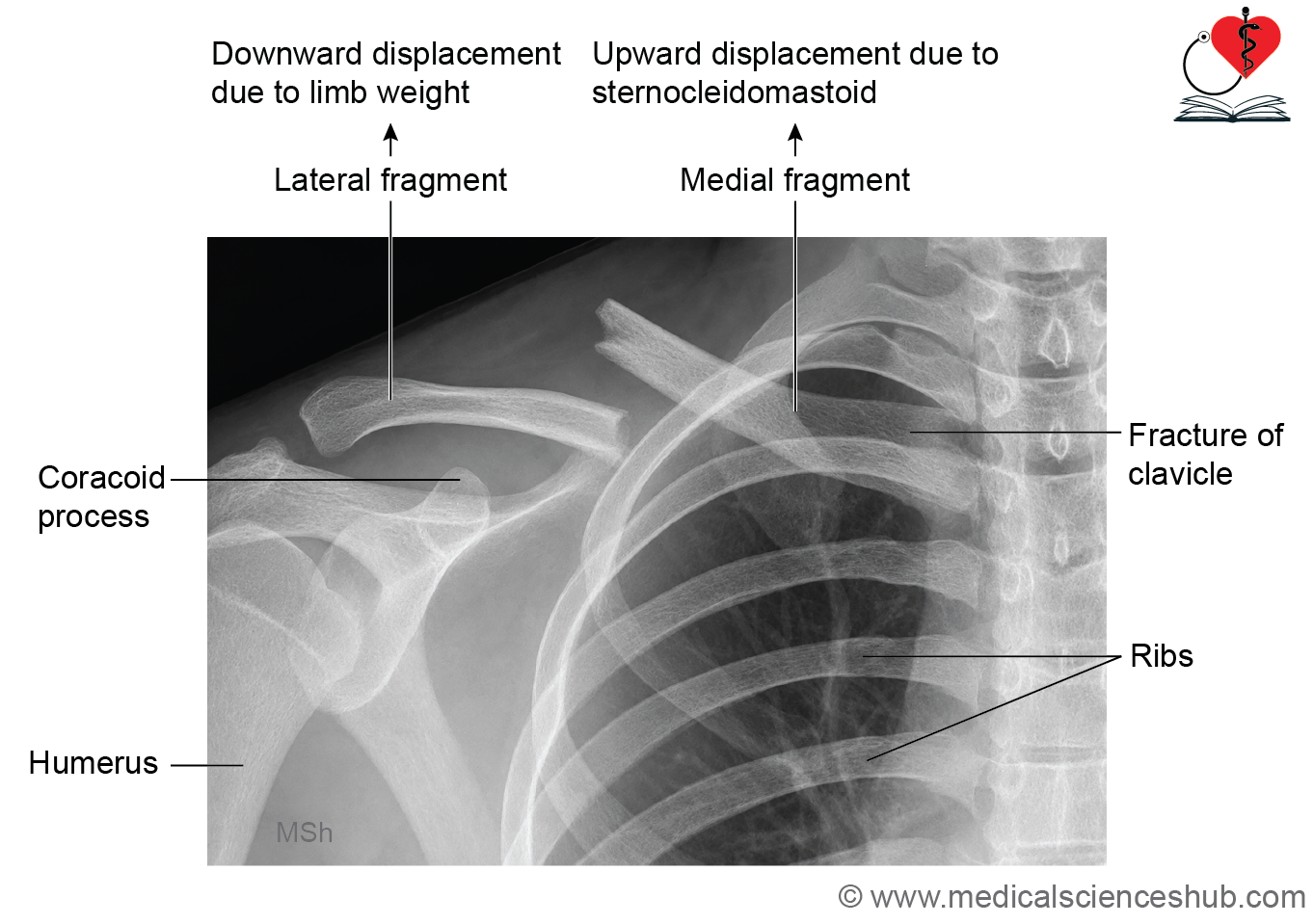
- The weakest part of the clavicle is the junction of the medial three-fifths and lateral two-fifths. This region is the most common site of clavicular fractures because it is structurally weaker than the adjacent parts.
- Clavicular fractures usually result from a fall on an outstretched hand, a direct blow to the shoulder, or high-energy trauma. The medial fragment is displaced superiorly by the pull of the sternocleidomastoid muscle. The lateral fragment is displaced inferiorly by the weight of the upper limb and the pull of surrounding muscles. The diagnosis is confirmed by radiographic examination, usually with plain radiographs.
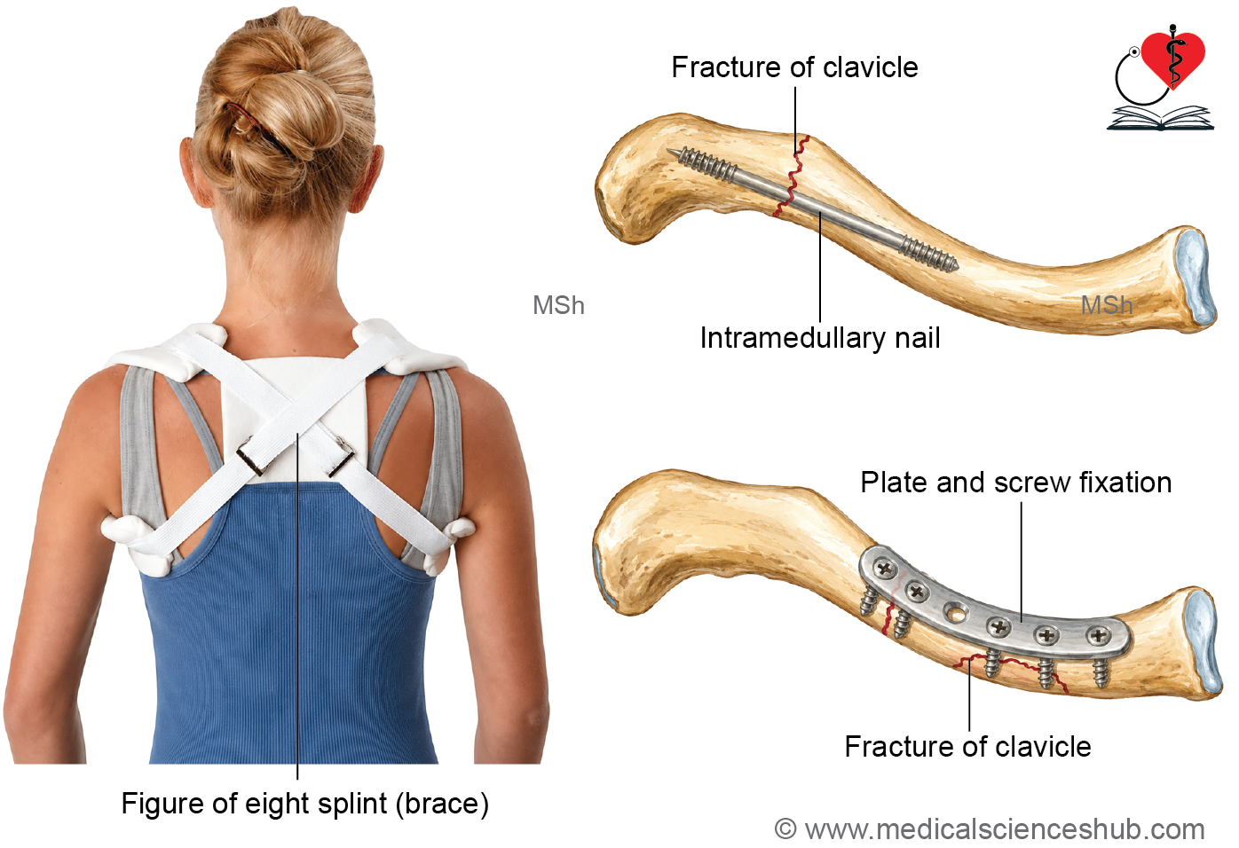
- Most fractures are treated conservatively using a sling or shoulder immobilizer, with or without a figure-of-eight brace. Surgical fixation with plates, screws, or intramedullary devices is indicated for displaced fractures, open fractures, neurovascular injury, skin compromise, or nonunion.
- Cleidocranial dysplasia is a congenital skeletal disorder characterized by absent or hypoplastic clavicles and abnormalities of the skull and teeth. Individuals with this condition can often approximate their shoulders in front of the chest.
- The weight of the upper limb is transmitted to the axial skeleton mainly through the coracoclavicular ligament, clavicle, sternoclavicular joint, and first rib.
Scapula
- The scapula is a large, flat, triangular bone that forms the posterior part of the shoulder girdle. It is commonly known as the shoulder blade.
- The scapula is located on the posterolateral aspect of the thoracic wall.
- It typically extends from the level of the second to the seventh ribs in the resting position.
- The scapula provides attachment for numerous muscles and plays an essential role in shoulder stability and upper limb movements.
Parts of Scapula
- The scapula consists of a body and three processes.
- The body is triangular. It has
- Two surfaces: the costal (anterior) surface and the posterior surface.
- Three borders: superior, medial, and lateral.
- Three angles: superior, inferior, and lateral.
- The lateral angle contains the glenoid cavity, which articulates with the head of the humerus to form the shoulder joint.
- It has three processes – the spine, acromion, and coracoid process.
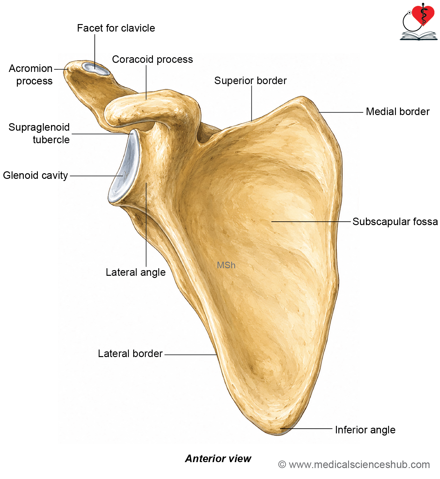
Figure H1.1.2: Hand-drawn diagram showing features of scapula – Anterior view (Click to see figure)
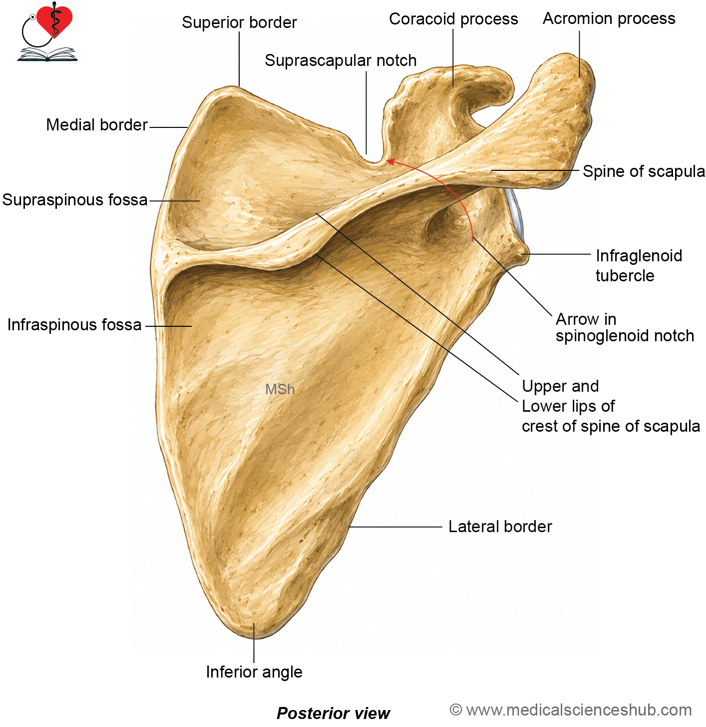
Figure H1.1.3: Hand-drawn diagram showing features of scapula – Posterior view (Click to see figure)
Anatomical Position and Side Determination
- Hold the scapula with its posterior surface facing backwards.
- The spine of the scapula should be visible, dividing the posterior surface into the supraspinous and infraspinous fossae.
- The pointed inferior angle must be directed downwards.
- The glenoid cavity should lie at the lateral angle and face forwards, slightly upwards, and laterally.
- The direction of the glenoid cavity is the most reliable feature for determining the side of the scapula.
Features of Scapula
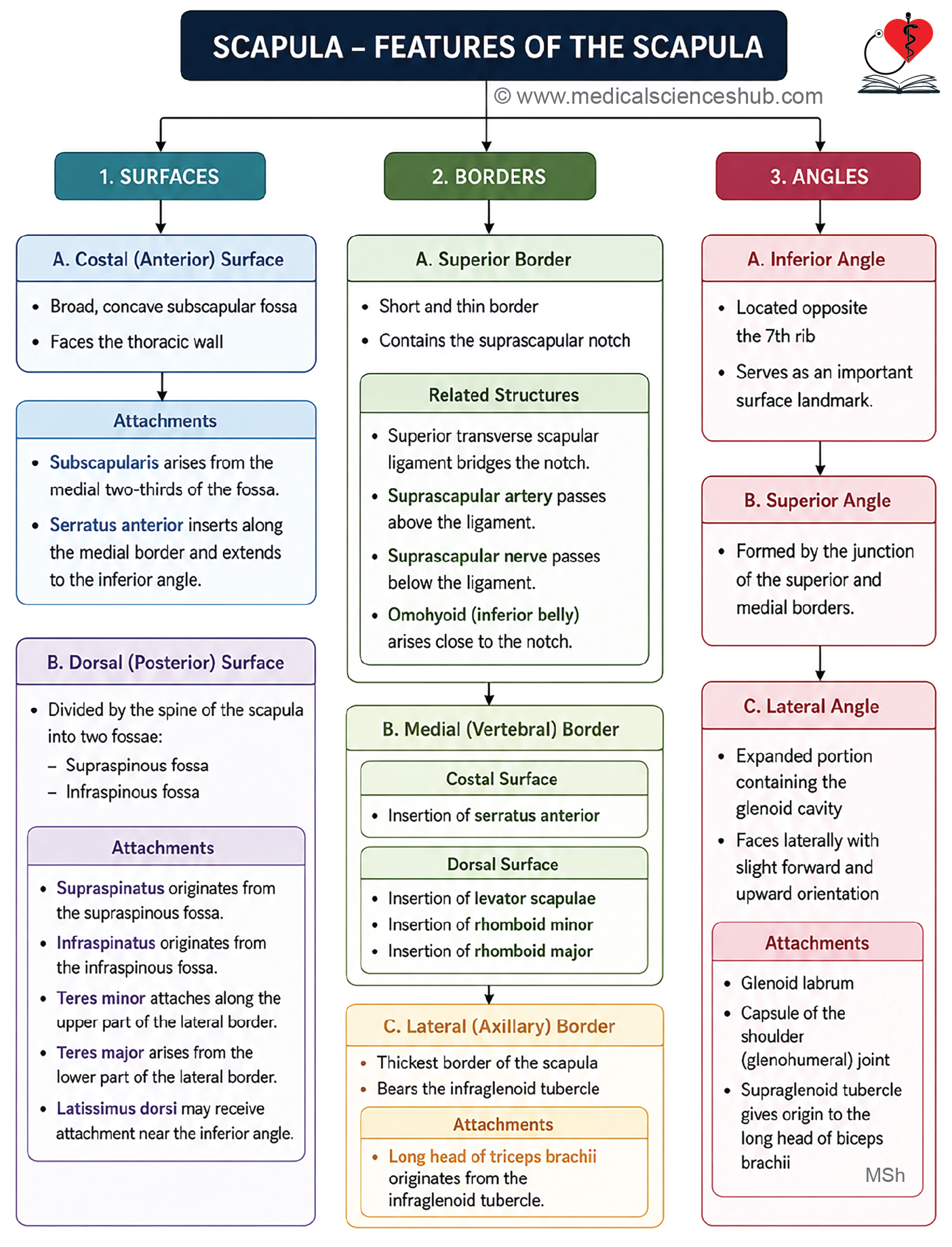
- The scapula is a flat, triangular bone that has two surfaces, namely the costal surface and the dorsal surface.
- It has three borders called the superior, medial, and lateral borders.
- The scapula also has three angles known as the superior, inferior, and lateral angles.
- It possesses three processes, namely the spine of the scapula, acromion process, and coracoid process.
Surfaces of Scapula
Costal surface
- The costal surface of the scapula is also known as the ventral surface or subscapular fossa.
- It is broad, concave, and faces anteromedially towards the thoracic wall.
- A rounded longitudinal ridge is present on the lateral part of this surface and is separated from the lateral border by a narrow groove.
- This ridge is more prominent in the upper part of the scapula and provides structural support.
- Four or five oblique ridges are seen on the costal surface and serve as attachment sites for muscle fibers.
Attachments
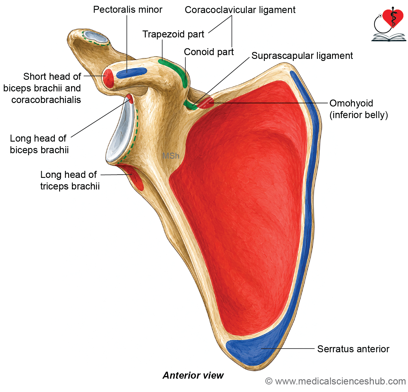
- The subscapularis muscle, which is multipennate in arrangement, arises from the medial two-thirds of the costal surface.
- A subscapular bursa lies between the neck of the scapula and the subscapularis muscle, reducing friction during shoulder movements.
- The lower five or six digitations of the serratus anterior muscle are inserted into the oval area near the inferior angle, while the remaining fibers attach along the ventral aspect of the medial border.
Dorsal surface
- The dorsal surface of the scapula is convex and faces posteriorly.
- A prominent spine of the scapula projects from the dorsal surface and divides it into two unequal fossae.
- The smaller supraspinous fossa lies above the spine, whereas the larger infraspinous fossa lies below it.
- These fossae provide broad surfaces for the attachment and origin of important shoulder muscles.
- The supraspinous and infraspinous fossae communicate laterally through the spinoglenoid notch.
- The spinoglenoid notch is situated lateral to the spine and posterior to the neck of the scapula.
- The suprascapular nerve and accompanying suprascapular vessels pass through this notch to reach the infraspinous fossa.
Attachments
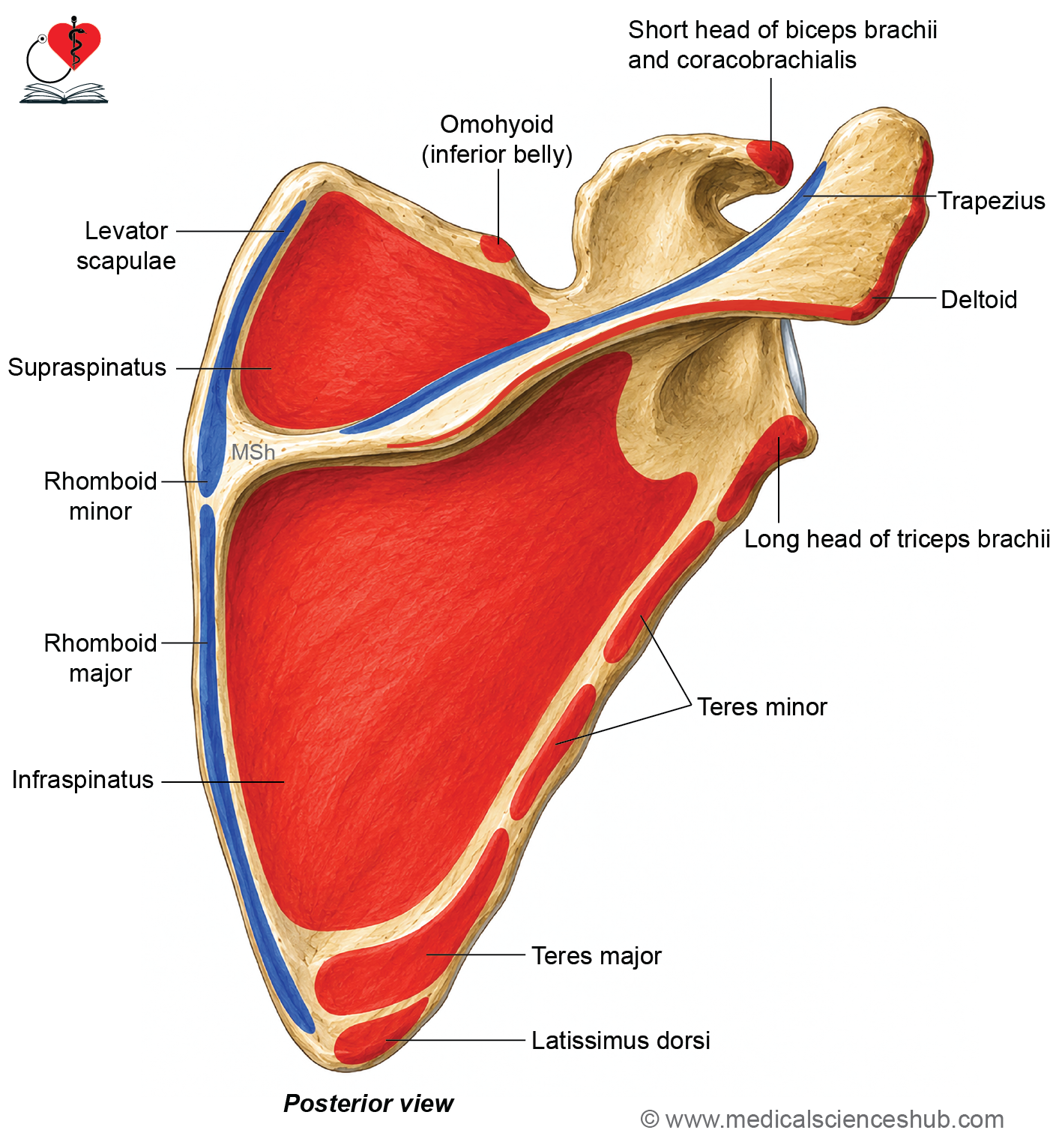
- The supraspinatus muscle arises from the medial two-thirds of the supraspinous fossa and the superior surface of the spine.
- The infraspinatus muscle originates from the medial two-thirds of the infraspinous fossa and the inferior surface of the spine.
- The teres minor muscle arises from the upper part of the lateral border, where a transverse groove transmits the circumflex scapular vessels.
- The teres major muscle originates from the lower part of the lateral border.
- A few fibers of the latissimus dorsi muscle may arise from the dorsal surface of the inferior angle of the scapula.
Borders of Scapula
• Scapula has three borders: Superior, medial, and lateral.
Superior border
- The superior border is the shortest and thinnest border of the scapula.
- It extends from the superior angle to the root of the coracoid process.
- Near the root of the coracoid process, the superior border presents the suprascapular notch.
Attachments
- The superior transverse scapular ligament bridges this notch and converts it into a suprascapular foramen.
- The suprascapular nerve passes through the foramen beneath the ligament to reach the supraspinous fossa.
- The suprascapular artery usually passes above the superior transverse scapular ligament.
- The inferior belly of the omohyoid muscle arises from the superior border close to the suprascapular notch.
Medial border
- The medial border, also called the vertebral border, is the longest and thinnest border of the scapula.
- It extends vertically from the superior angle to the inferior angle.
Attachments
- The serratus anterior muscle is inserted along the costal surface of the medial border.
- Its upper fibers attach between the superior angle and the root of the spine, the middle fibers attach below the spine, and the lower fibers insert near the inferior angle.
- On the dorsal surface, the levator scapulae muscle is inserted between the superior angle and the root of the spine.
- The rhomboid minor muscle is inserted opposite the root of the spine.
- The rhomboid major muscle is inserted from the spine to the inferior angle.
Lateral border
- The lateral border, also called the axillary border, is the thickest border of the scapula and extends from the lateral angle to the inferior angle.
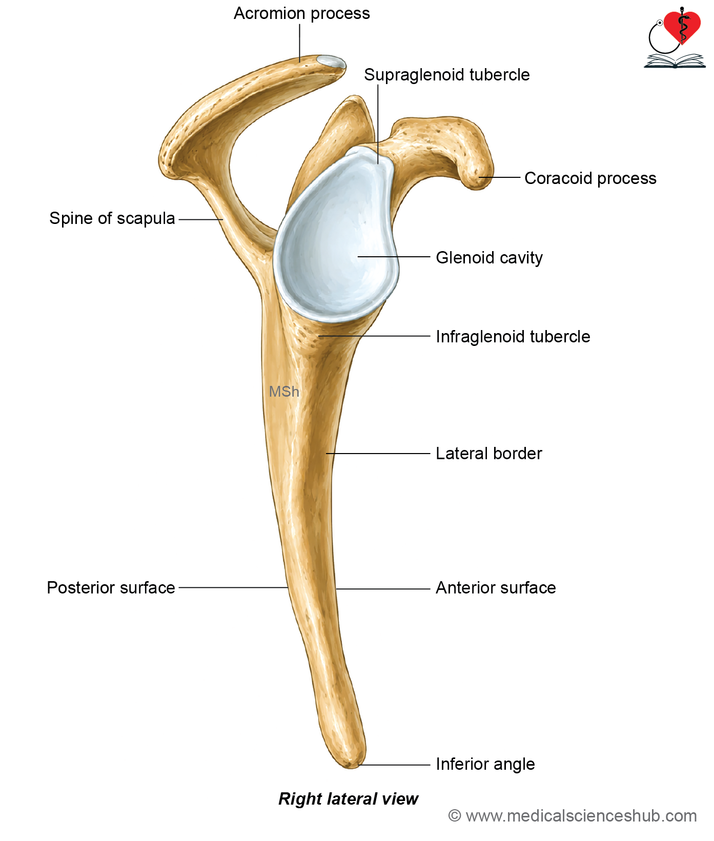
- The infraglenoid tubercle is a rough elevation located immediately below the glenoid cavity.
Attachments
- The long head of the triceps brachii muscle originates from the infraglenoid tubercle.
- The teres minor and teres major muscles arise from the dorsal surface of the lateral border.
Angles of Scapula
- Scapula has three angles: Superior, inferior, and lateral.
Inferior angle
- The inferior angle is the lowest and most pointed part of the scapula.
- It usually lies opposite the seventh rib or the seventh intercostal space in the anatomical position.
Attachments
- On its costal surface, the inferior angle provides insertion for the lower five or six digitations of the serratus anterior muscle.
- Its dorsal surface may give origin to a few fibers of the latissimus dorsi muscle.
Superior angle
- The superior angle is formed by the junction of the superior and medial borders of the scapula.
- It is covered by the trapezius muscle and serves as an important anatomical landmark.
Lateral angle
- The lateral angle forms the expanded head of the scapula and supports the glenoid cavity.
- A narrow neck of the scapula connects the lateral angle to the body of the bone.
- The glenoid cavity is a shallow, pear-shaped articular surface that faces laterally, slightly anteriorly, and slightly superiorly.
- It articulates with the head of the humerus to form the glenohumeral joint.
Attachments
- The supraglenoid tubercle, located above the glenoid cavity, gives origin to the long head of the biceps brachii muscle.
- The glenoid labrum, a fibrocartilaginous rim, is attached to the margin of the glenoid cavity and deepens the articular surface.
- The fibrous capsule of the shoulder joint is attached around the margin of the glenoid cavity, external to the glenoid labrum, and encloses the intracapsular origin of the long head of the biceps brachii tendon.
Processes of Scapula
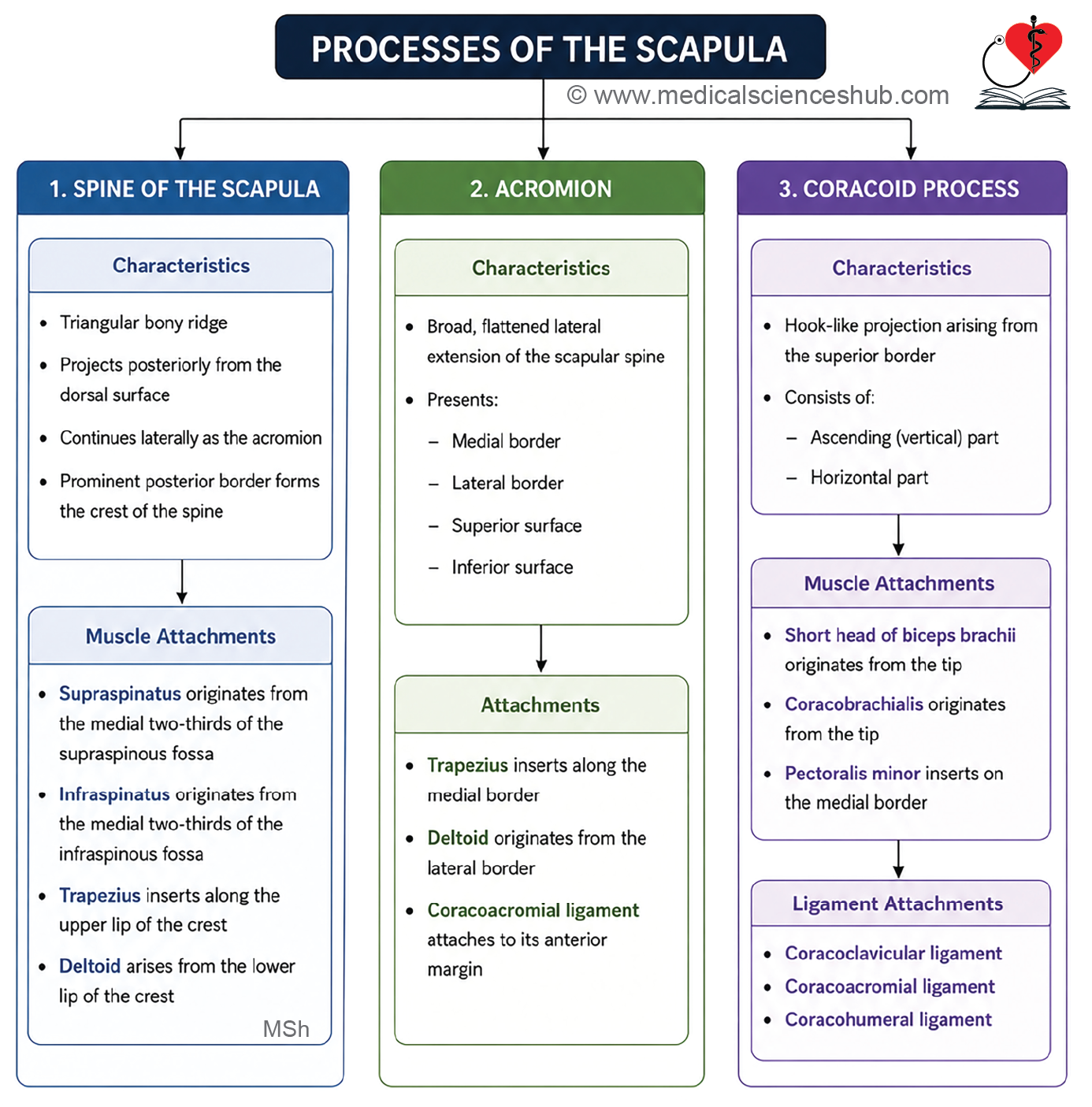
- Scapula has three processes: Spine, acromion, and coracoid processes.
Spine of scapula
- The spine of the scapula is a prominent triangular, shelf-like projection on the dorsal surface of the scapula.
- Its anterior border is firmly fused with the body of the scapula.
- The posterior border forms the crest of the spine, which has upper and lower lips separated by an intermediate rough area.
- Near the medial border, the two lips diverge to enclose a small triangular area.
- The lateral border is rounded and ends near the spinoglenoid notch.
- The upper surface of the spine is concave and continuous with the supraspinous fossa, whereas the lower surface is convex and continuous with the infraspinous fossa.
Attachments
- The supraspinatus muscle originates from the supraspinous fossa and the superior surface of the spine.
- The infraspinatus muscle arises from the infraspinous fossa and the inferior surface of the spine.
- The trapezius muscle is inserted into the upper lip of the crest, while the deltoid muscle originates from its lower lip.
Acromion process
- The acromion process is a broad, flattened projection that extends laterally from the spine of the scapula.
- It forms the highest point of the shoulder and contributes to the contour of the shoulder region.
- The acromion has a tip, medial and lateral borders, and superior and inferior surfaces.
- Its lateral border is continuous with the lower lip of the scapular spine, whereas the medial border continues with the upper lip.
- The medial border bears an oval articular facet for articulation with the lateral end of the clavicle, forming the acromioclavicular joint.
- The superior surface is rough, subcutaneous, and easily palpable, while the inferior surface is smooth.
Attachments
- The trapezius muscle is inserted into the medial border of the acromion.
- The middle fibers of the deltoid muscle originate from its lateral border.
- The coracoacromial ligament extends from the tip of the acromion to the coracoid process, forming the coracoacromial arch.
- The capsule of the acromioclavicular joint is attached around the margin of the articular facet.
Coracoid process
- The coracoid process is a strong, hook-like projection that arises from the superior part of the lateral angle of the scapula.
- It consists of a broad ascending part and a horizontal part that projects anterolaterally.
- The ascending part is thick and bears a smooth anterior surface and a rough posterior surface.
- The horizontal part has superior and inferior surfaces, medial and lateral borders, and a pointed tip.
Attachments
- The short head of the biceps brachii and the coracobrachialis muscle arise together from the tip of the coracoid process.
- The pectoralis minor muscle is inserted into the medial border of its horizontal part.
- The coracoclavicular ligament attaches to the coracoid process through its conoid and trapezoid parts.
- The conoid ligament attaches near the junction of the ascending and horizontal parts, whereas the trapezoid ligament attaches to the superior surface of the horizontal part.
- The coracoacromial ligament attaches to the lateral border, and the coracohumeral ligament attaches near the root of the coracoid process.
Ossification
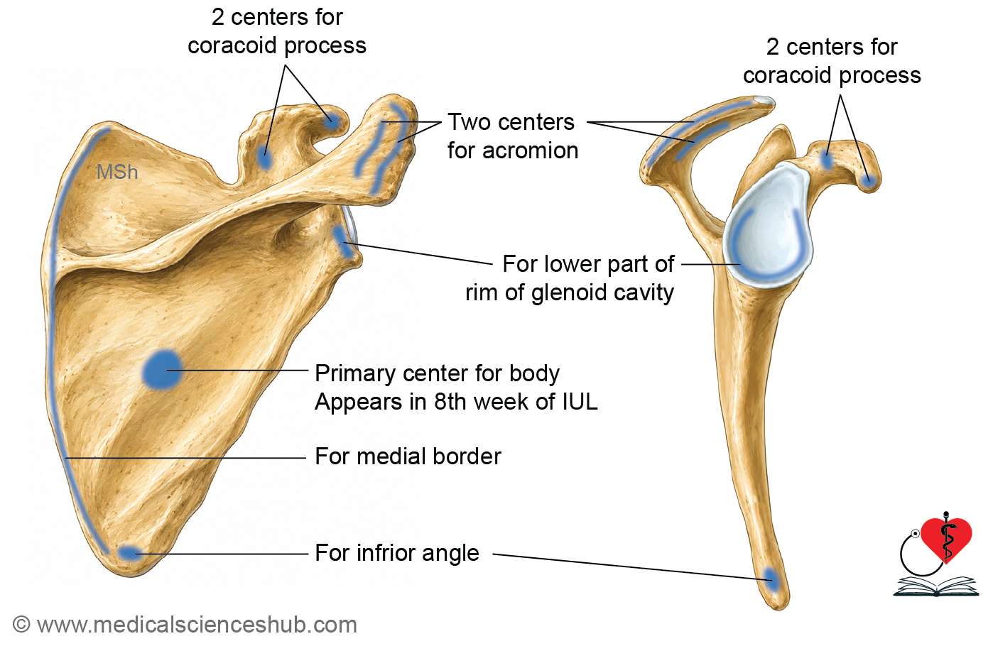
- The scapula develops from one primary ossification center and seven secondary ossification centers.
- The primary center for the body of the scapula appears during the eighth week of intrauterine life.
- The seven secondary centers include two for the coracoid process, two for the acromion, one for the medial border, one for the inferior angle, and one for the inferior part of the glenoid cavity.
- The first secondary center appears in the coracoid process during the first year after birth.
- Most remaining secondary centers appear around puberty.
- The coracoid process usually fuses with the scapular body by about 15 years, and complete fusion is generally achieved by 20 years of age.
CLINICAL INTEGRATION
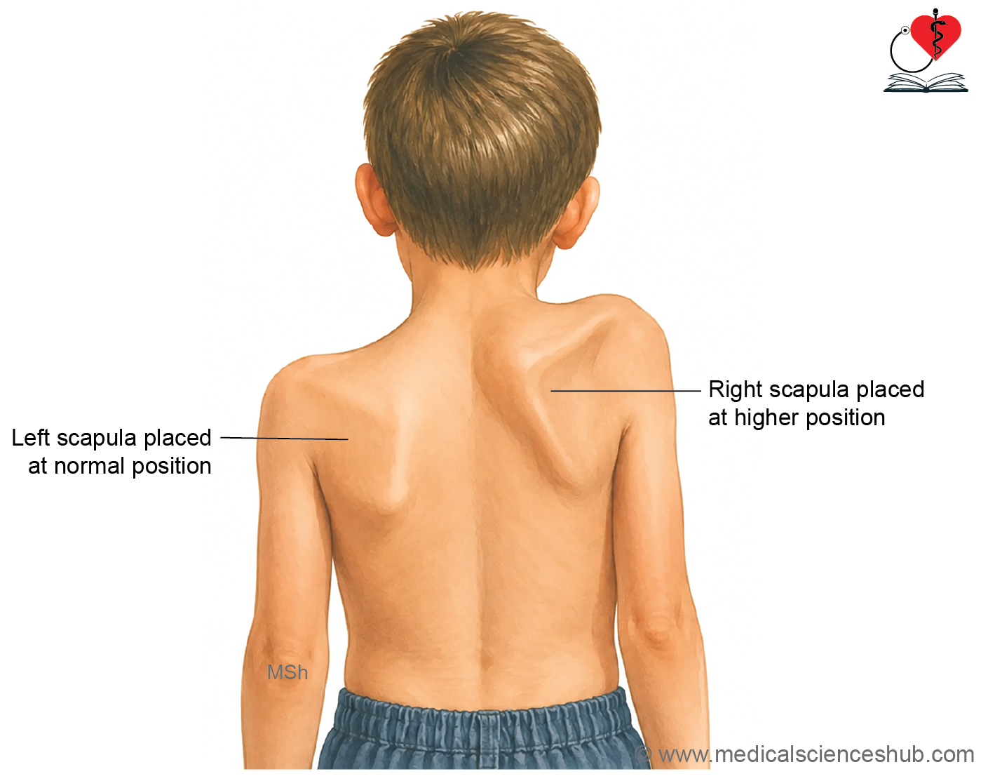
- Sprengel deformity is a congenital anomaly in which the scapula remains abnormally elevated on the posterior thoracic wall. It results from failure of the scapula to descend from the cervical region to its normal position during embryonic development. The affected scapula is usually smaller, rotated, and may restrict movements of the shoulder.
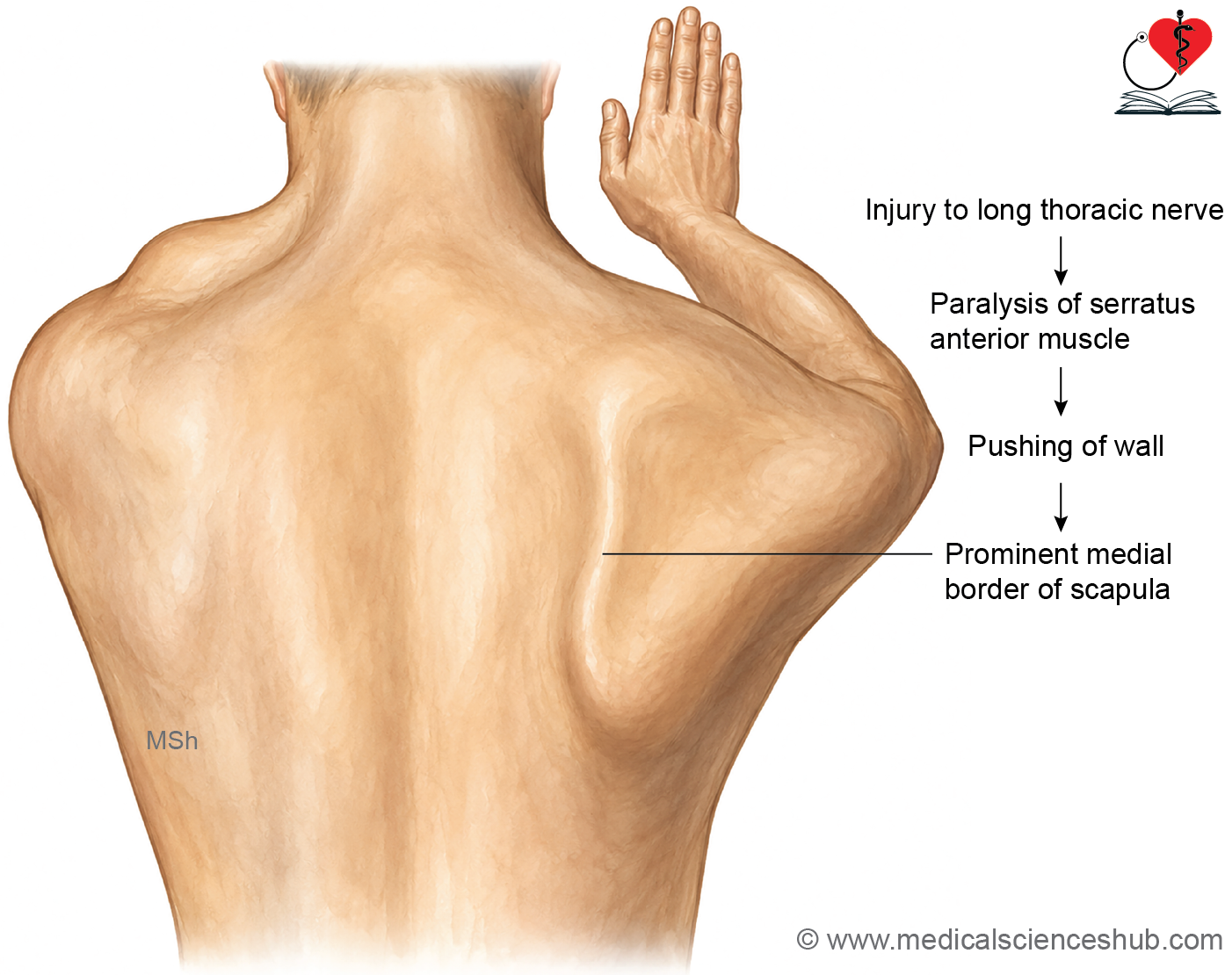
- Winging of the scapula commonly occurs due to paralysis of the serratus anterior muscle following injury to the long thoracic nerve. In this condition, the medial border and inferior angle of the scapula become prominent, especially when the patient pushes against a wall or performs a punching movement.
- Fractures of the scapula are uncommon and usually result from severe blunt trauma, such as road traffic accidents. Most scapular fractures are managed conservatively because the surrounding muscles provide good support and promote healing.
Important Questions
- Explain: Clavicle is a modified long bone.
- List the peculiarities of clavicle.
- Name the muscles attached to the medial border of scapula.
📝 Test Your Knowledge – Practice MCQs
Attempt the chapter MCQ quiz and assess your understanding of key concepts.
Click to see: Medical Disclaimer and Copyright Notice