
Competencies
- AN8.1: Features of individual bones (upper limb): Identify the given bone, its side, important features and keep it in anatomical position.
- AN8.2: Features of individual bones (upper limb): Identify and describe joints formed by the given bone.
- AN8.4: Features of individual bones (upper limb): Demonstrate important muscle attachment on the given bone. This chapter includes description of humerus.
Humerus
- The humerus is the bone of the arm and is the longest and strongest bone of the upper limb.
- It extends between the shoulder joint and the elbow joint.
- The humerus is divided into the upper end, shaft, and lower end.
- The upper end consists of the head, anatomical neck, greater tubercle, lesser tubercle, and intertubercular sulcus.
- The rounded head forms approximately one-third of a sphere and articulates with the glenoid cavity of the scapula.
- The shaft is cylindrical in its upper part and triangular in its lower part.
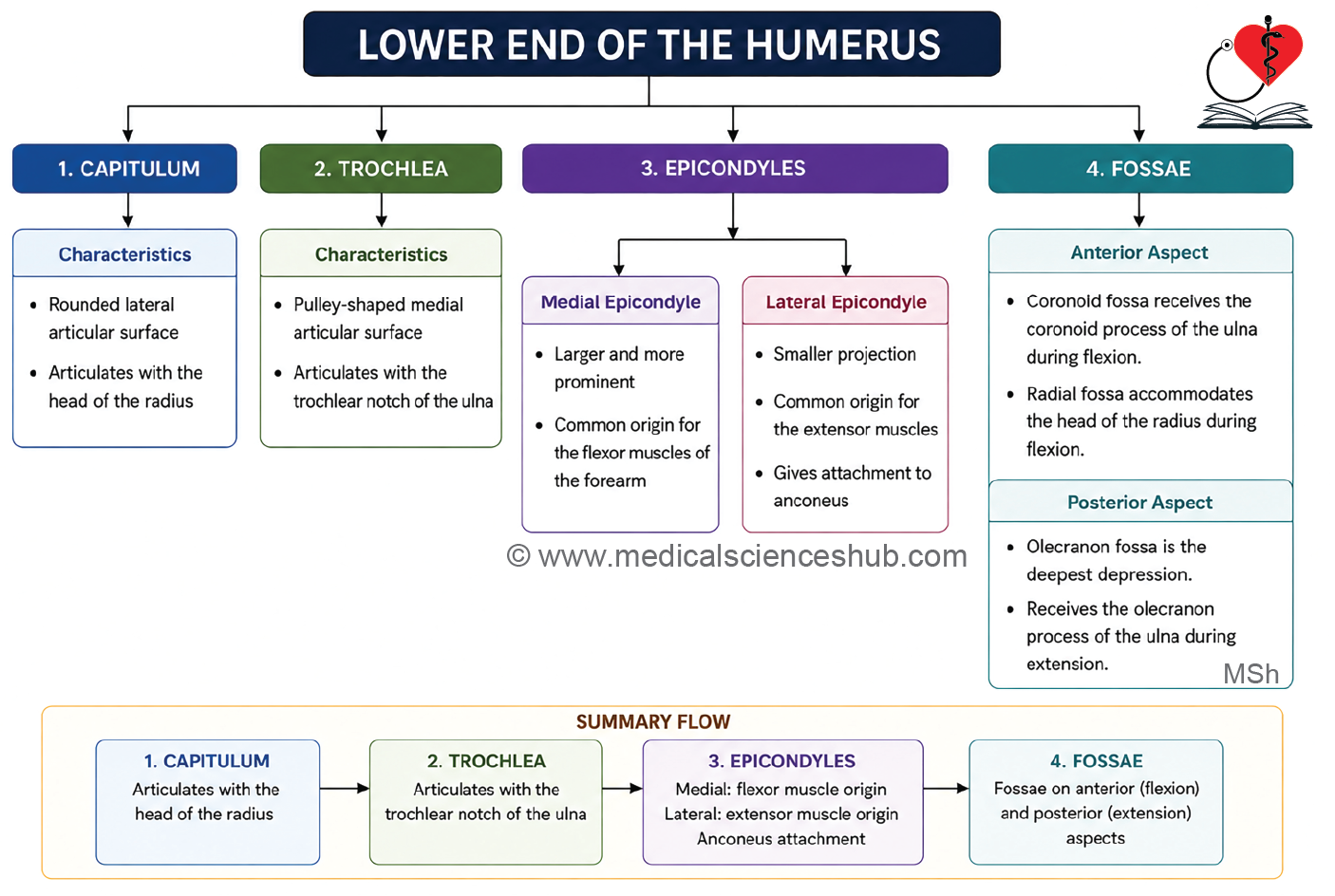
- The lower end includes the capitulum, trochlea, radial fossa, coronoid fossa, olecranon fossa, and medial and lateral epicondyles.
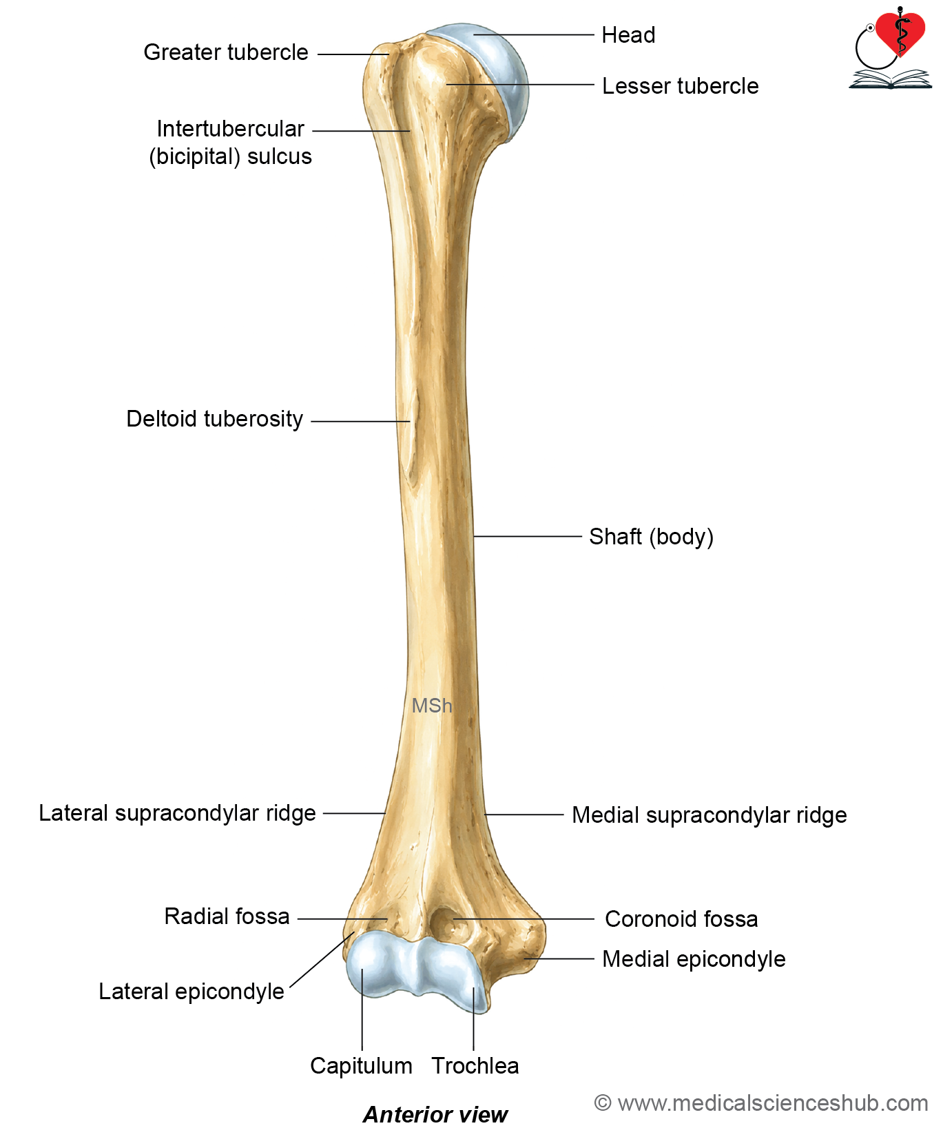
Figure H1.2.1: Hand-drawn diagram showing features of humerus – Anterior view (Click to see figure)
Side Determination and Anatomical Position
- Hold the humerus vertically with the rounded head directed upward, medially, and slightly backward.
- Ensure that the lesser tubercle faces anteriorly.
- Confirm that the medial epicondyle projects medially and lies on the side opposite the bone.
- These anatomical features help determine the correct side and anatomical position of the humerus accurately.
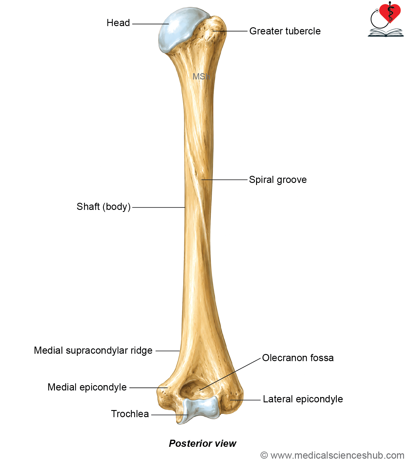
Figure H1.2.2: Hand-drawn diagram showing features of humerus – Posterior view (Click to see figure)
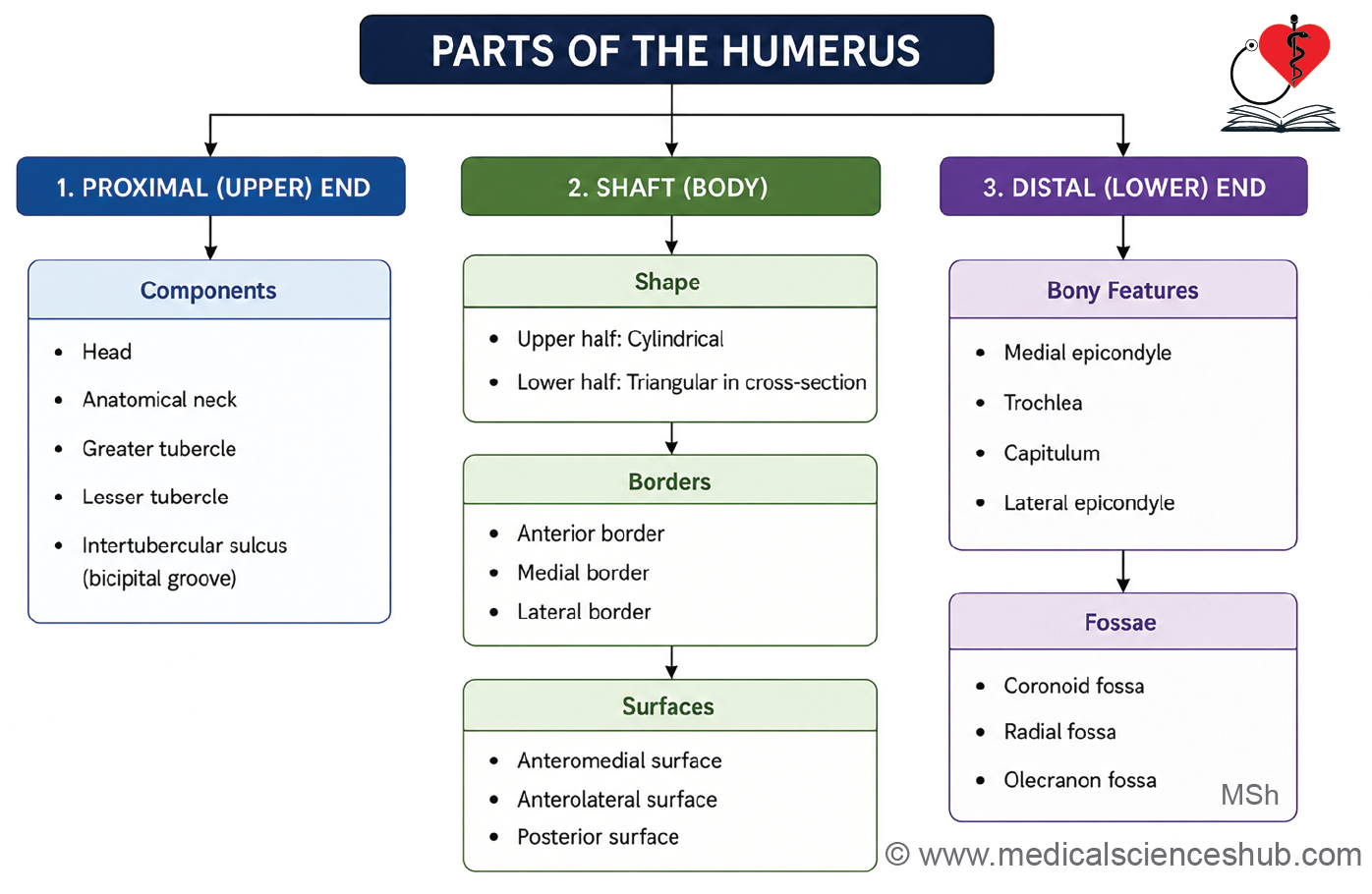
Features of Humerus
- Humerus is having these parts: upper end, shaft, and lower end
Upper End
- Upper end has head, neck, two tubercles (greater and lesser), and intertubercular sulcus.
Head
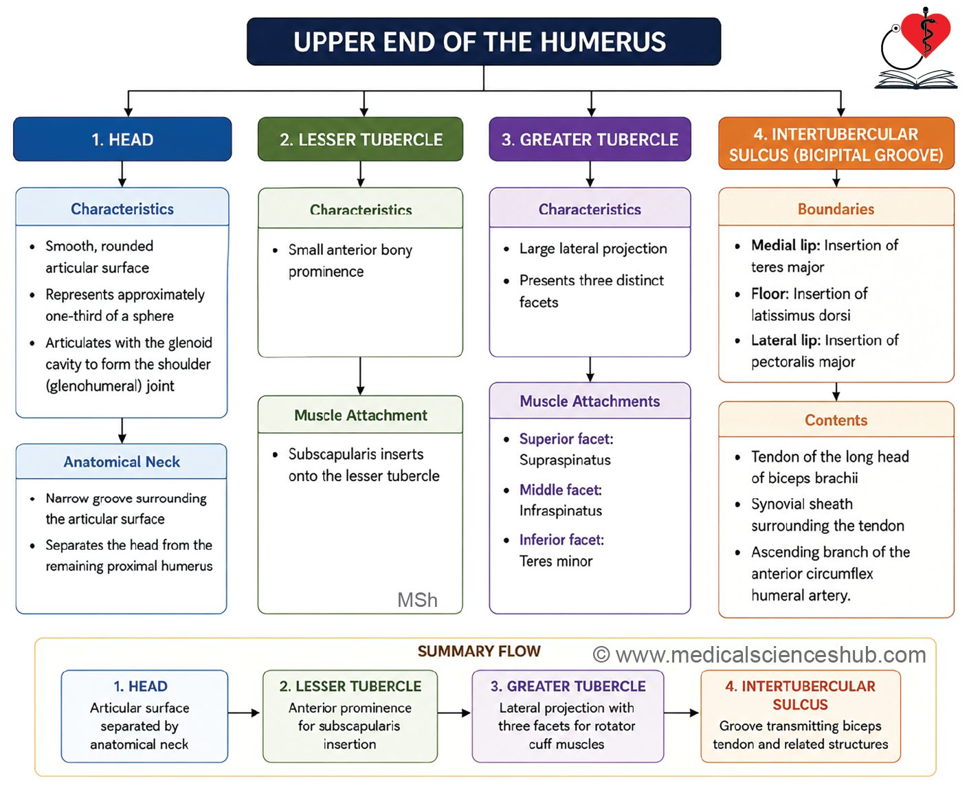
- The head of the humerus is smooth, globular, and forms approximately one-third of a sphere.
- It is directed medially, upward, and slightly posteriorly.
- The articular surface is covered by hyaline cartilage, which reduces friction during joint movements.
- The head articulates with the glenoid cavity of the scapula to form the shoulder joint.
Attachment
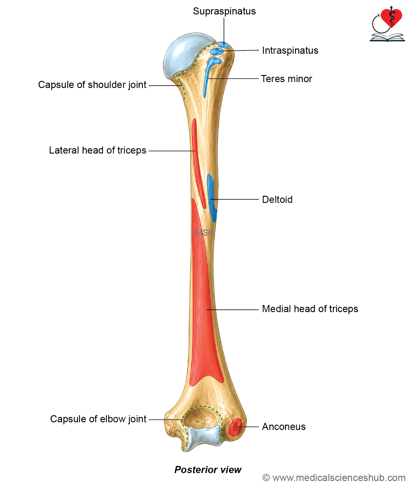
- The capsule of the shoulder joint is attached around the anatomical neck of the humerus.
- Medially, the capsule extends approximately 2 centimetres inferior to the anatomical neck onto the shaft.
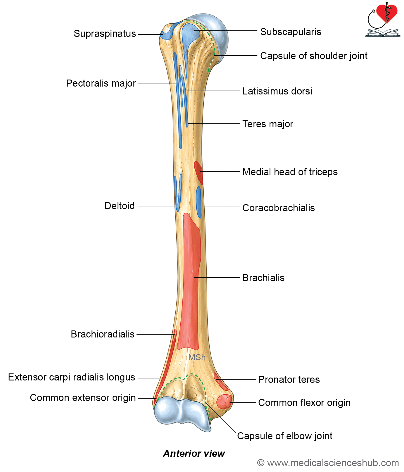
Lesser tubercle
The lesser tubercle is a small anterior projection located at the proximal end of the humerus.
Attachments
- It provides the insertion for the subscapularis muscle.
- The insertion of the subscapularis extends about 1 centimetre below the lesser tubercle onto the shaft.
Greater tubercle
- The greater tubercle is a prominent lateral projection at the proximal end of the humerus.
- It bears three distinct facets for muscle attachment.
Attachment
- The superior facet receives the insertion of the supraspinatus muscle.
- The middle facet provides attachment to the infraspinatus muscle.
- The inferior facet receives the insertion of the teres minor muscle, which extends approximately 2.5 centimetres onto the shaft.
- Together, these muscles form part of the rotator cuff, which stabilizes the shoulder joint and facilitates its movements.
Intertubercular sulcus (bicipital groove)
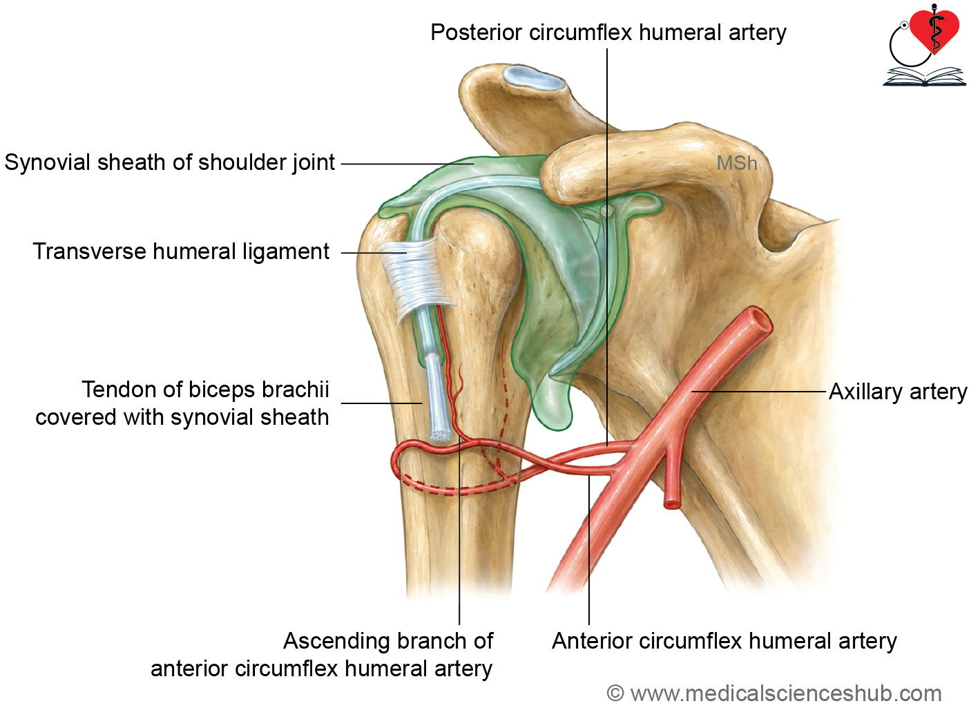
The intertubercular sulcus is a deep vertical groove between the greater and lesser tubercles on the anterior aspect of the humerus.
Content
It transmits the tendon of the long head of the biceps brachii, its synovial sheath, and the ascending branch of the anterior circumflex humeral artery.
Parts
- Intertubercular sulcus has medial lip, floor, and lateral lips.
Attachments
- The teres major inserts on the medial lip.
- The latissimus dorsi inserts on the floor.
- The pectoralis major inserts on the lateral lip.
- These muscle attachments contribute to powerful movements of the shoulder joint.
Shaft
- Shaft or body of the humerus is cylindrical in the upper half and triangular in lower half.
- Shaft has
- Three borders: Anterior, medial, and lateral.
- Three surfaces: Anterolateral, anteromedial, and posterior.
Borders
Anterior border
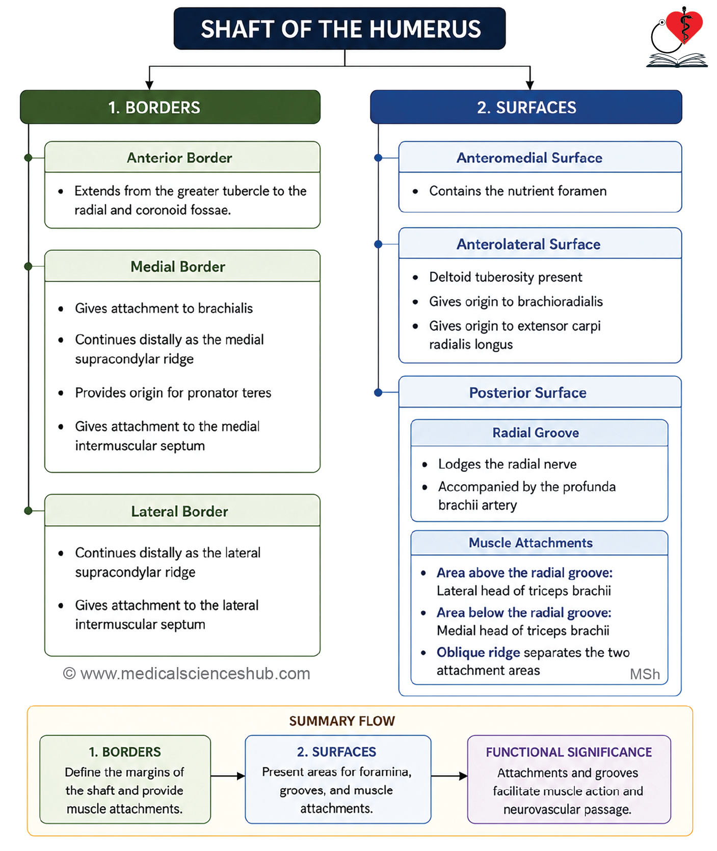
- The anterior border is divided into upper, middle, and lower parts.
- Its upper part forms the lateral lip of the intertubercular sulcus.
- The middle part forms the anterior margin of the deltoid tuberosity.
- The lower part is smooth and rounded and terminates above the radial fossa.
Medial border
- The medial border extends from the lesser tubercle to the medial epicondyle.
- Its upper part forms the medial lip of the intertubercular sulcus, while its lower part forms the medial supracondylar ridge.
Attachments
- The coracobrachialis muscle inserts into the middle of the medial border.
- The pronator teres muscle originates from the medial supracondylar ridge.
- The medial intermuscular septum is attached to the medial supracondylar ridge.
Lateral border
- The lateral border extends from the posterior aspect of the greater tubercle to the lateral epicondyle.
- Its lower part forms the lateral supracondylar ridge, which provides attachment for the lateral intermuscular septum.
Surfaces
Anteromedial surface
- The shaft of the humerus has three surfaces: anteromedial, anterolateral, and posterior.
- The anteromedial surface lies between the anterior and medial borders.
- Its upper narrow part forms the floor of the intertubercular sulcus.
- The nutrient foramen is located near the middle of this surface and is directed downward, indicating that the upper end is the growing end of the humerus.
Attachments
- The latissimus dorsi muscle inserts into the floor of the intertubercular sulcus.
- The brachialis muscle originates from the distal half of both the anteromedial and anterolateral surfaces.
Anterolateral surface
- The anterolateral surface lies between the anterior and lateral borders.
- It bears the deltoid tuberosity, a rough V-shaped elevation for muscle attachment.
Attachments
- The deltoid muscle inserts into the deltoid tuberosity.
- The brachioradialis and extensor carpi radialis longus muscles originate from the lateral supracondylar ridge and the adjacent distal anterolateral surface.
Posterior surface
- The posterior surface lies between the medial and lateral borders.
- Its upper part shows an oblique ridge that extends downward and laterally.
- The radial groove runs obliquely across the middle of the posterior surface.
Attachments
- The lateral head of the triceps brachii originates above the radial groove, whereas the medial head originates below it.
- The radial groove transmits the radial nerve and the profunda brachii vessels, making this region clinically important in humeral shaft fractures.
Lower End
- The lower end of the humerus is flattened anteroposteriorly and expanded from side to side.
- It forms the articular and non-articular components of the elbow joint.
Capitulum
- The capitulum is a rounded, convex articular surface located laterally.
- It articulates with the head of the radius during elbow movements.
Trochlea (trochileia means block of pulley, in Latin)
- The trochlea is a pulley-shaped medial articular surface that extends onto the anterior, inferior, and posterior aspects of the distal humerus.
- It articulates with the trochlear notch of the ulna.
- A shallow groove separates the trochlea from the capitulum.
- The medial margin of the trochlea projects farther distally than the lateral margin, contributing to the carrying angle of the elbow.
Medial epicondyle
- The medial epicondyle is a large, palpable bony projection on the medial side.
- The ulnar nerve passes behind the medial epicondyle in a groove, making it vulnerable to injury.
- The common flexor tendon of the superficial forearm flexor muscles originates from the anterior aspect of the medial epicondyle.
Lateral epicondyle
- The lateral epicondyle is a smaller rough projection on the lateral side.
- The common extensor tendon of the forearm originates from its anterolateral surface.
- The anconeus muscle originates from the posterior surface of the lateral epicondyle.
Fossae at lower end of humerus
- The distal end of the humerus contains three fossae that facilitate elbow movements.
- The coronoid fossa is a shallow anterior depression located above the trochlea.
- It receives the coronoid process of the ulna during elbow flexion.
- The radial fossa is a small anterior depression situated above the capitulum.
- It accommodates the head of the radius during elbow flexion.
- The olecranon fossa is a large, deep posterior depression located above the trochlea.
- It receives the olecranon process of the ulna during elbow extension.
- These fossae increase the range of elbow movement by accommodating adjacent bony processes.
Attachments
- The fibrous capsule of the elbow joint is attached around the margins of all three fossae.
- Medially, it is attached near the medial margin of the trochlea, and laterally near the lateral margin of the capitulum.
Important Points
- The head of the humerus is considerably larger than the glenoid cavity of the scapula.
- This size difference provides a wide range of shoulder movements but reduces the inherent stability of the joint.
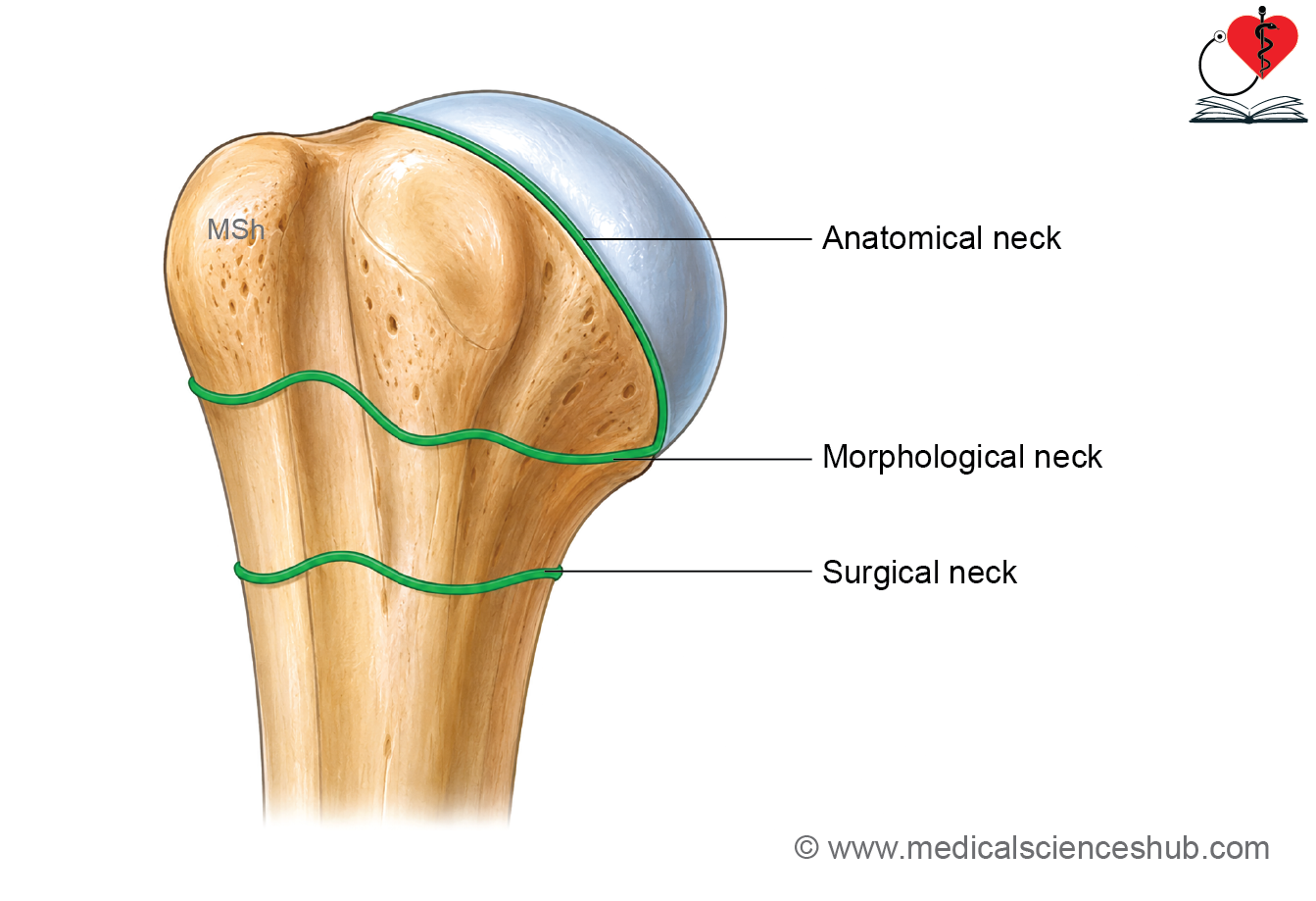
- The humerus has three necks: anatomical neck, surgical neck, and morphological neck.
- The anatomical neck is the narrow groove that separates the head from the tubercles. It serves as the attachment site for the fibrous capsule of the shoulder joint.
- The surgical neck is the constricted region located just distal to the greater and lesser tubercles. It is a common site of humeral fractures. The axillary nerve and the anterior and posterior circumflex humeral vessels closely surround the surgical neck and are at risk of injury following fractures.
- The morphological neck represents the junction between the upper epiphysis and the shaft. It lies approximately 0.5 centimetre above the surgical neck and passes through the lower parts of the greater and lesser tubercles.
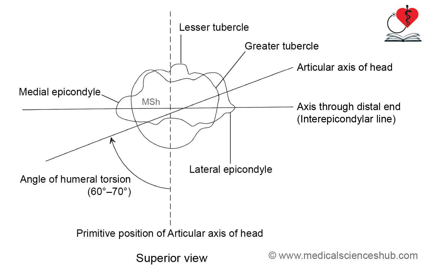
- The angle of humeral torsion is the angle between the proximal and distal articular axes of the humerus and normally measures about 60° to 70°.
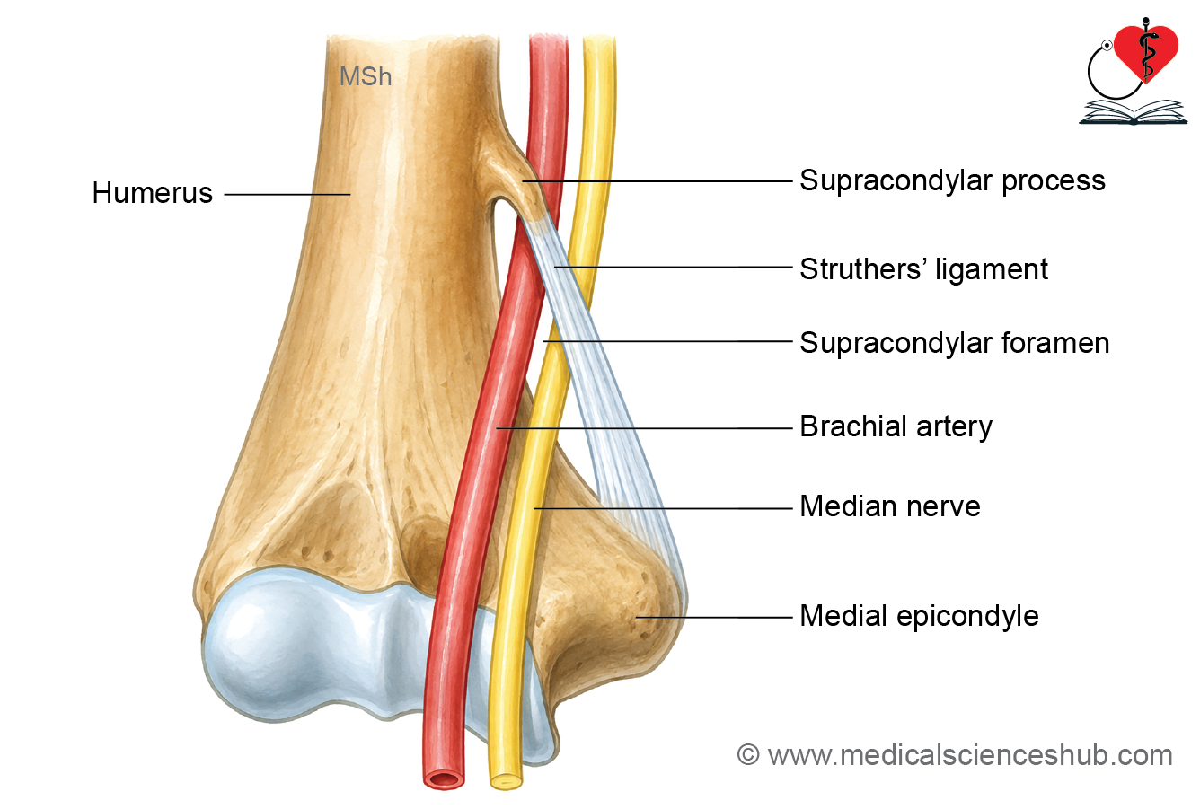
- A supracondylar process is an uncommon hook-like bony projection on the anteromedial aspect of the distal humerus, approximately 5 centimetres above the medial epicondyle.
- A fibrous Struthers ligament may extend from the supracondylar process to the medial epicondyle. Together, these structures may form a tunnel that transmits the median nerve and brachial artery.
- Compression within this tunnel may produce neurovascular symptoms and reduce blood supply to the forearm and hand.
Ossification
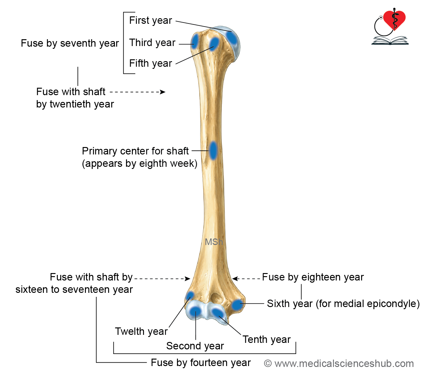
- The humerus ossifies from one primary and seven secondary ossification centers.
- The primary ossification center for the shaft appears during the eighth week of intrauterine life.
- Three secondary centers develop at the proximal end.
- The center for the head appears by the first year.
- The center for the greater tubercle appears by the third year.
- The center for the lesser tubercle appears by the fifth year.
- These proximal centers fuse with each other by the seventh year.
- They unite with the shaft by approximately 20 years of age.
- Four secondary ossification centers develop at the distal end.
- The center for the capitulum and lateral part of the trochlea appears by the second year.
Clinical anatomy
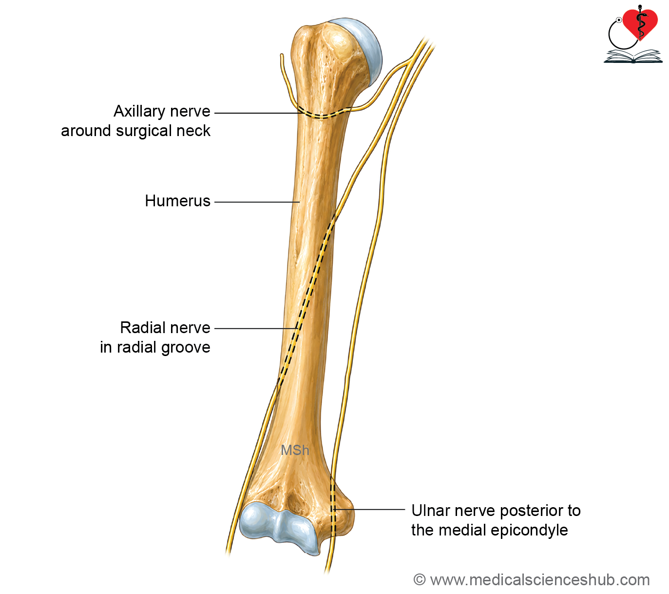
- The humerus is closely related to three major nerves. The axillary nerve winds around the surgical neck of the humerus. The radial nerve passes through the radial groove on the posterior surface of the shaft. The ulnar nerve runs posterior to the medial epicondyle.
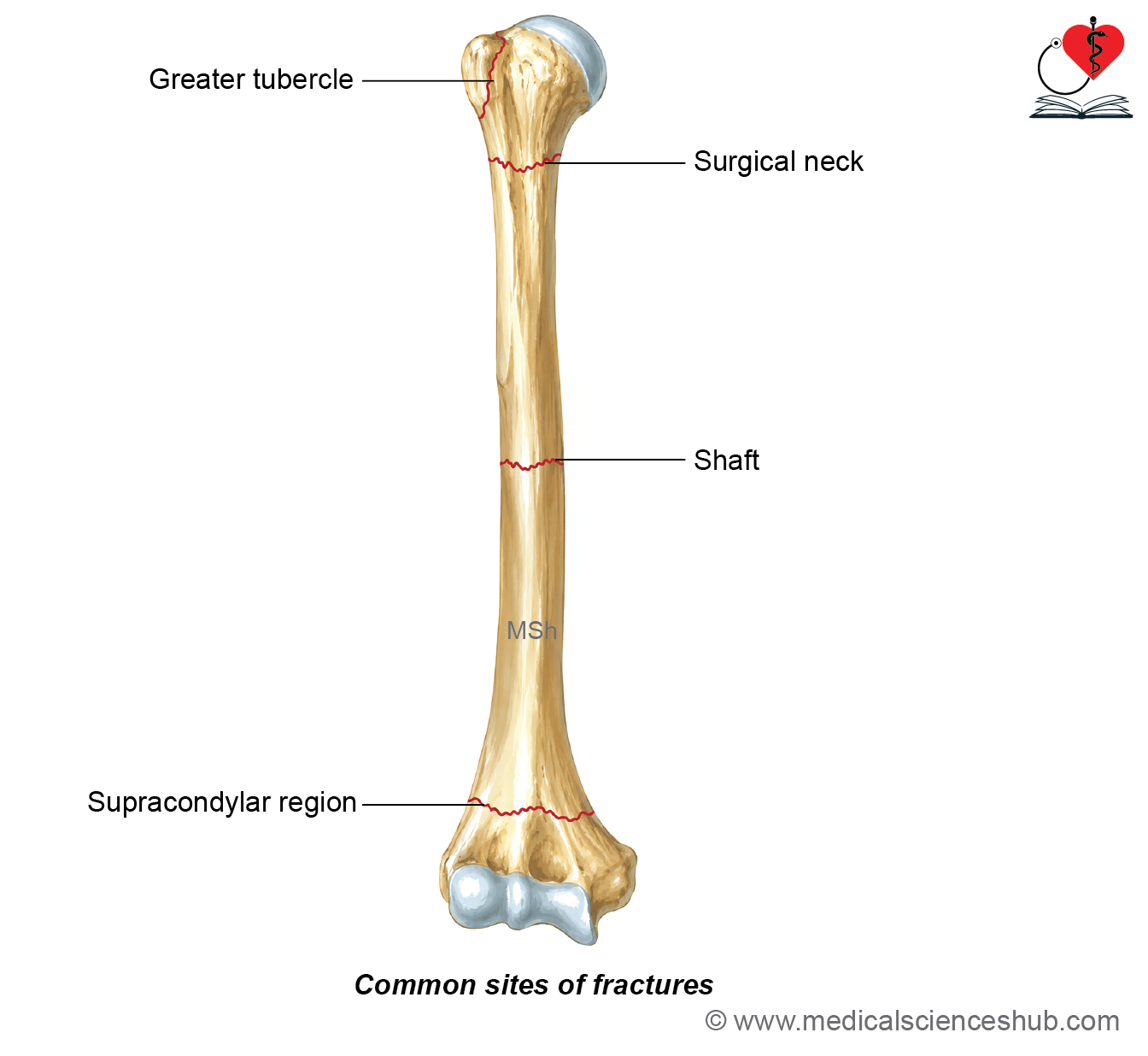
- Common fracture sites include the surgical neck, shaft, and supracondylar region of the humerus. Fracture of the surgical neck may injure the axillary nerve, causing weakness of the deltoid muscle and impaired shoulder abduction. Fracture of the shaft may damage the radial nerve, resulting in wrist drop. Fracture of the medial epicondyle may injure the ulnar nerve, producing sensory and motor deficits in the hand. In adolescents, an unfused medial epicondyle may be mistaken for a fracture on radiographs.
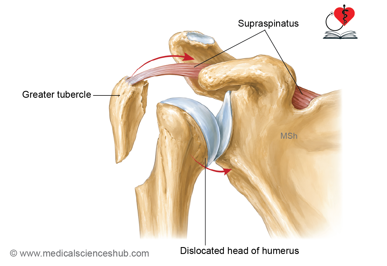
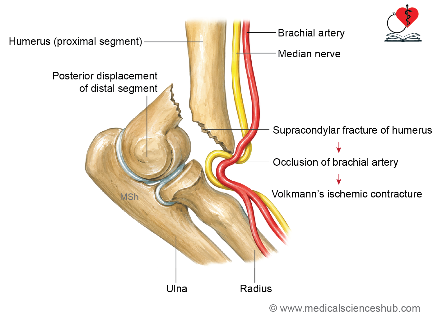
- In a supracondylar fracture, the distal fragment is commonly displaced posteriorly. Injury to the brachial artery in supracondylar fractures may lead to Volkmann ischemic contracture, causing permanent flexion deformity of the wrist and fingers. An avulsion fracture of the greater tubercle results from traction by the attached rotator cuff muscles.
- Fractures of the humeral shaft may be transverse, oblique, spiral, comminuted, greenstick, or open.
- The head of the humerus most commonly dislocates anteroinferiorly beneath the coracoid process (subcoracoid dislocation).
Important Questions
- Write a short note on lower end of humerus.
- Supracondylar fracture of humerus.
- List the nerve closely related to humerus.
📝 Test Your Knowledge – Practice MCQs
Attempt the chapter MCQ quiz and assess your understanding of key concepts.
Click to see: Medical Disclaimer and Copyright Notice