
Competencies
- AN8.1: Features of individual bones (upper limb): Identify the given bone, its side, important features, and keep it in anatomical position.
- AN8.2: Features of individual bones (upper limb): Identify and describe joints formed by the given bone.
- AN8.3: Enumerate peculiarities of clavicle.
- AN8.4: Features of individual bones (upper limb): Demonstrate important muscle attachment on the given bone. This chapter includes description of scapula and clavicle.
Radius
- The radius forms the lateral skeletal framework of the forearm in the anatomical position. The term radius is a Latin word meaning ‘ray’.
- Among the bones of the lower limb, the radius corresponds developmentally to the tibia, which lies on the medial side of the leg.
Parts
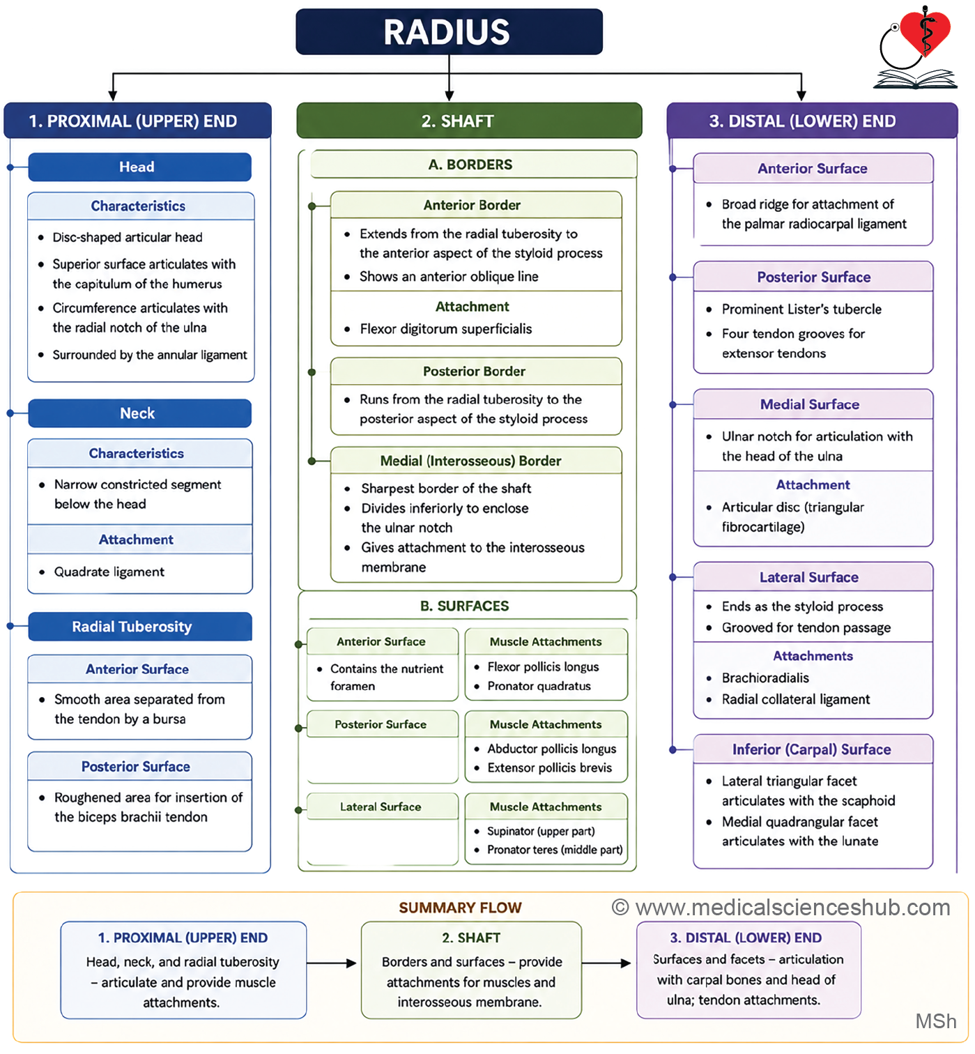
- The radius consists of an upper end, a shaft, and a lower end.
- The upper end includes a disc-shaped head, a narrow neck, and the radial tuberosity.
- The shaft is long, curves laterally, and possesses a sharp medial interosseous border for attachment of the interosseous membrane.
- The lower end is expanded and bears a laterally projecting styloid process and the dorsal tubercle (Lister’s tubercle) on its posterior surface.
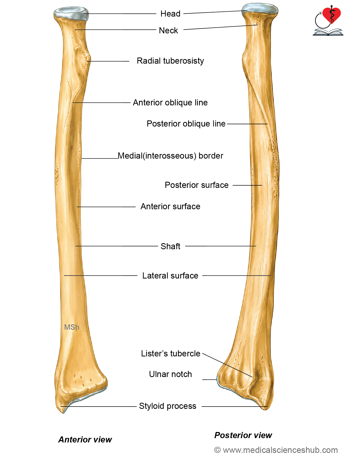
Figure H1.3.1: Hand-drawn diagram showing features of radius (Click to see figure)
Side Determination and Anatomical Position
- Hold the radius with the disc-shaped head directed upward and the expanded lower end directed downward.
- Keep the anterior concavity of the shaft facing forward and the dorsal tubercle (Lister’s tubercle) facing posteriorly.
- The styloid process should project laterally. Its direction identifies the side of the bone, with the styloid process pointing toward the corresponding upper limb.
Features of Radius
- Radius has upper end, shaft, and lower end.
Upper End
- The upper end of radius has head, neck, and radial tuberosity.
Head
- The head of the radius is disc-shaped and covered by hyaline cartilage over its articular surfaces.
- Its superior concave articular facet articulates with the capitulum of the humerus, forming the humeroradial component of the elbow joint.
- The smooth articular circumference articulates with the radial notch of the ulna and is enclosed by the annular ligament, forming the superior radioulnar joint.
Neck
- The neck is the narrow, cylindrical segment immediately below the head of the radius.
- The lower margin of the annular ligament surrounds the neck and stabilizes the superior radioulnar joint.
Attachments
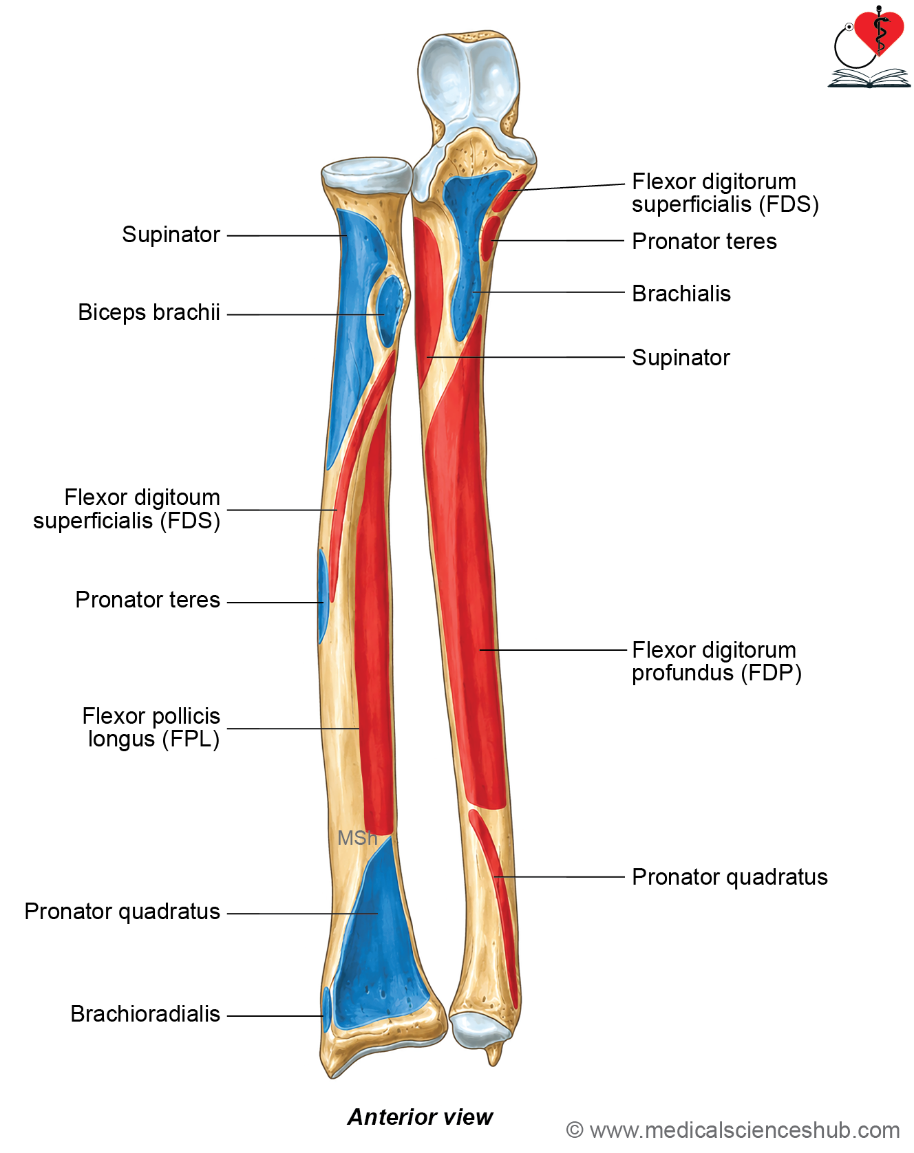
- The supinator muscle is inserted onto the anterior, lateral, and posterior aspects of the neck, extending onto the proximal shaft.
- The quadrate ligament is attached to the medial surface of the neck.
Radial tuberosity
- The radial tuberosity is a rough bony prominence located just distal to the neck on the medial aspect of the proximal radius.
Shaft
- The shaft of the radius is narrow and cylindrical proximally and gradually broadens distally.
- It has three borders (anterior, posterior, and interosseous) and three surfaces (anterior, posterior, and lateral).
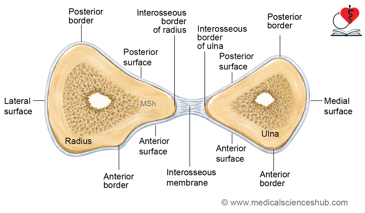
Borders
Anterior border
- The anterior border extends from the anterolateral aspect of the radial tuberosity to the styloid process.
- In its proximal part, the border descends obliquely downward and laterally, forming the anterior oblique line.
- The distal part becomes sharp and crest-like and is easily palpable beneath the skin.
Attachment
- The radial head of the flexor digitorum superficialis originates from the anterior oblique line, making it an important site for muscle attachment.
Posterior border
- The posterior border is distinct only along the middle part of the shaft.
- It extends from the posterior aspect of the radial tuberosity to the styloid process.
- Its proximal oblique segment forms the posterior oblique line, which runs downward and laterally.
Medial (interosseous) border
- The interosseous border is the sharpest border of the radius and begins below the radial tuberosity on its posteromedial aspect.
- Distally, it divides into two ridges, enclosing a small triangular area just proximal to the ulnar notch.
- Attachments
- The interosseous membrane is attached to the lower three-fourths of the interosseous border and to the posterior margin of the triangular area.
- The pronator quadratus is inserted into the distal one-fourth of the anterior surface of the radius and the adjacent triangular area near the lower end.
Surfaces
- Radius has three surfaces: Anterior, posterior, and lateral.
Anterior surface
- The anterior surface lies between the anterior and interosseous borders.
- It is narrow proximally, broad distally, and slightly concave.
- A nutrient foramen is present in its upper part and is directed proximally because the distal end is the growing end of the radius.
- The nutrient artery usually arises from the anterior interosseous artery.
Attachments
- The flexor pollicis longus originates from the proximal two-thirds of this surface.
- The pronator quadratus is inserted into its distal one-fourth and the adjacent triangular area near the lower end.
Posterior surface
- The posterior surface lies between the posterior and interosseous borders.
Attachments
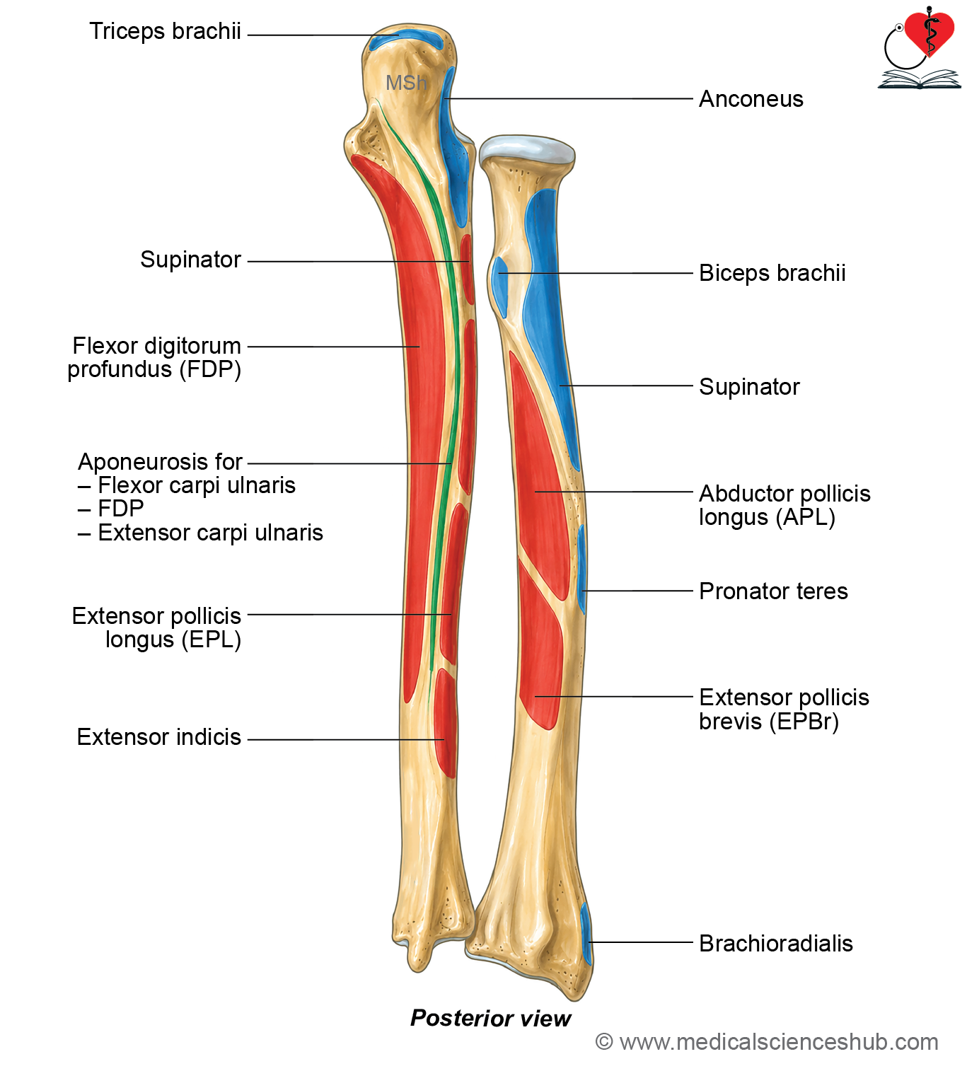
- The abductor pollicis longus and extensor pollicis brevis originate from this surface.
Lateral surface
- The lateral surface lies between the anterior and posterior borders of the radial shaft.
- It is convex laterally, with a rough oblique ridge marking the point of maximum convexity.
- Superiorly, the lateral surface is broader and continues with the neck of the radius.
Attachments
- The supinator is inserted into the upper part of this surface, whereas the pronator teres is inserted into its middle part.
Lower End
- Lower end of radius is broad and expanded. It has 5 surfaces: Anterior, posterior, medial, lateral, and inferior.
Anterior surface of lower end
- The anterior surface of the lower end is separated from the shaft by a prominent transverse ridge.
Attachments
- The palmar radiocarpal ligament is attached to this ridge, contributing to the stability of the wrist joint.
Posterior surface of lower end
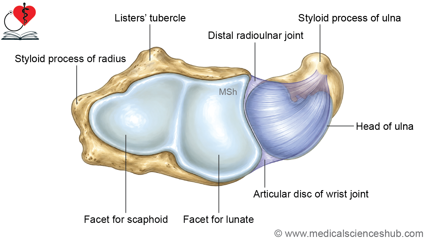
- The posterior surface of the lower end of the radius is broad and contains a small dorsal tubercle with four shallow grooves for extensor tendons.
- The dorsal tubercle acts as a pulley, changing the direction of the tendon of the extensor pollicis longus.
- The lateral groove on the posterior aspect of the styloid process transmits the tendons of abductor pollicis longus and extensor pollicis brevis.
- The second groove transmits the tendons of extensor carpi radialis longus and extensor carpi radialis brevis.
- The oblique groove medial to the dorsal tubercle transmits the tendon of extensor pollicis longus.
- The medial groove transmits the tendons of extensor digitorum and extensor indicis, along with the posterior interosseous nerve and the terminal part of the anterior interosseous artery.
Medial surface of lower end
- The medial surface of the lower end of the radius contains the ulnar notch, which articulates with the head of the ulna to form the inferior radioulnar joint.
- A smooth ridge between the ulnar notch and the inferior articular surface provides attachment for the base of the triangular articular disc of the inferior radioulnar joint.
Lateral surface of lower end
- The lateral surface forms the styloid process, which projects distally beyond the rest of the bone.
- A groove on the lateral aspect of the styloid process transmits the tendons of abductor pollicis longus and extensor pollicis brevis.
- The radial artery passes medial to the styloid process of the radius.
Attachments
- The tendon of brachioradialis inserts at the base of the styloid process.
- The radial collateral ligament of the wrist is attached to the tip of the styloid process.
Inferior surface of lower end
- The inferior surface of the lower end of the radius is concave, triangular, and covered by articular cartilage, forming part of the wrist joint.
- A faint ridge divides this surface into a lateral triangular facet for the scaphoid and a medial quadrangular facet for the lunate.
Attachment
- The capsule of the wrist joint is attached to the anterior and posterior margins of the inferior articular surface.
Ossification
- The radius develops from one primary and two secondary ossification centers.
- The primary ossification center appears in the eighth week of intrauterine life and forms the shaft.
- A secondary ossification center appears at the lower end during the first year after birth and fuses with the shaft between 17 and 19 years of age.
- Another secondary ossification center appears at the upper end around the fifth year and fuses with the shaft between 14 and 17 years of age.
- Because the lower end fuses later than the upper end, the lower end of the radius is the growing end of the bone.
CLINICAL INTEGRATION
- Fractures of radius are classified into fractures of the proximal end, shaft, and distal end.
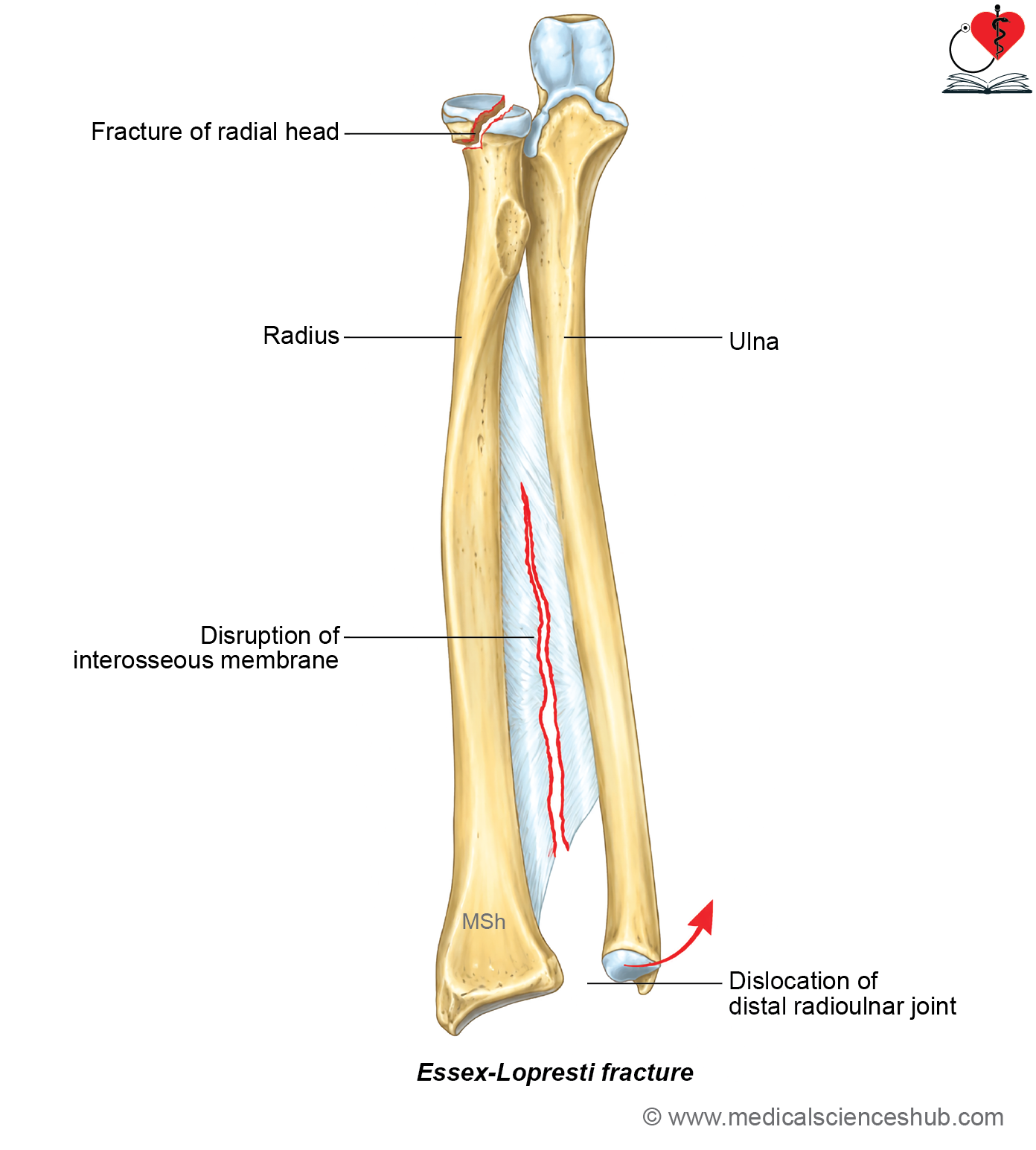
- Essex-Lopresti fracture: An Essex-Lopresti fracture consists of a fracture of the radial head with disruption of the interosseous membrane and dislocation of the distal radioulnar joint.
- Distal radius fractures include Colles’, Smith’s, Barton’s, and Chauffeur’s fractures.
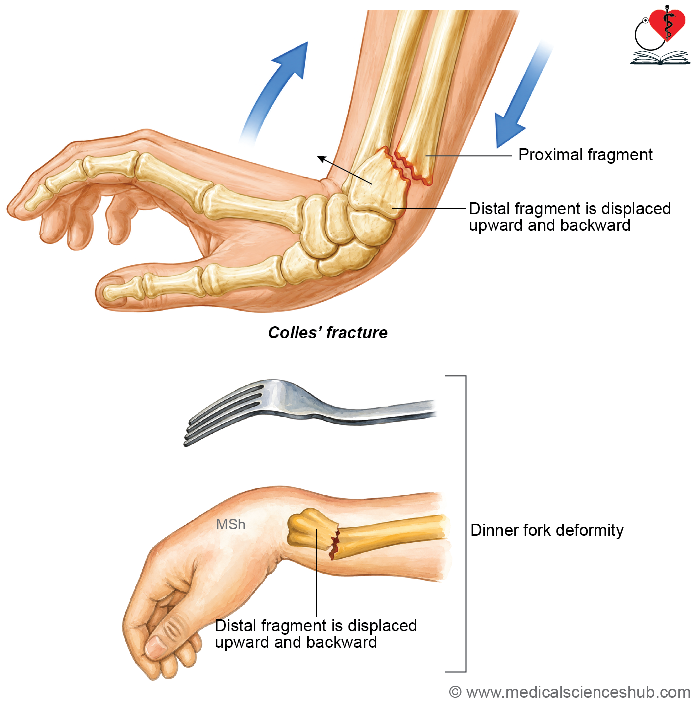
- Colles’ fracture: A Colles’ fracture occurs about 2 centimetres proximal to the distal end of the radius, with the distal fragment displaced posteriorly and proximally. It usually results from a fall on an outstretched hand. The characteristic posterior displacement produces a dinner-fork deformity.
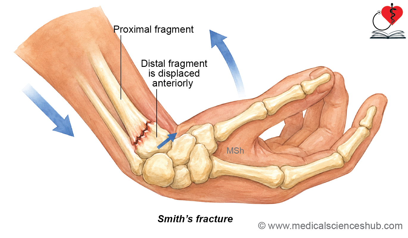
- Smith’s fracture: A Smith’s fracture is the reverse of a Colles’ fracture, with anterior displacement of the distal fragment, commonly following a fall on a flexed wrist.
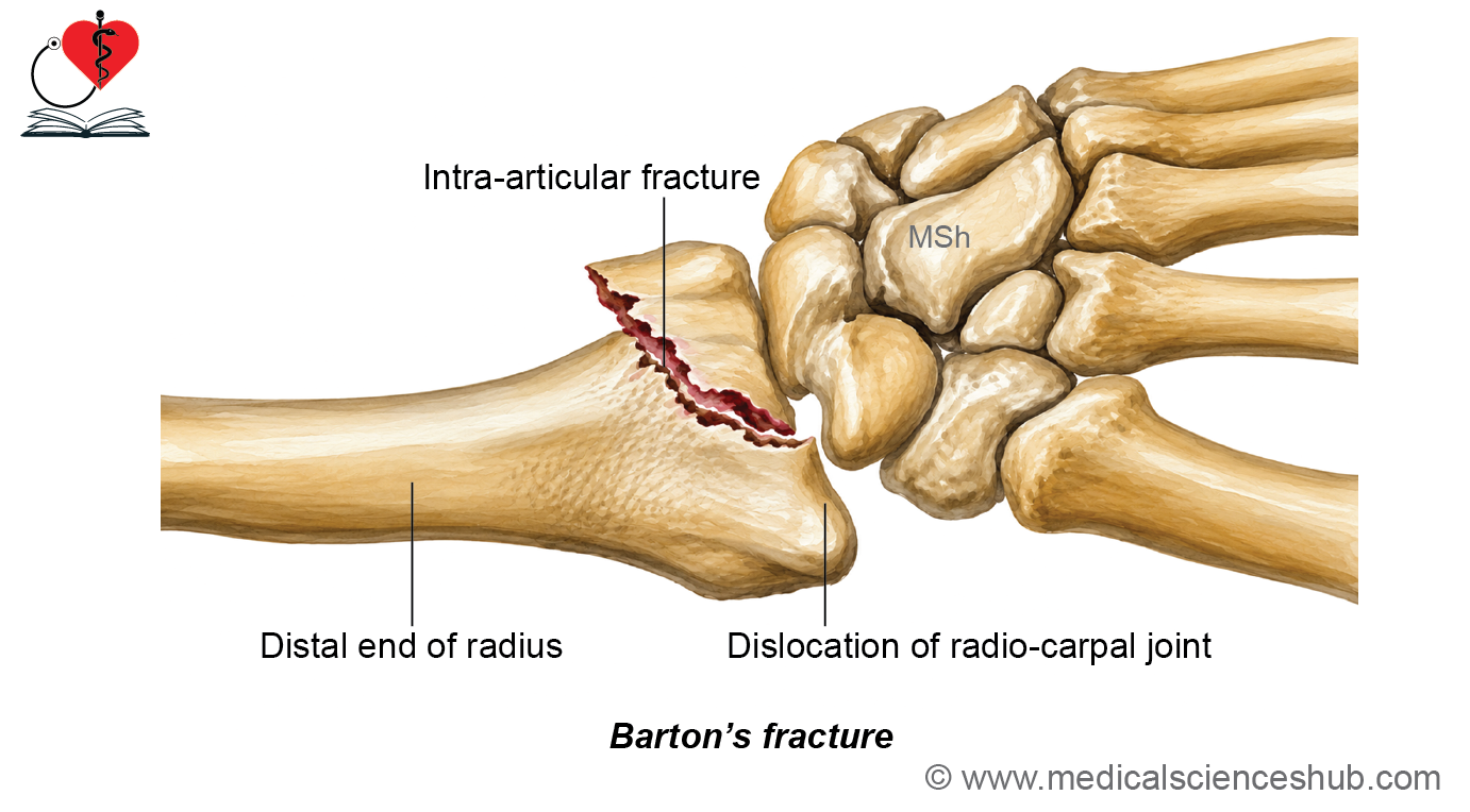
- Barton’s fracture: A Barton’s fracture is an intra-articular fracture of the distal radius associated with dislocation or subluxation of the radiocarpal joint.
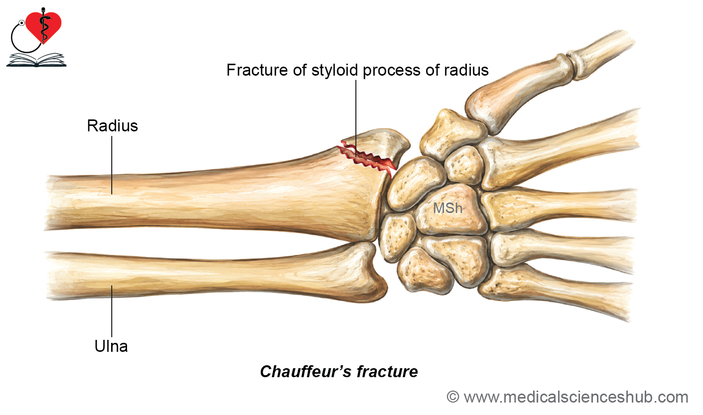
- Chauffeur’s fracture or Hutchinson fracture: A Chauffeur’s fracture is an isolated fracture of the radial styloid process caused by compression of the scaphoid against the styloid process.
- Pulled elbow (subluxation of head of radius): Pulled elbow is a subluxation of the head of the radius that usually occurs in young children after a sudden longitudinal traction or forceful pull on the hand or forearm.
Ulna
- The ulna is the medial bone of the forearm, whereas the radius is the lateral bone.
- The ulna is homologous to the fibula of the lower limb, although the fibula is located laterally in the leg.
- The ulna is classified as a long bone and forms the principal skeletal support of the forearm, especially at the elbow joint.
Parts
- The ulna consists of an upper end, a shaft, and a lower end.
- The upper end is expanded and includes the hook-shaped olecranon process, the smooth trochlear notch, and the crest-like coronoid process.
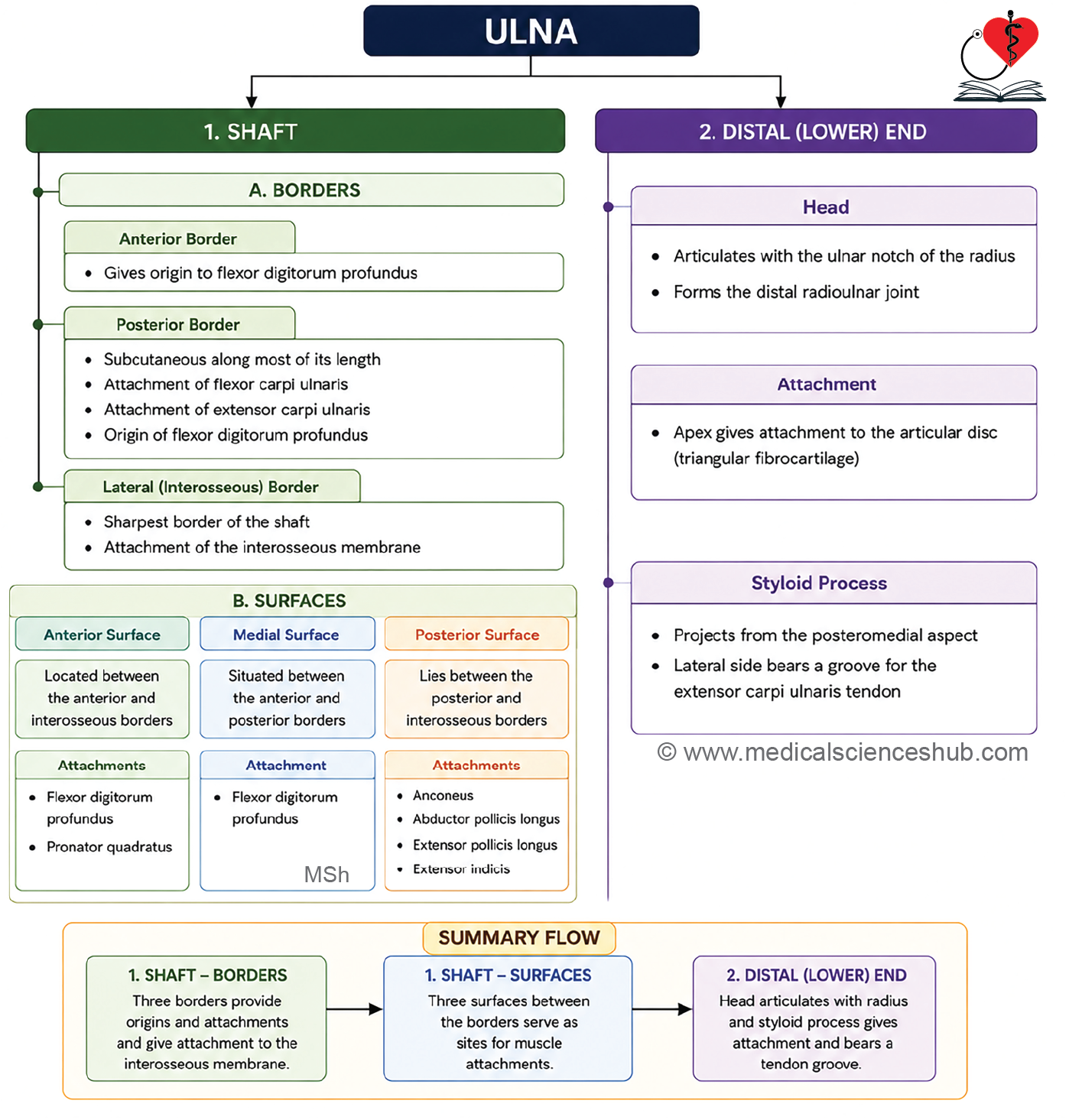
- The shaft is cylindrical and possesses a sharp lateral border, known as the interosseous border, for attachment of the interosseous membrane.
- The lower end consists of the head and the styloid process.
- The styloid process projects posteroinferiorly from the head of the ulna.
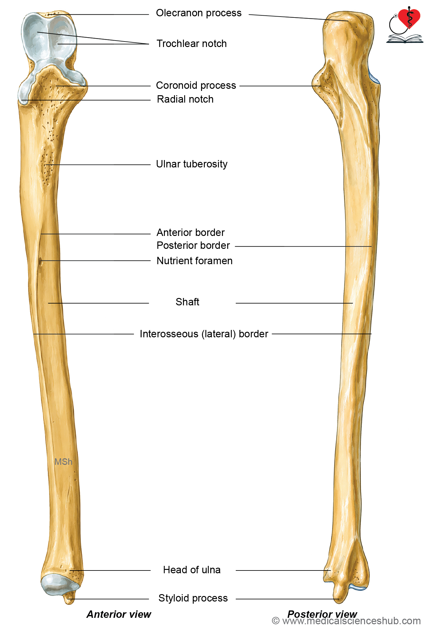
Figure H1.3.2: Hand-drawn diagram showing features of ulna (Click to see figure)
Side Determination and Anatomical Position
- Hold the ulna vertically with the hook-shaped olecranon process directed upward.
- Ensure that the trochlear notch faces anteriorly.
- The sharp interosseous border should face laterally.
- The side of the bone is identified by the direction of the interosseous border. In the right ulna, the interosseous border faces to the right, whereas in the left ulna, it faces to the left.
Features of Ulna
- During pronation and supination, the ulna remains relatively stable and serves as the fixed bone around which the radius rotates.
- The ulna is divided into three parts: the upper end, the shaft, and the lower end.
Upper End
• Upper end of ulna has coronoid and olecranon processes, trochlear and radial notches.
Processes
Olecranon process
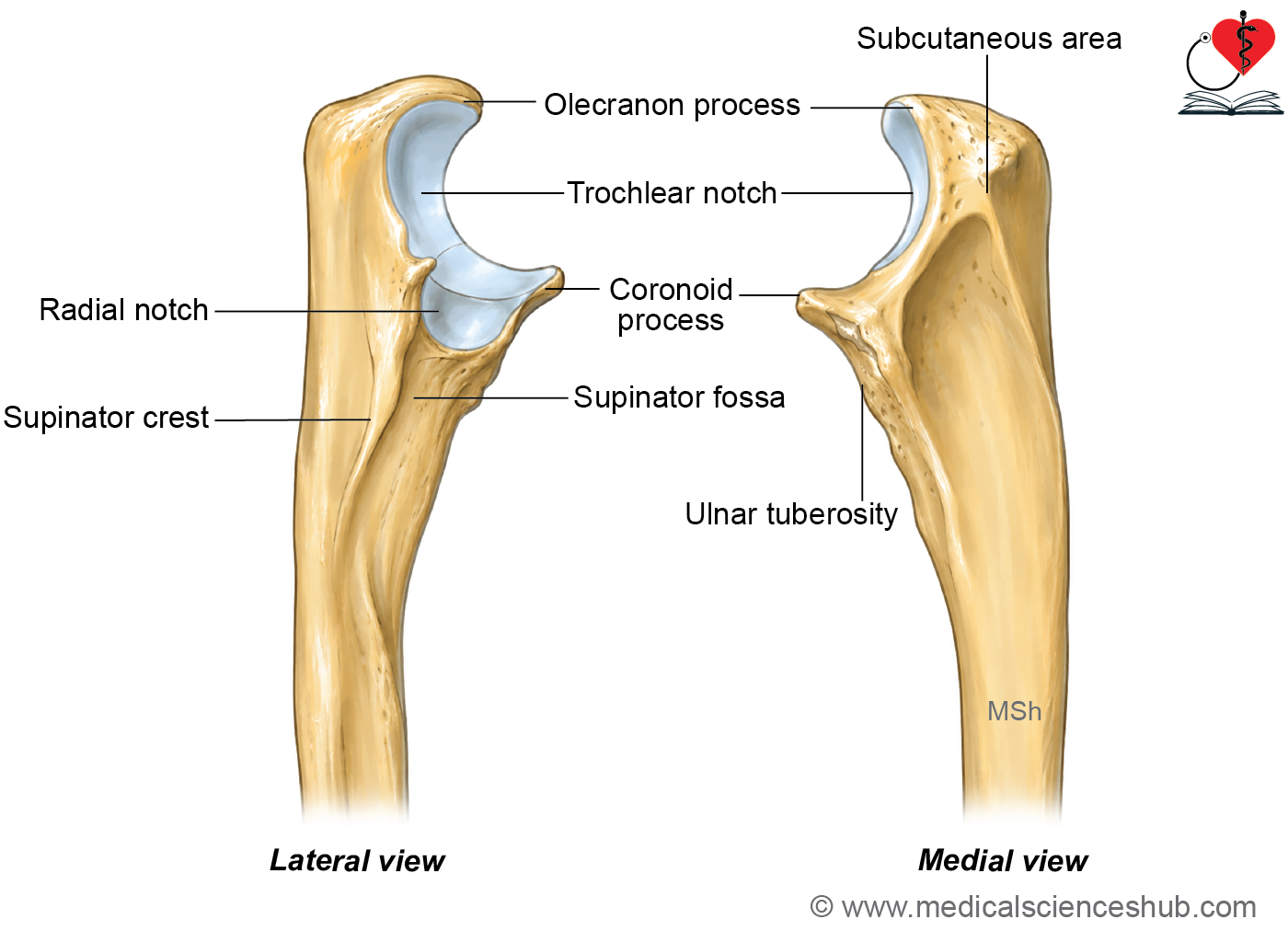
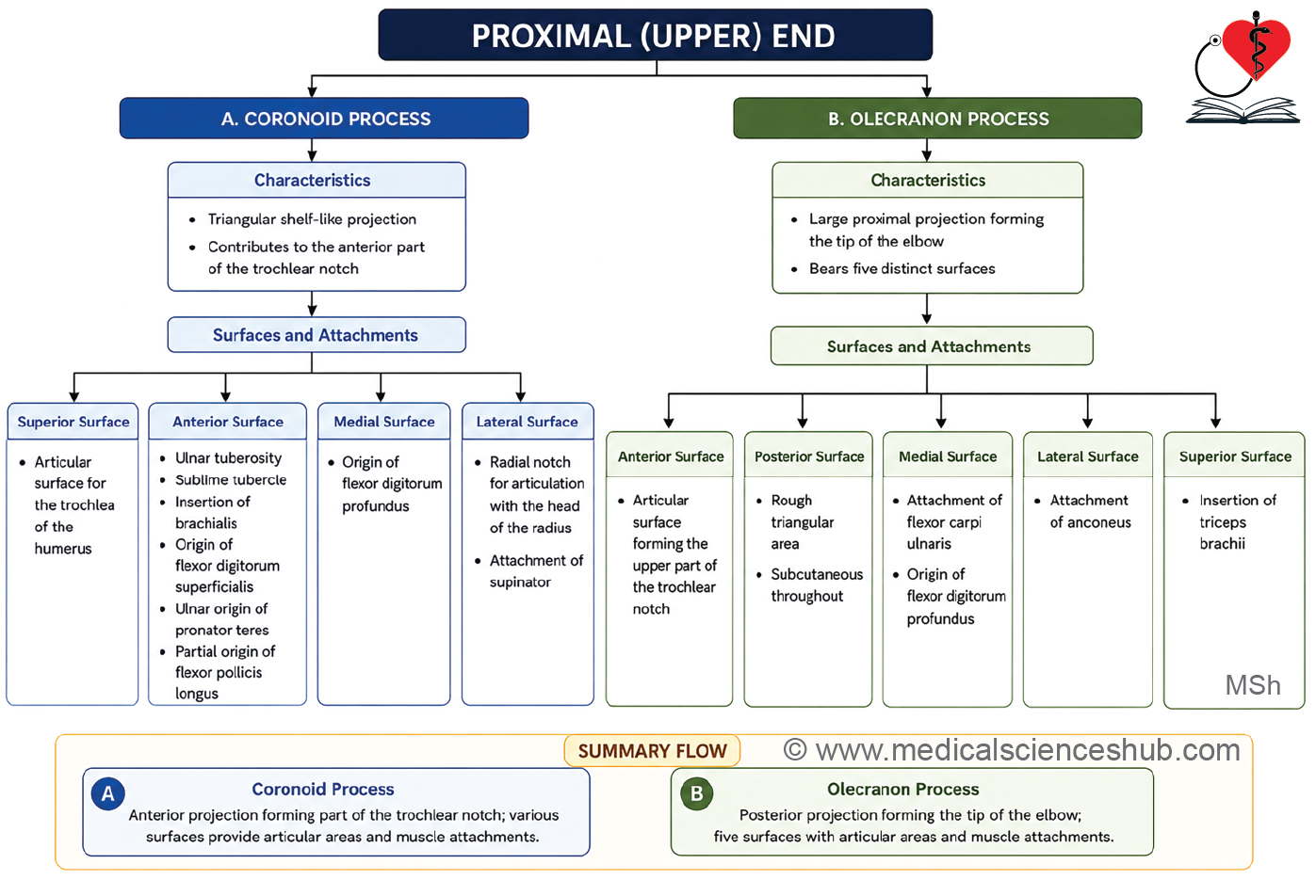
- The olecranon process is a beak-shaped projection at the upper end of the ulna.
- It has five surfaces: superior, inferior, anterior, medial, and lateral.
- The anterior surface is concave and covered by hyaline cartilage. Together with the upper surface of the coronoid process, it forms the trochlear notch, which articulates with the trochlea of the humerus to form the humeroulnar component of the elbow joint.
- The posterior surface is rough, triangular, subcutaneous, and easily palpable. It is covered by the subcutaneous olecranon bursa.
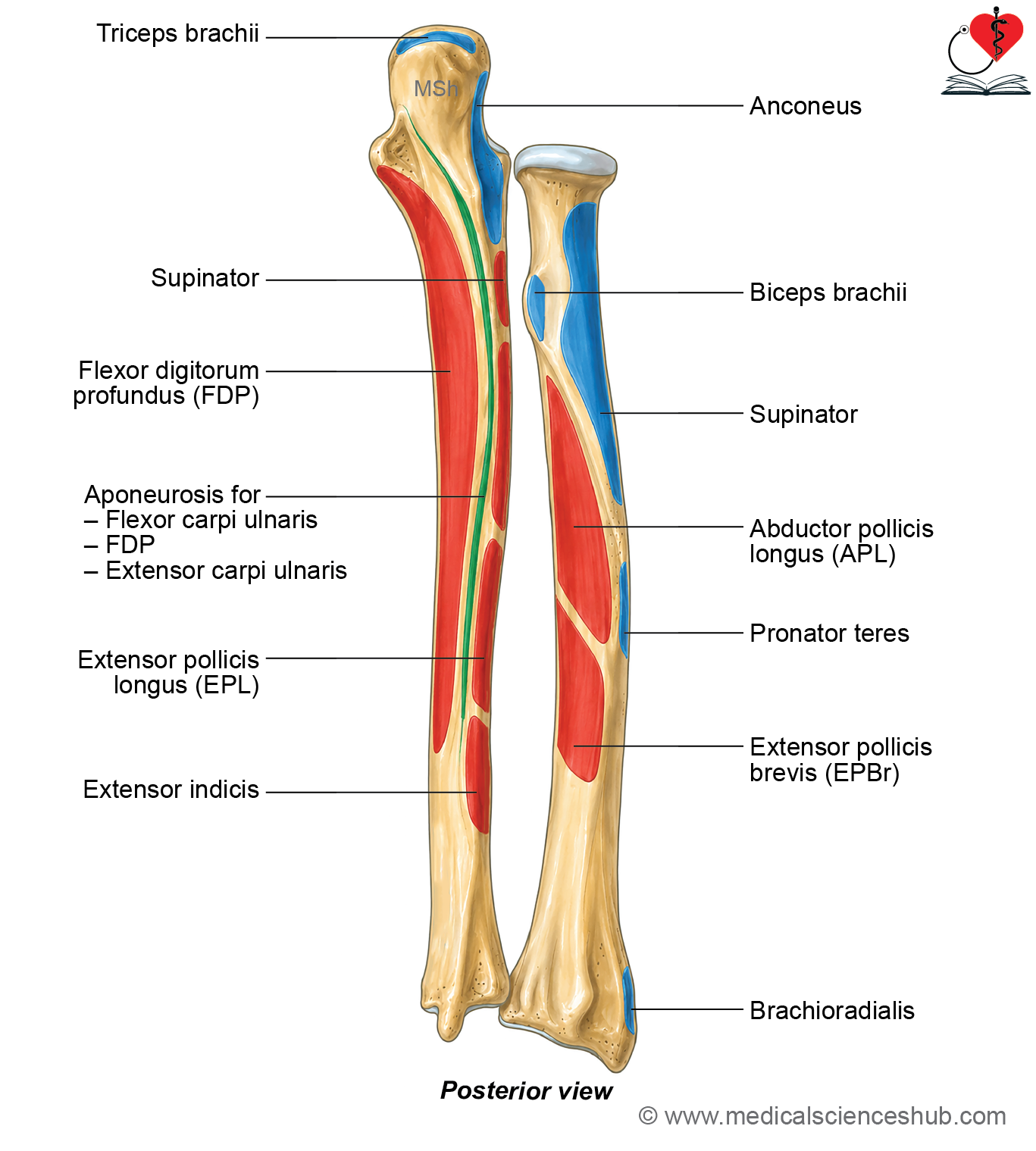
- The medial surface is smooth and continuous with the medial surface of the shaft. It provides attachment to the elbow joint capsule, posterior and oblique bands of the ulnar collateral ligament, and gives origin to flexor carpi ulnaris and flexor digitorum profundus.
- The lateral surface is smooth and continuous with the posterior surface of the shaft. It provides attachment to the joint capsule, radial collateral ligament, and insertion of the anconeus muscle.
- The superior surface has a rough posterior part for insertion of the triceps brachii tendon and a sharp anterior margin for attachment of the joint capsule.
- A synovial bursa lies between the tendon of triceps brachii and the elbow joint capsule, reducing friction during movement.
Coronoid process
- The coronoid process is a shelf-like projection extending anteriorly from the upper part of the shaft of the ulna.
- It has four surfaces: superior, anterior, medial, and lateral.
- The superior surface, together with the anterior surface of the olecranon process, forms the trochlear notch, which articulates with the trochlea of the humerus.
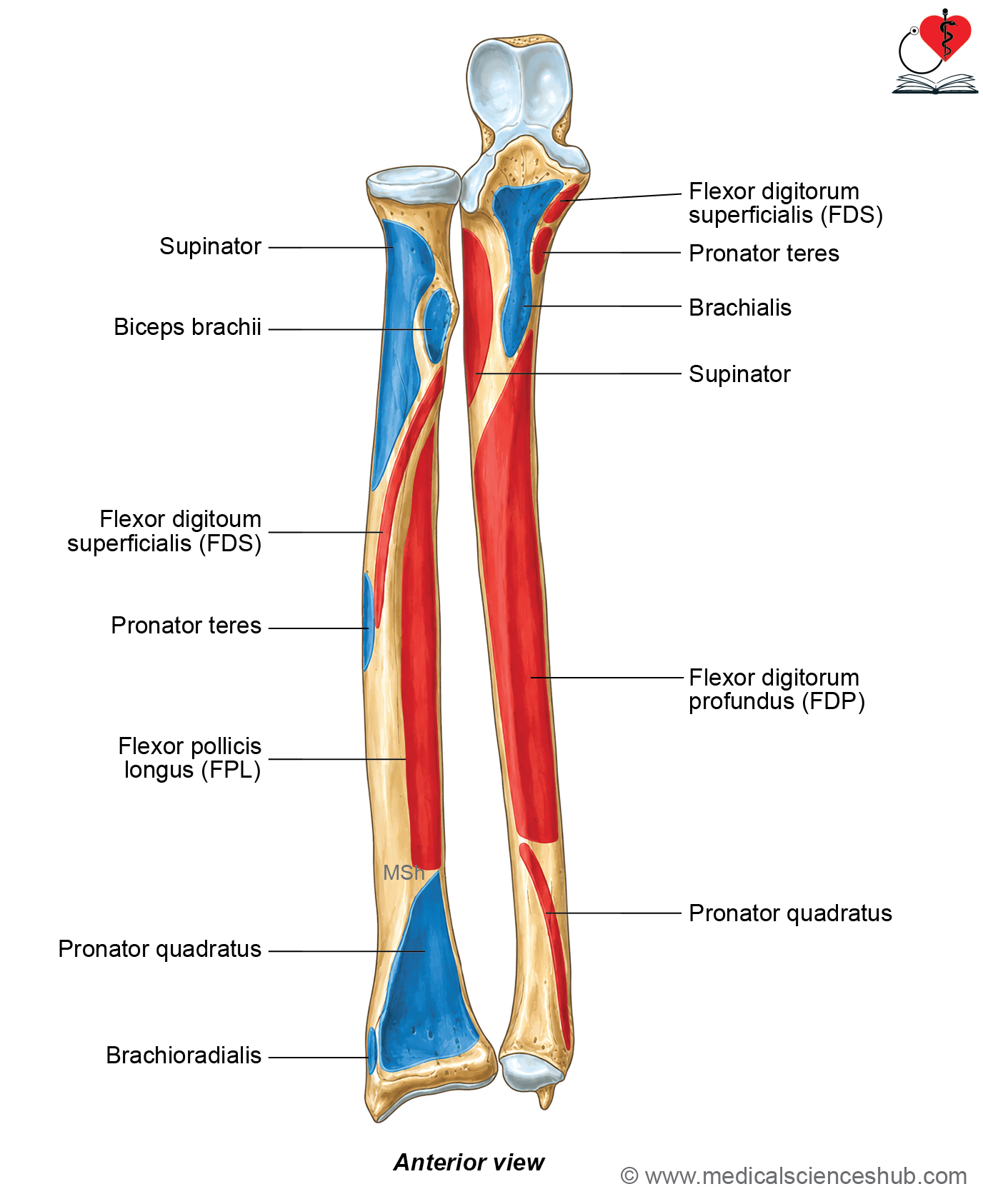
- The anterior surface is triangular. Its rough lower part forms the ulnar tuberosity, and a sublime tubercle is present on its medial margin. The anterior surface receives the insertion of brachialis. The medial margin provides attachment to the anterior and oblique bands of the ulnar collateral ligament and gives origin to flexor digitorum superficialis, pronator teres, and the ulnar head of flexor pollicis longus.
- The medial surface is continuous with the medial surface of the shaft and gives origin to flexor digitorum profundus.
- The lateral surface contains the radial notch, which articulates with the head of the radius. The annular ligament attaches to the anterior and posterior margins of the radial notch.
- Below the radial notch is the supinator fossa, bounded posteriorly by the supinator crest. These areas provide origin for the deep fibers of the supinator muscle.
Shaft of Ulna
- The shaft of the ulna is cylindrical and gradually narrows from above downward.
- It has three borders: anterior, posterior, and lateral (interosseous) border.
- It also has three surfaces: anterior, posterior, and medial.
Borders
- The anterior border extends from the ulnar tuberosity to the base of the styloid process. It gives origin to the flexor digitorum profundus from its upper three-fourths.
- The posterior border extends from the apex of the triangular posterior surface of the olecranon process to the posterior aspect of the styloid process. It is subcutaneous throughout its length and can be easily palpated. The posterior border gives origin to flexor carpi ulnaris, extensor carpi ulnaris, and flexor digitorum profundus through a common aponeurosis.
- The lateral (interosseous) border is sharp in its upper part and becomes less distinct inferiorly. It extends from the supinator crest to the medial side of the styloid process. The interosseous membrane is attached along the lateral (interosseous) border.
Surfaces
Anterior surface
- The anterior surface of the shaft of the ulna lies between the anterior and lateral (interosseous) borders.
- A nutrient foramen is present in the upper part of this surface. It is directed upward because the lower end of the ulna is the growing end of the bone.
- An oblique ridge is present in the lower one-fourth of the anterior surface.
Attachments
- The upper three-fourths of the anterior surface provide origin to the flexor digitorum profundus muscle.
- The oblique ridge in the lower one-fourth gives origin to the pronator quadratus muscle.
Medial surface
- The medial surface lies between the anterior and posterior borders of the shaft.
Attachments
- The upper three-fourths of the medial surface also give origin to the flexor digitorum profundus, contributing to its broad attachment on the ulna.
Posterior surface
- The posterior surface lies between the posterior and lateral (interosseous) borders.
- An oblique line extending from the supinator crest to the posterior border divides the posterior surface into a smaller upper area and a larger lower area.
- A vertical ridge further subdivides the larger lower area into a medial part adjacent to the posterior border and a lateral part adjacent to the interosseous border.
Attachments
- The anconeus muscle is inserted into the lateral surface of the olecranon process and the upper one-fourth of the posterior surface of the ulnar shaft.
- The larger lateral part of the posterior surface gives origin, from above downward, to the abductor pollicis longus, extensor pollicis longus, and extensor indicis muscles.
Lower End
- It is slightly expanded and has head and styloid process.
Head
- The head of the ulna forms the expanded lower end of the bone.
- Its lateral surface is smooth, convex, and covered with articular cartilage. It articulates with the ulnar notch of the distal radius to form the inferior radioulnar joint.
- The inferior surface has a lateral semilunar articular area that articulates with the superior surface of the triangular articular disc.
- A rough groove on the medial part of the inferior surface provides attachment to the apex of the triangular articular disc.
- The anterior and posterior margins of the head provide attachment to the capsule of the wrist joint.
Styloid process of ulna
The styloid process of the ulna projects downward from the posteromedial aspect of the lower end of the bone.
Attachments
- The tendon of extensor carpi ulnaris passes through a groove located posteriorly between the head and the styloid process.
- The ulnar collateral ligament of the wrist is attached to the tip of the styloid process.
Ossification
- The ulna develops from one primary and two secondary ossification centers.
- The primary ossification center appears in the eighth week of intrauterine life and forms the shaft.
- A secondary ossification center for the olecranon process appears around the ninth year and fuses with the shaft by approximately 18 years of age.
- Another secondary ossification center appears at the lower end around the sixth year and fuses with the shaft by approximately 20 years of age.
- Because the lower end fuses later than the upper end, the lower end of the ulna is the growing end of the bone.
CLINICAL INTEGRATION
- Fractures of the ulna commonly involve the shaft, proximal end, or may occur in association with injuries of the radius and elbow joint.
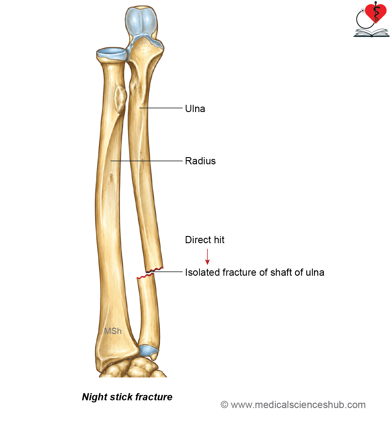
- A nightstick fracture is an isolated fracture of the shaft of the ulna without an associated radial fracture. It usually results from a direct blow to the forearm, such as when a person raises the forearm to protect the head from trauma.
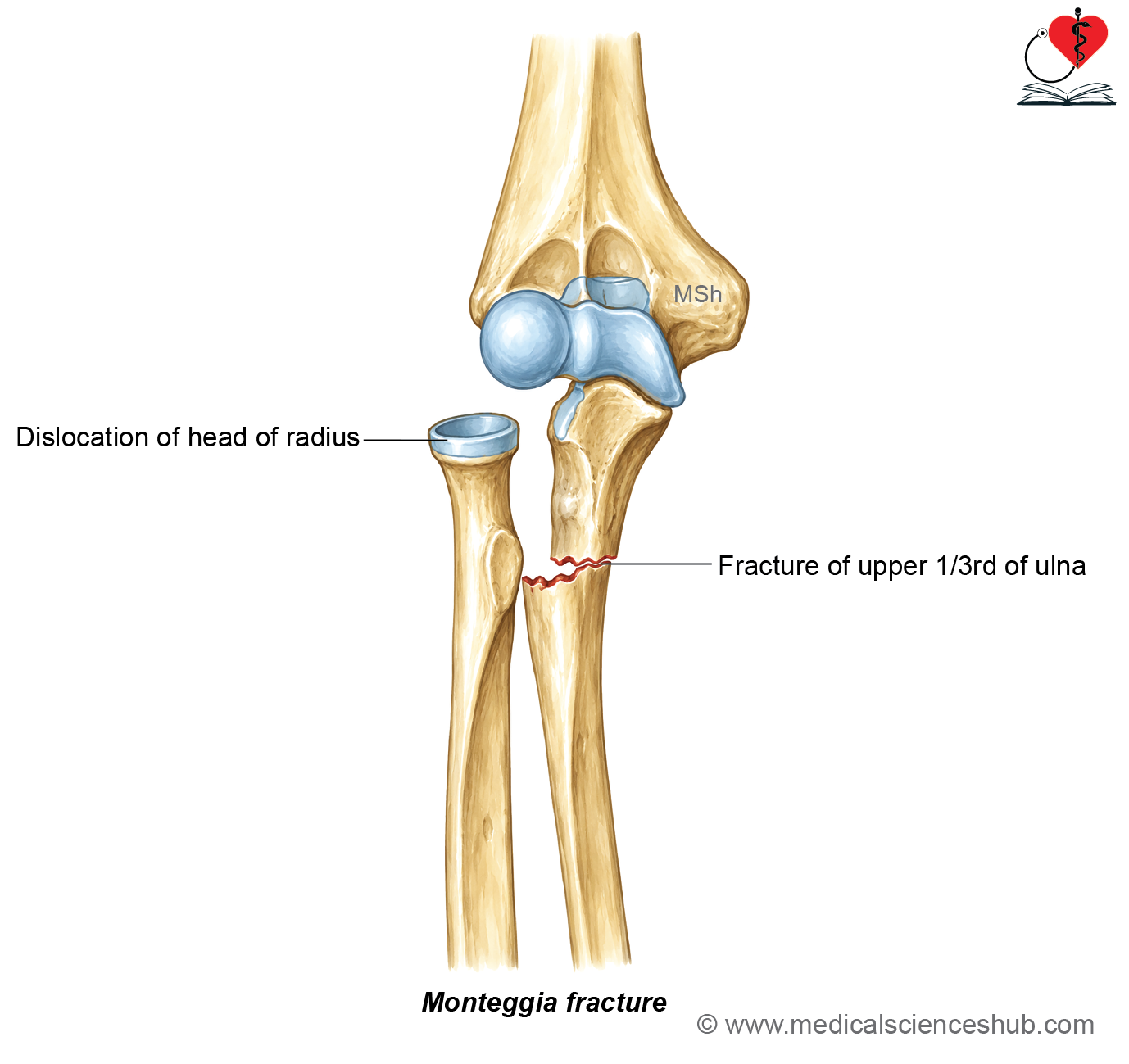
- A Monteggia fracture consists of a fracture of the upper one-third of the ulna associated with dislocation of the head of the radius at the proximal radioulnar joint. This injury requires prompt recognition because delayed treatment may impair elbow function.
- A Hume fracture is characterized by a fracture of the olecranon accompanied by anterior dislocation of the head of the radius. Although uncommon, it represents a severe injury around the elbow.
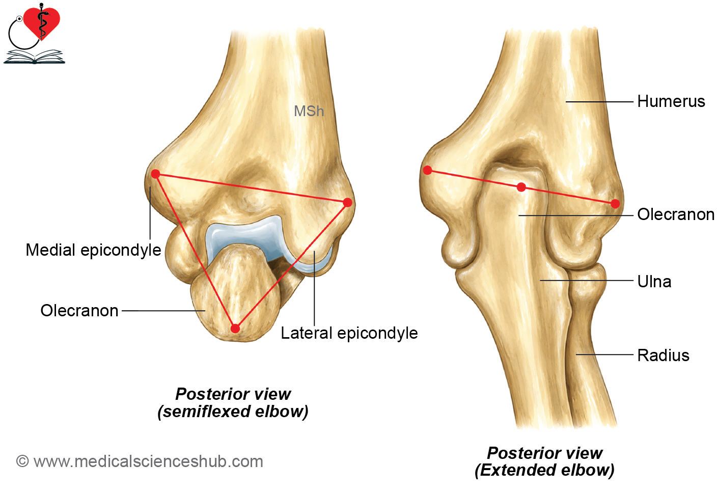
- The alignment of the elbow can be assessed using the three-point bony relationship formed by the medial epicondyle, lateral epicondyle, and olecranon. In a fully extended elbow, these three bony landmarks lie in a straight horizontal line. In a flexed elbow, they form a triangular arrangement. Normal variation exists, and the triangle is not always equilateral or identical in both elbows. Alteration of this relationship may indicate fracture or dislocation around the elbow.
- Madelung deformity results from premature growth disturbance of the distal radial growth plate, leading to abnormal growth of the radius. It is associated with relative overgrowth of the ulna, widening of the interosseous space, bowing of the radius, and subluxation of the distal radioulnar joint.
📝 Test Your Knowledge – Practice MCQs
Attempt the chapter MCQ quiz and assess your understanding of key concepts.
Click to see: Medical Disclaimer and Copyright Notice