
Competencies
- AN10.10: Describe and identify the deltoid and rotator cuff muscles.
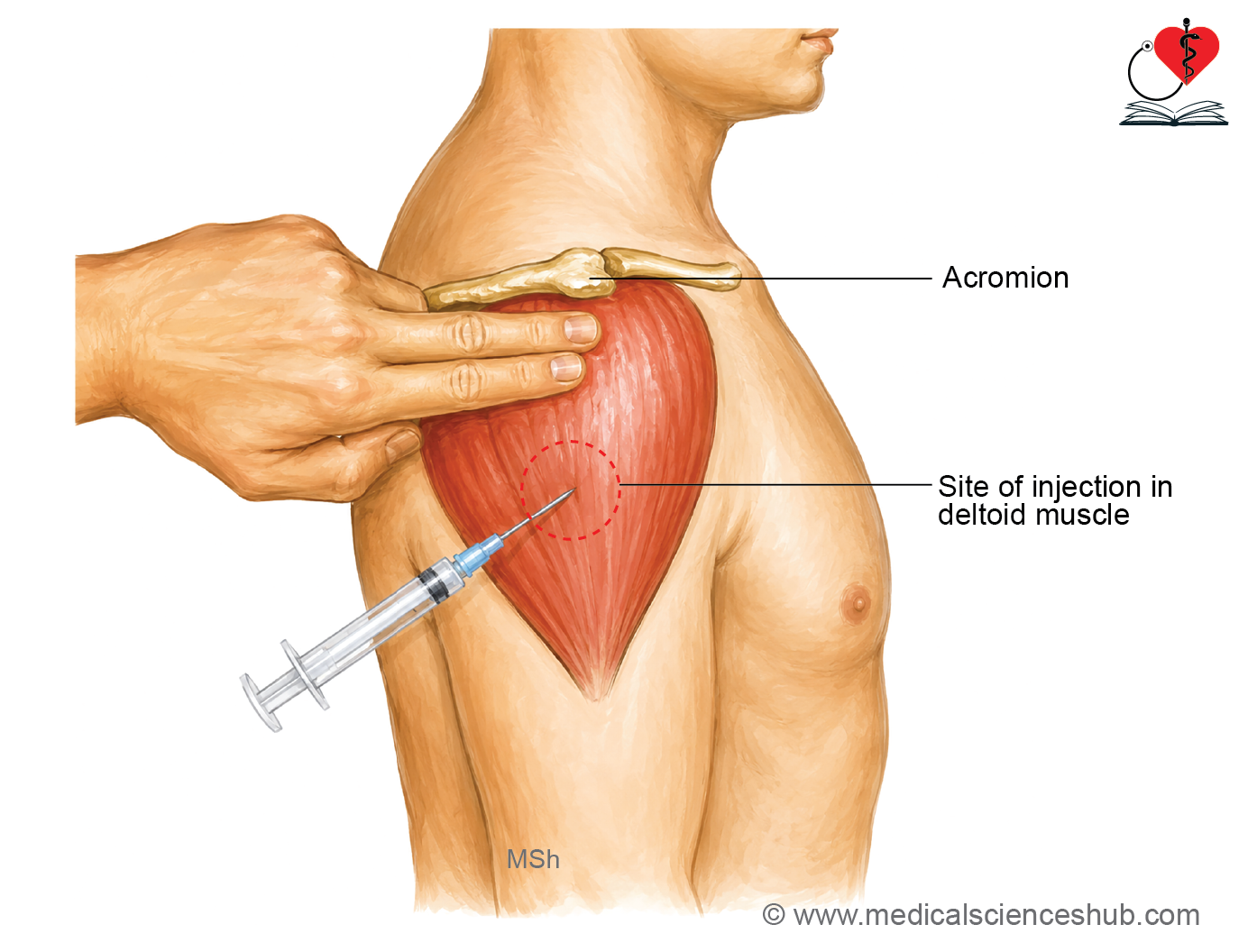
- AN10.13: Explain anatomical basis of injury to axillary nerve during intramuscular injections.
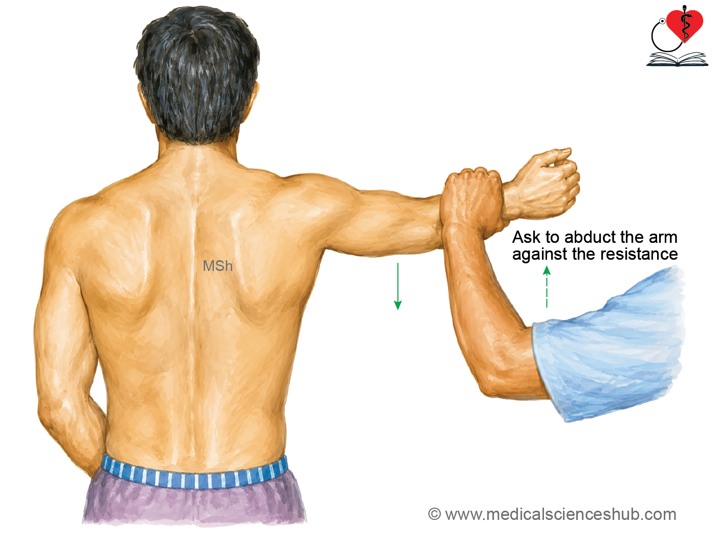
- AN13.7: ………..,Testing of muscles: deltoid, …………… .
- This chapter includes muscles of rotator cuff, deltoid muscle, and axillary nerve.
Introduction
- The scapular region comprises the scapula, adjacent soft tissues, and the muscles surrounding the shoulder blade.
- It contains the scapulohumeral (intrinsic shoulder) muscles, which originate from the scapula and insert onto the humerus.
- The muscles of the scapular region are:
- Deltoid
- Subscapularis
- Supraspinatus
- Infraspinatus
- Teres minor
- Teres major
- The skin over the scapular and shoulder region receives cutaneous innervation from:
- Lateral supraclavicular nerves (C3, C4)
- Upper lateral cutaneous nerve of the arm (branch of the axillary nerve)
- Posterior (dorsal) rami of the upper thoracic spinal nerves.
Muscles of Scapular Region
Deltoid muscle
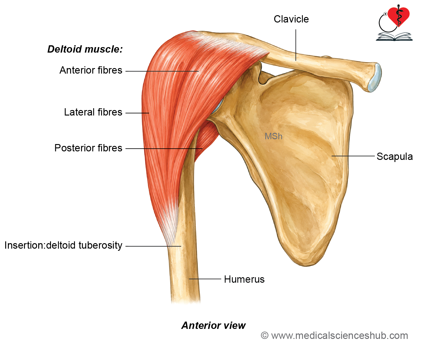
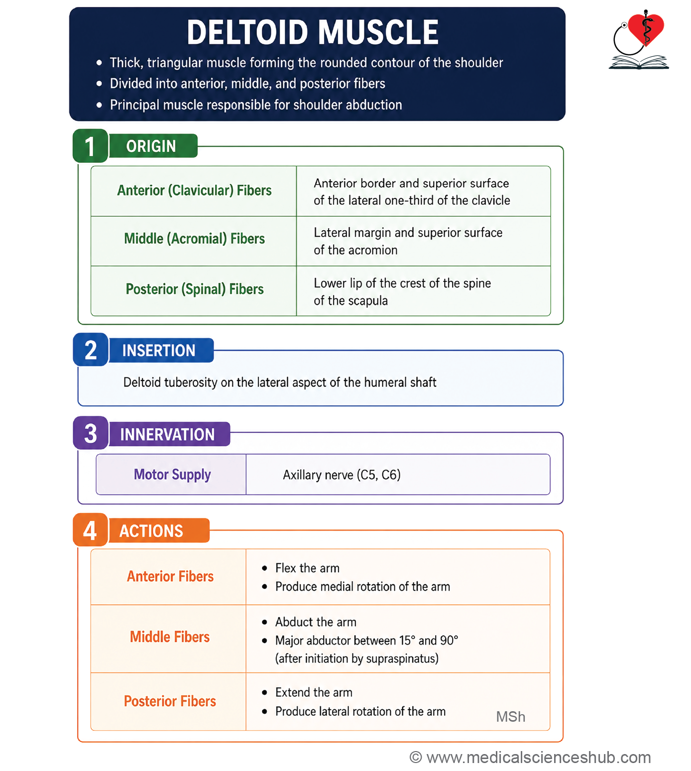
- The deltoid muscle is a large, triangular muscle that forms the rounded contour of the shoulder.
- It is named after the Greek letter delta (Δ) because of its triangular shape.
- The deltoid consists of three distinct parts:
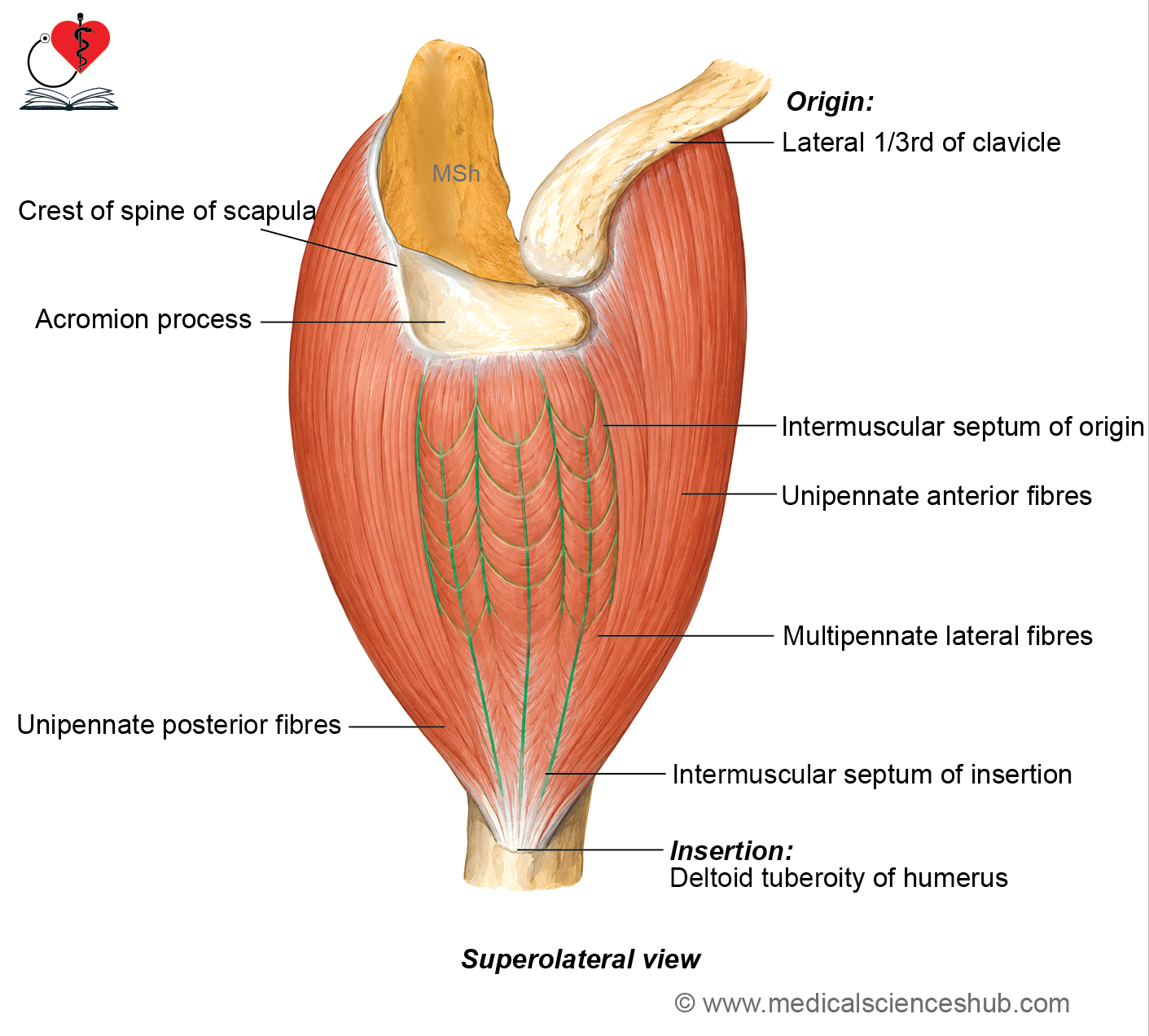
- Anterior (clavicular) part: Unipennate
- Middle (acromial) part: Multipennate
- Posterior (spinal) part: Unipennate
Origin
- Deltoid originates as follows:
- The anterior part arises from the anterior border and superior surface of the lateral one-third of the clavicle.
- The middle part arises from the lateral margin of the acromion and the attached intermuscular septa.
- The posterior part arises from the lower lip of the crest of the spine of the scapula.
Insertion
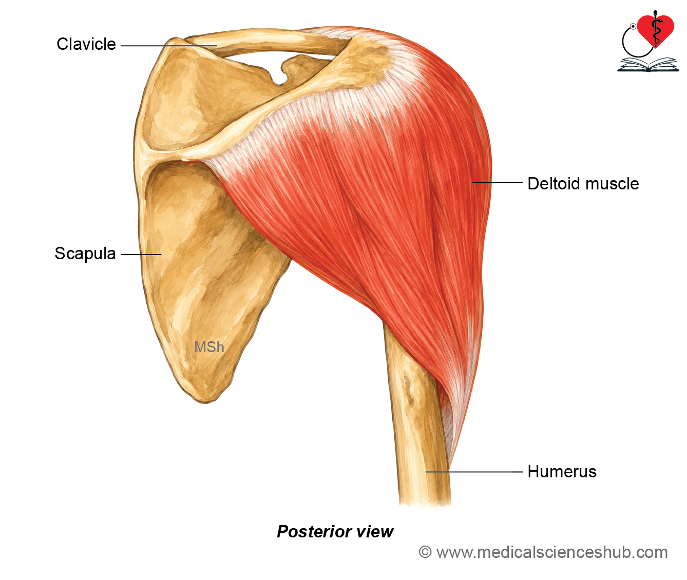
All deltoid fibers converge inferiorly to form a strong tendon that inserts into the V-shaped deltoid tuberosity on the lateral surface of the humeral shaft.
Innervation
The deltoid is supplied by the axillary nerve (C5, C6).
Actions
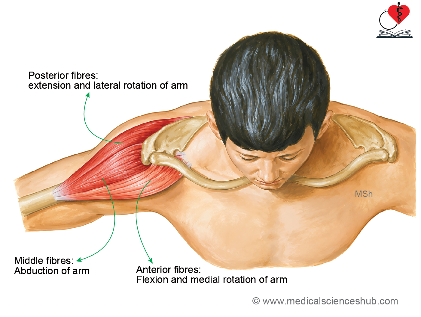
- The anterior (clavicular) fibers produce flexion and medial rotation of the arm and assist in horizontal adduction.
- The middle (acromial) fibers are the chief abductors of the arm and produce abduction from approximately 15° to 90°.
- The posterior (spinal) fibers produce extension and lateral rotation of the arm and assist in horizontal abduction.
CLINICAL INTEGRATION
Intramuscular injection
- The deltoid muscle is a common site for intramuscular (IM) injections because it is easily accessible and well vascularized. To avoid injury to the axillary nerve, the injection should be administered into the central, thickest part of the muscle, well below the acromion.
Paralysis of the Deltoid
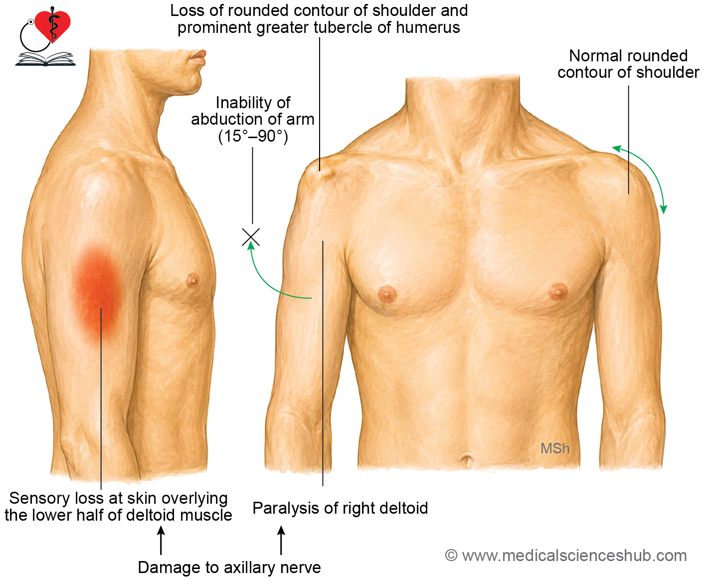
- Injury to the axillary nerve causes paralysis or marked weakness of the deltoid muscle. Clinical features include:
- Inability or marked weakness in abducting the arm beyond the initial 15°, particularly up to 90°.
- Weakness of shoulder flexion and medial rotation due to loss of anterior deltoid function.
- Flattening of the normal rounded contour of the shoulder because of deltoid muscle wasting.
- Loss of sensation over the skin covering the lower part of the deltoid (the regimental badge area), supplied by the upper lateral cutaneous nerve of the arm.
Rotator Cuff Muscles
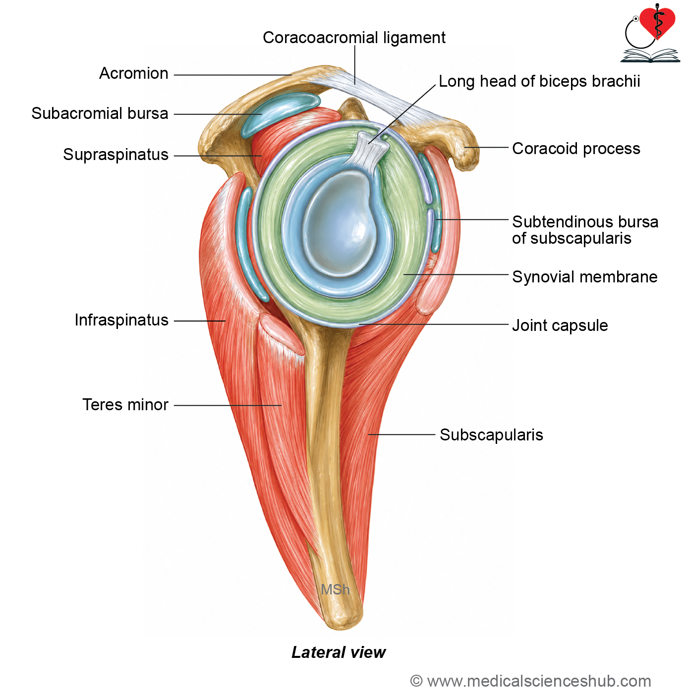
- The rotator cuff is a musculotendinous (fibrotendinous) cuff that surrounds the capsule of the shoulder joint and acts as a dynamic stabilizer of the glenohumeral joint.
- It is formed by the flattened tendons of four muscles:
- Subscapularis
- Supraspinatus
- Infraspinatus
- Teres minor
- These tendons blend firmly with the fibrous capsule of the shoulder joint, reinforcing it on all sides except inferiorly.
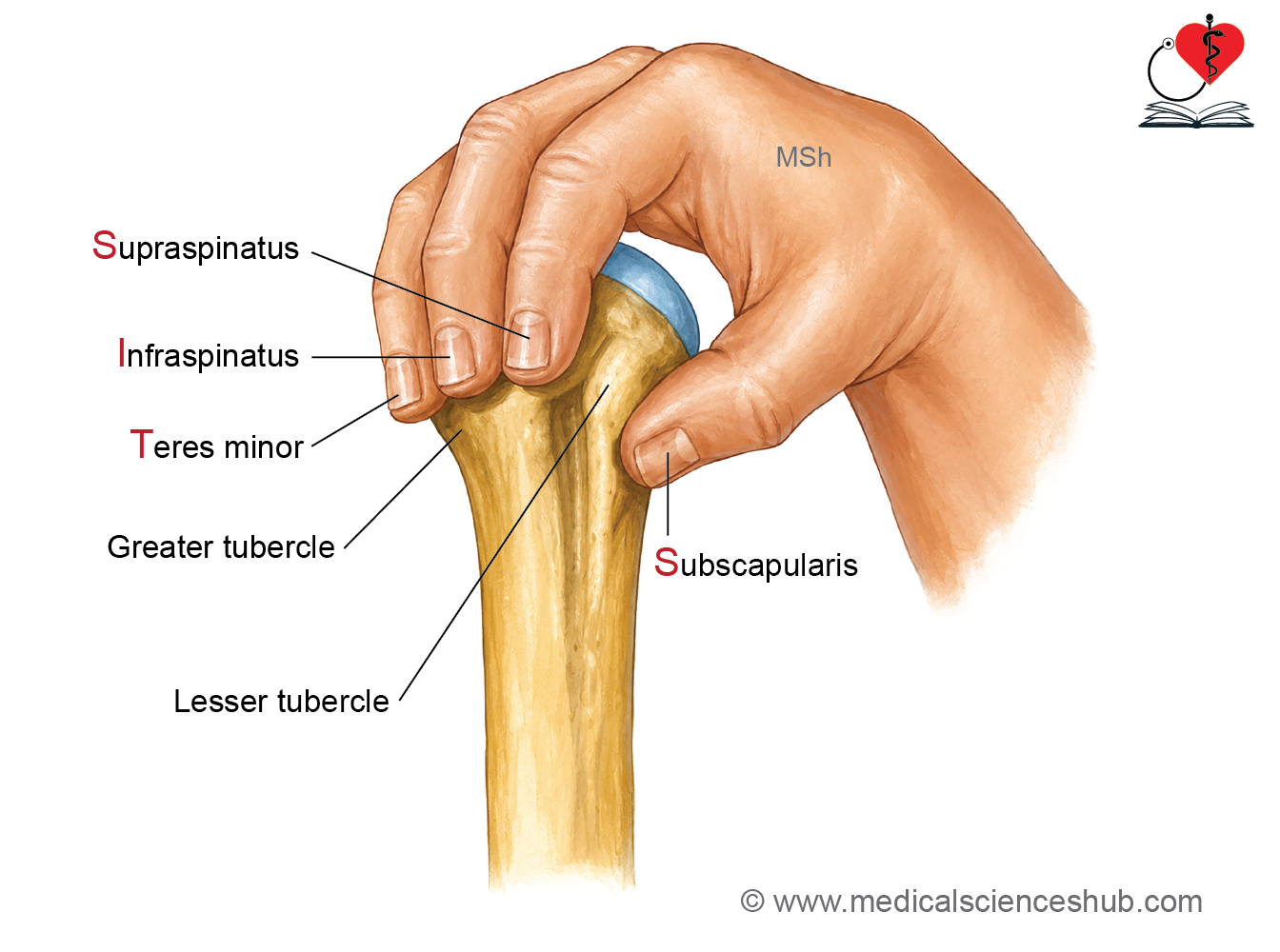
- The rotator cuff tendons insert onto the greater and lesser tubercles of the humerus:
- Subscapularis inserts on the lesser tubercle.
- Supraspinatus, infraspinatus, and teres minor insert on the greater tubercle.
- Besides producing shoulder movements, the rotator cuff muscles stabilize the head of the humerus within the glenoid cavity during arm movements.
Subscapularis
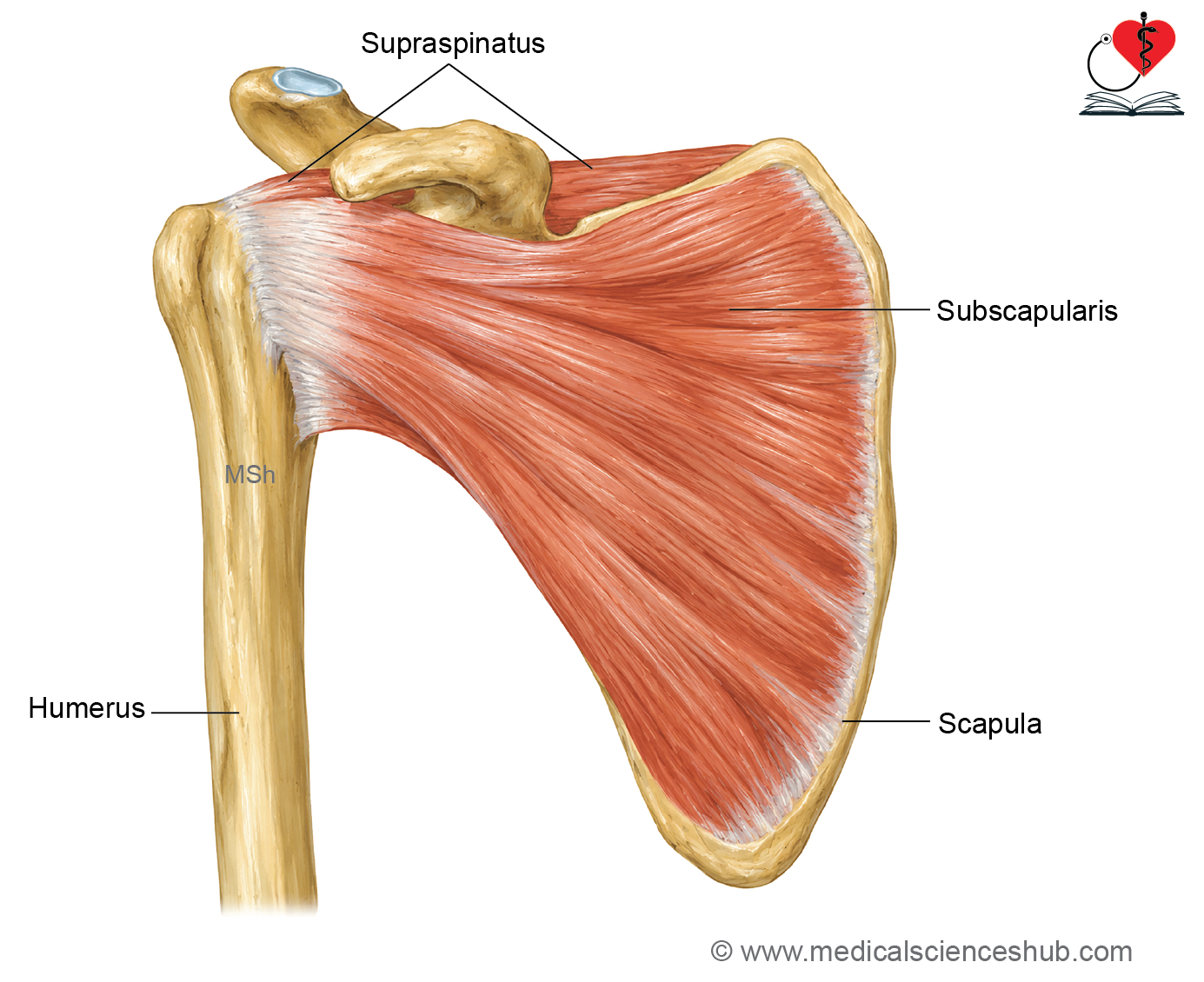
The subscapularis is a large, multipennate, triangular muscle that occupies the anterior (costal) surface of the scapula and forms part of the rotator cuff.
Origin
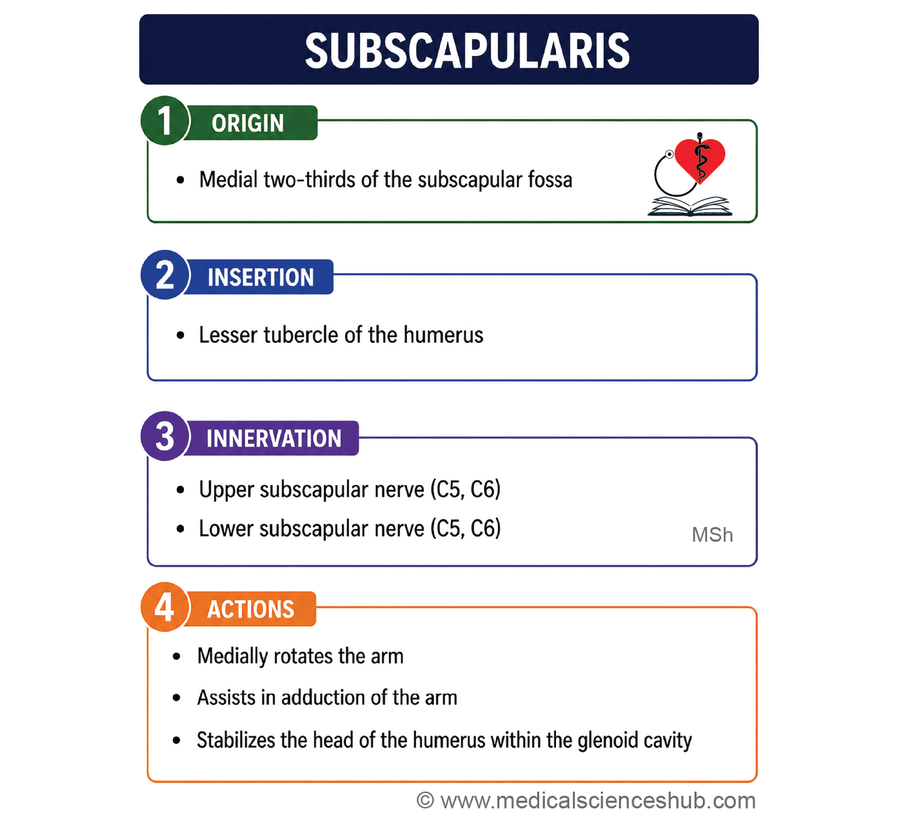
- It arises from the medial two-thirds of the subscapular fossa on the costal surface of the scapula.
Direction of fibers
- The muscle fibers converge laterally to form a strong tendon.
- The tendon is separated from the neck of the scapula by the subscapular bursa, which commonly communicates with the cavity of the shoulder joint.
Insertion
The tendon inserts onto the lesser tubercle of the humerus and blends with the anterior part of the shoulder joint capsule.
Innervation
The muscle is supplied by the upper and lower subscapular nerves (C5, C6).
Actions
- It is the chief medial rotator of the arm. It assists in adduction and extension of the arm from a flexed position.
- As a rotator cuff muscle, it stabilizes the head of the humerus within the glenoid cavity during shoulder movements.
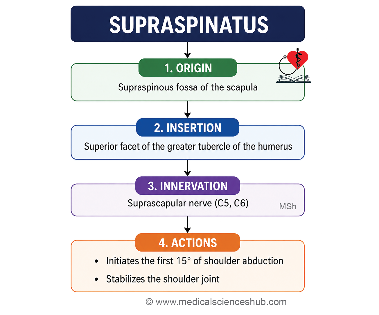
Supraspinatus
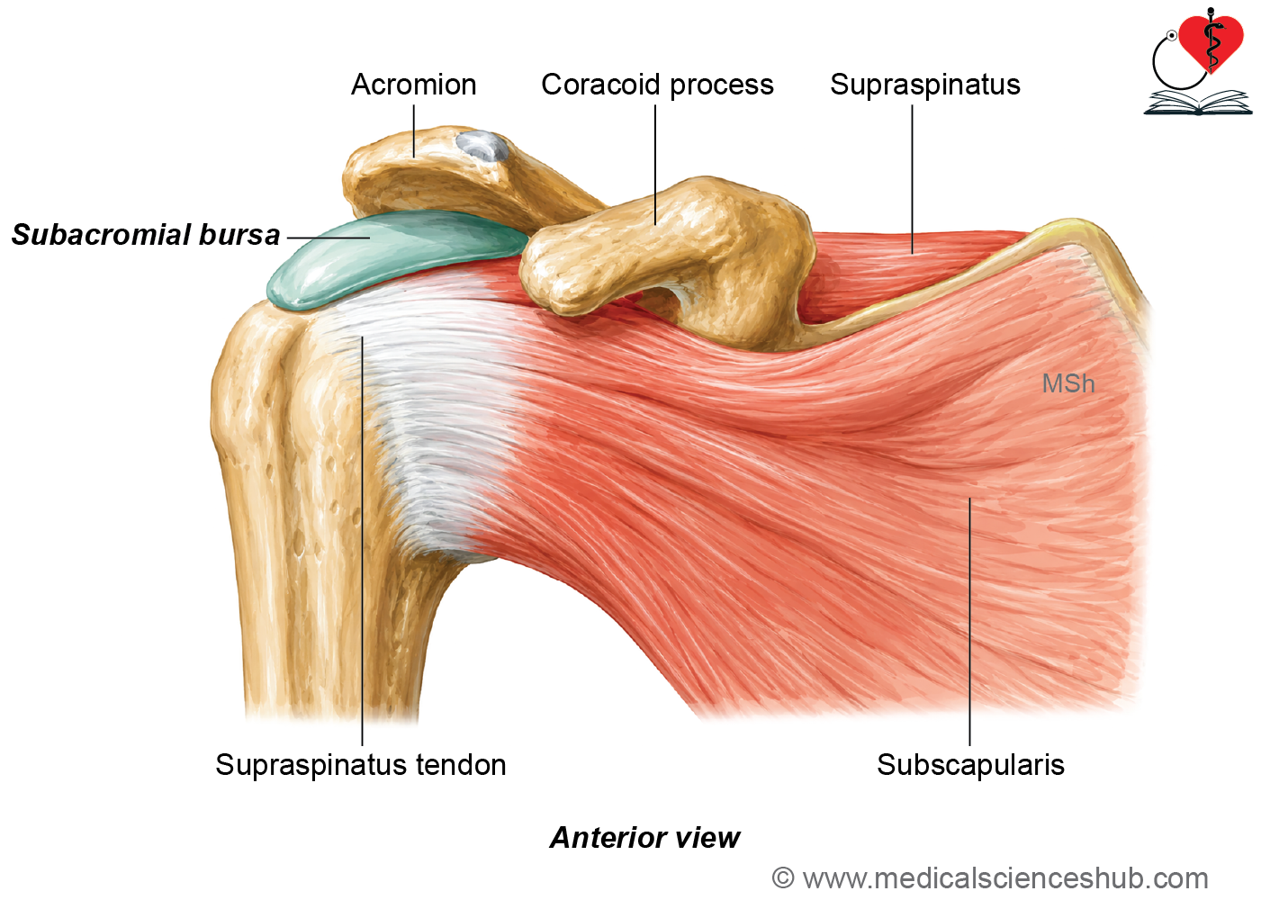
The supraspinatus is a small rotator cuff muscle that occupies the supraspinous fossa of the scapula.
Origin
- It arises from the medial two-thirds of the supraspinous fossa, including the upper surface of the spine of the scapula.
Direction of fibers
- The muscle fibers pass horizontally and laterally to form a tendon.
- The tendon passes beneath the coracoacromial arch and is separated from it by the subacromial bursa, which reduces friction during shoulder movements.
Insertion
- The tendon inserts onto the superior impression of the greater tubercle of the humerus and blends with the capsule of the shoulder joint.
Innervation
The muscle is supplied by the suprascapular nerve (C5, C6).
Actions
- It initiates the first 15° of arm abduction.
- It works with the deltoid muscle to continue abduction up to 90°.
- As a rotator cuff muscle, it stabilizes the head of the humerus within the glenoid cavity during shoulder movements.
Infraspinatus
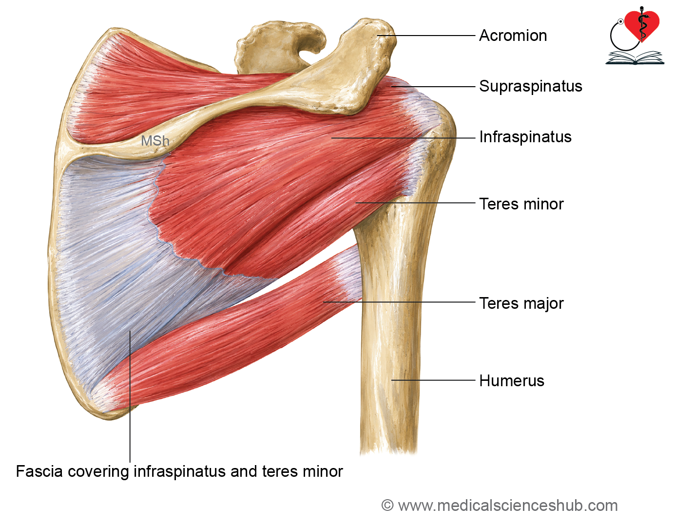
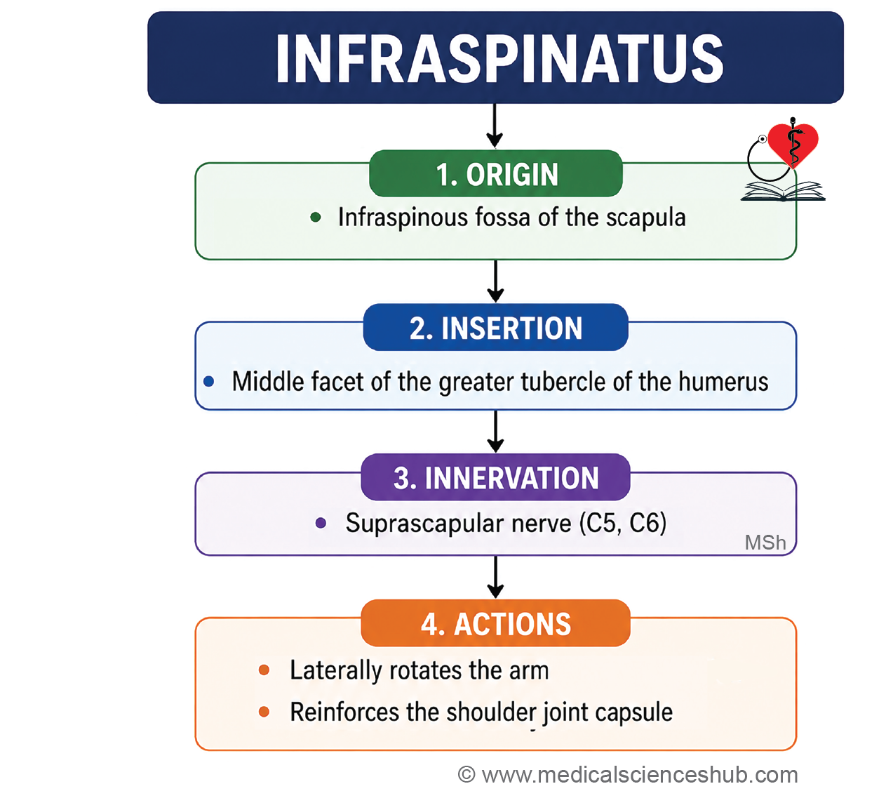
The infraspinatus is a triangular rotator cuff muscle that occupies the infraspinous fossa on the posterior surface of the scapula.
Origin
- It arises from the medial two-thirds of the infraspinous fossa, including the inferior surface of the spine of the scapula.
Direction of fibers
- The muscle fibers converge laterally to form a tendon that passes posterior to the shoulder joint.
Insertion
The tendon inserts onto the middle impression of the greater tubercle of the humerus and blends with the posterior part of the shoulder joint capsule.
Innervation
The muscle is supplied by the suprascapular nerve (C5, C6).
Actions
- It is a powerful lateral rotator of the arm.
- It assists in horizontal abduction of the arm.
- As a rotator cuff muscle, it helps stabilize the head of the humerus within the glenoid cavity during shoulder movements.
Teres Minor
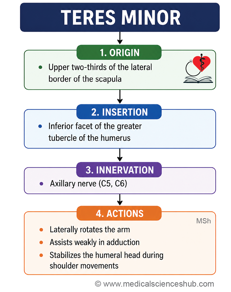
The teres minor is a small, rounded rotator cuff muscle located along the lateral border of the scapula.
Origin
- It arises from the upper two-thirds of the dorsal surface of the lateral border of the scapula.
- The circumflex scapular artery passes between the upper and lower parts of its origin, an important identification feature during dissection.
Direction of fibers
- The muscle fibers run obliquely upward and laterally toward the humerus.
Insertion
The tendon inserts onto the inferior (lowest) impression of the greater tubercle of the humerus and blends with the posterior capsule of the shoulder joint.
Innervation
The muscle is supplied by the axillary nerve (C5, C6).
Actions
- It acts as a lateral rotator of the arm.
- It assists in adduction of the arm.
- As a rotator cuff muscle, it helps stabilize the head of the humerus within the glenoid cavity during shoulder movements.
Teres major muscle
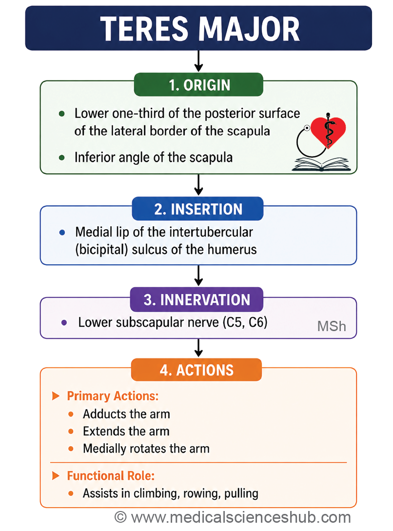
The teres major is a scapulohumeral muscle located along the inferior part of the posterior scapular region. Unlike the teres minor, it is not a rotator cuff muscle.
Origin
- It arises from the lower one-third of the dorsal surface of the lateral border and the inferior angle of the scapula.
Direction of fibers
- The muscle fibers run upward, forward, and laterally.
- The tendon passes anterior to the shoulder joint, accompanying the latissimus dorsi, to reach the intertubercular (bicipital) groove of the humerus.
Insertion
- The tendon inserts onto the medial lip of the intertubercular (bicipital) groove of the humerus.
Innervation
The muscle is supplied by the lower subscapular nerve (C5, C6).
Actions
- It produces medial rotation and adduction of the arm.
- It also assists in extension of the arm, particularly from a flexed position.
- It works synergistically with the latissimus dorsi during movements such as climbing, rowing, and pulling.
CLINICAL INTEGRATION
- Rupture of the supraspinatus tendon: A tear of the supraspinatus tendon causes difficulty or inability to initiate the first 15° of arm abduction. After abduction is initiated, the deltoid muscle may continue the movement.
- Subacromial bursitis: The subacromial bursa lies between the supraspinatus tendon and the coracoacromial arch, reducing friction during shoulder movements. Inflammation of this bursa is known as subacromial bursitis and commonly causes shoulder pain, especially during abduction.
- Dawbarn’s sign: In subacromial bursitis, tenderness is elicited by pressing below the acromion while the arm is adducted. The tenderness diminishes or disappears when the arm is abducted because the inflamed subacromial bursa moves beneath the acromion.
- Supraspinatus tendinitis: This condition refers to inflammation of the supraspinatus tendon, usually resulting from repetitive overhead activities or degenerative changes. It presents with painful shoulder abduction, particularly between 60° and 120° (painful arc syndrome).
Subscapular Spaces
- The subscapular (intermuscular) spaces are anatomical gaps formed between the scapulohumeral muscles in the scapular region.
- These spaces provide important passageways for nerves and blood vessels between the axilla, scapular region, and posterior arm.
- There are three intermuscular spaces in the scapular region:
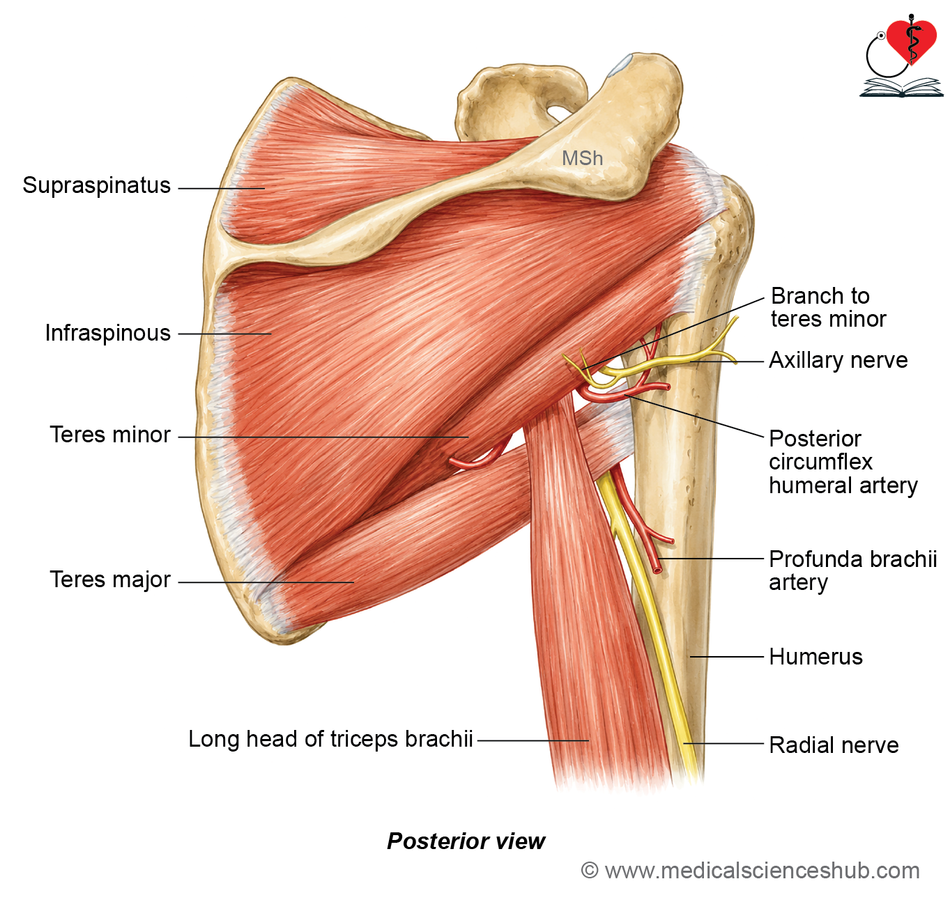
- Quadrangular space
- Upper triangular space(medial triangular space)
- Lower triangular space(triangular interval)
- Knowledge of these spaces is clinically important because they transmit major neurovascular structures and serve as landmarks during surgical procedures involving the shoulder and axilla.
Quadrangular space
Boundaries
- Superior: Subscapularis (anteriorly), capsule of the shoulder joint, and teres minor (posteriorly)
- Inferior: Teres major
- Medial: Long head of the triceps brachii
- Lateral: Surgical neck of the humerus
Contents
- Axillary nerve
- Posterior circumflex humeral artery and accompanying veins
Clinical importance
- The axillary nerve and posterior circumflex humeral vessels pass through the quadrangular space to reach the deltoid region.
- Compression or injury within this space may result in quadrangular space syndrome, producing shoulder pain, weakness of the deltoid and teres minor, and sensory loss over the regimental badge area.
Upper triangular space
Boundaries
- Superior: Teres minor
- Inferior: Teres major
- Lateral: Long head of the triceps brachii
Contents
- Circumflex scapular artery (a branch of the subscapular artery)
Lower triangular space
Boundaries
- Superior: Teres major
- Medial: Long head of the triceps brachii
- Lateral: Shaft of the humerus (or lateral head of the triceps in the proximal part)
Contents
- Radial nerve
- Profunda brachii (deep brachial) artery and accompanying veins
Clinical importance
The triangular interval serves as the passage through which the radial nerve and profunda brachii vessels enter the posterior compartment of the arm.
Injury or compression in this region may affect the radial nerve, leading to weakness of the extensor muscles of the arm and forearm.
Axillary Nerve
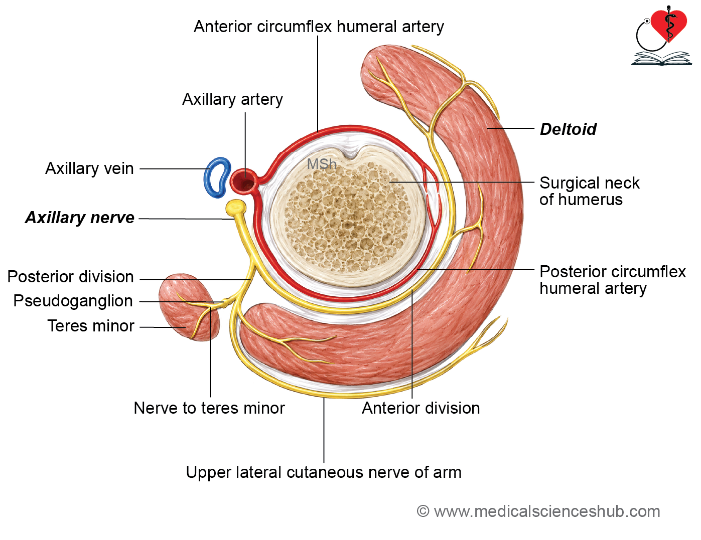
- The axillary nerve, also known as the circumflex nerve, winds around the surgical neck of the humerus.
- Root value: C5, C6
Beginning
It arises from the posterior cord of the brachial plexus.
Course and relations
In the axilla
- The axillary nerve originates from the posterior cord and passes posterolateral to the third part of the axillary artery.
- It lies on the subscapularis muscle before entering the quadrangular space.
In the quadrangular space
- The nerve leaves the axilla through the quadrangular space accompanied by the posterior circumflex humeral vessels.
Relations within the quadrangular space
- Superior: Subscapularis, capsule of the shoulder joint, and teres minor
- Inferior: Teres major
- Medial: Long head of the triceps brachii
- Lateral: Surgical neck of the humerus
Termination
Within the quadrangular space, the axillary nerve divides into anterior and posterior branches.
Branches
Articular branch
- Supplies the shoulder (glenohumeral) joint.
Anterior branch
- Accompanies the posterior circumflex humeral vessels around the surgical neck of the humerus.
- Supplies:
- Deltoid muscle (mainly the anterior and middle parts)
- Skin over the anteroinferior part of the deltoid
Posterior branch
- Passes around the posterior border of the deltoid.
- Supplies:
- Teres minor
- Posterior part of the deltoid
- Continues as the upper lateral cutaneous nerve of the arm, which supplies the skin over the lower half of the deltoid (regimental badge area).
- The nerve to teres minor characteristically contains a pseudoganglion, an anatomical enlargement without neuronal cell bodies.
CLINICAL INTEGRATION
- Axillary Nerve Palsy
- Axillary nerve palsy results from injury to the axillary nerve, leading to weakness of the deltoid and teres minor muscles.
- Common causes
- Fracture of the surgical neck of the humerus
- Anterior dislocation of the shoulder joint
- Improper intramuscular injection placed too high in the deltoid region
- Compression of the nerve within the quadrangular space (less common)
- Clinical features
- Weakness of arm abduction, especially from 15° to 90°, due to paralysis of the deltoid muscle. The supraspinatus can still initiate the first 15° of abduction.
- Flattening of the normal rounded contour of the shoulder because of deltoid muscle wasting.
- Weakness of lateral rotation of the arm due to involvement of the teres minor.
- Regimental badge anesthesia, characterized by loss of sensation over the skin covering the lower half of the deltoid, supplied by the upper lateral cutaneous nerve of the arm.
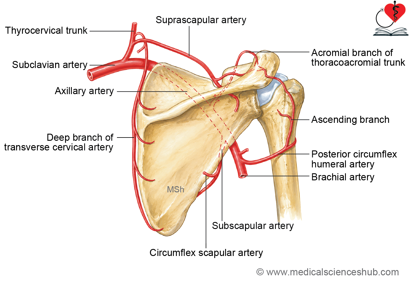
Anastomosis Around Scapula
- The scapular arterial anastomosis is a network of arteries that provides collateral circulation to the scapular region and shoulder.
- It maintains an adequate blood supply when the axillary artery is occluded proximal to the origin of the subscapular artery.
- The arterial network is divided into:
- Anastomosis around the body of the scapula
- Anastomosis around the acromion process
Anastomosis Around the Body of Scapula
The arterial network is formed by the following vessels:
Branches of the first part of the subclavian artery
- Suprascapular artery (branch of the thyrocervical trunk)
- Deep branch of the transverse cervical artery (dorsal scapular artery, where present)
Branch of the third part of the axillary artery
- Circumflex scapular artery, a branch of the subscapular artery
Anastomosis Over the Acromion Process
The arterial network over the acromion is formed by:
Branch of the first part of the subclavian artery
Acromial branch of the suprascapular artery
Branch of the second part of the axillary artery
Acromial branch of the thoracoacromial artery
Branch of the third part of the axillary artery
Acromial branch of the posterior circumflex humeral artery
CLINICAL INTEGRATION
- The scapular anastomosis provides an alternative route for blood flow between the subclavian and axillary arteries.
- It becomes particularly important when the axillary artery is gradually obstructed proximal to the origin of the subscapular artery, helping to preserve the blood supply to the upper limb.
Important Questions
- Write a short note on deltoid muscle.
- Write a short note on rotator cuff muscles.
- Write a short note on quadrangular and triangular spaces of upper limb.
- Write a short note on axillary nerve.
- Write short note on arterial anastomosis around the scapula.
📝 Test Your Knowledge – Practice MCQs
Attempt the chapter MCQ quiz and assess your understanding of key concepts.
Click to see: Medical Disclaimer and Copyright Notice