
Competencies
- AN13.3: Identify and describe the type, articular surfaces, capsule, synovial membrane, ligaments, relations, movements, blood and nerve supply of elbow joint, proximal and distal radioulnar joints, wrist joint, and first carpometacarpal joint.
- AN13.4: Describe sternoclavicular joint, acromioclavicular joint, carpometacarpal joint and metacarpophalangeal joint.
Elbow Joint
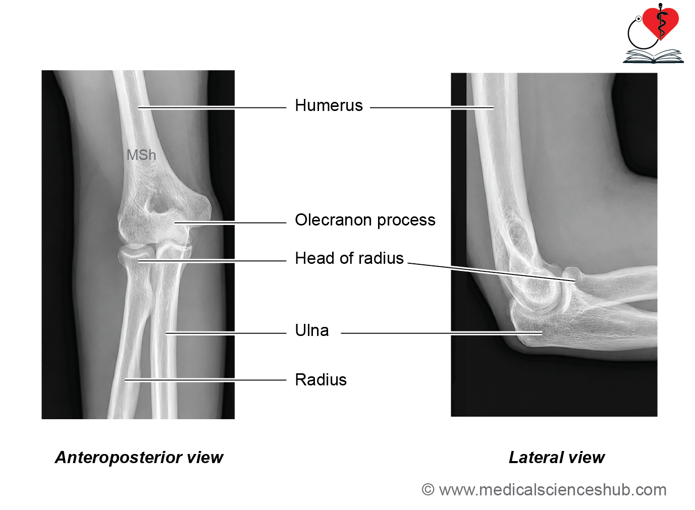
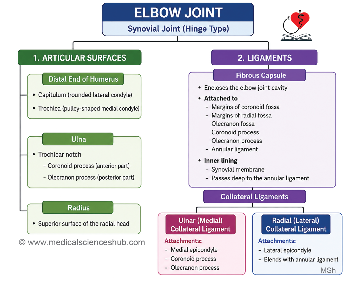
The elbow joint is formed by the articulation of the distal end of the humerus with the proximal ends of the radius and ulna. It provides stability while allowing flexion and extension of the forearm.
Type of joint
- The elbow is a synovial hinge joint, permitting movement mainly in a single plane, namely flexion and extension.
- It is also classified as a compound joint because three bones participate in its formation: the humerus, radius, and ulna.
Articular surfaces
Superior Articular Surface (Humerus)
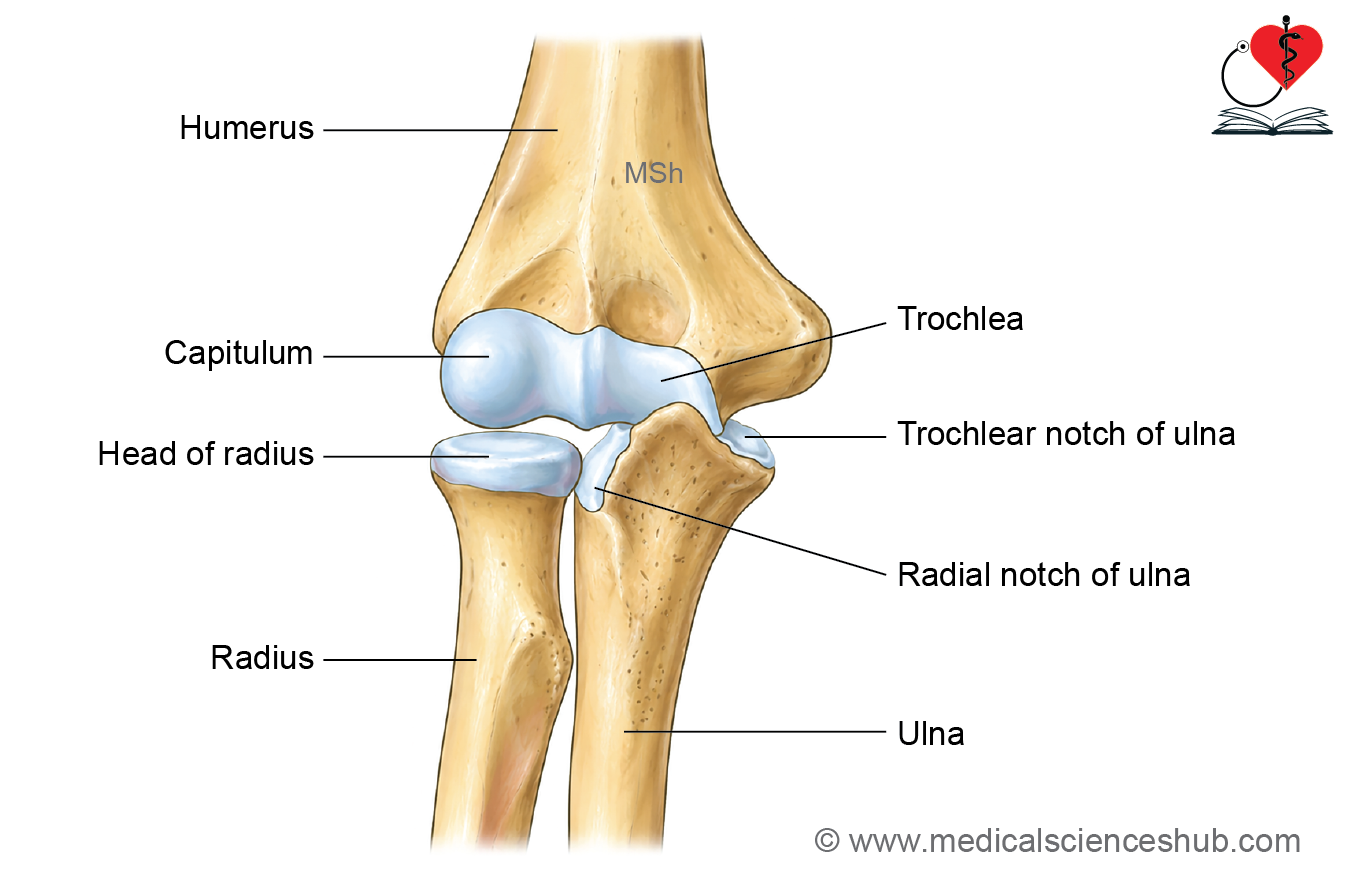
- The distal end of the humerus contributes two articular areas:
- Capitulum: A rounded, convex structure located on the lateral side. It articulates with the head of the radius.
- Trochlea: A spool-shaped or pulley-like medial articular surface that articulates with the trochlear notch of the ulna.
Inferior Articular Surfaces
Trochlear Notch of Ulna
- The trochlear notch is formed by the:
- Upper surface of the coronoid process, and
- Anterior surface of the olecranon process.
- It receives the trochlea of the humerus, forming the humeroulnar articulation.
Head of Radius
- The head of the radius is circular and disc-shaped.
- Its superior surface articulates with the capitulum of the humerus, forming the humeroradial articulation.
Ligaments
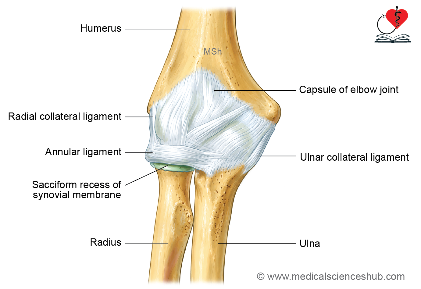
The elbow joint is reinforced by a strong fibrous capsule and two collateral ligaments that provide stability during movement.
Capsular ligaments
- The capsular ligament is a fibrous capsule that surrounds the elbow joint and encloses its synovial cavity.
Attachments
Superior Attachment
- Attached to the distal humerus around the articular margins, including:
- Medial epicondyle
- Margins of the coronoid, radial, and olecranon fossae
- Lateral margin of the capitulum
- The capsule encloses the non-articular fossae of the humerus.
Inferior Attachment
- Attached to the margins of the trochlear notch of the ulna.
- Laterally, it blends with the annular ligament of the superior radioulnar joint.
Synovial Membrane
- The inner surface of the capsule is lined by a synovial membrane.
- On the inferolateral side, the synovial membrane extends beneath the annular ligament to the neck of the radius.
- As a result, the synovial cavity of the elbow joint communicates with that of the superior radioulnar joint.
- The synovial membrane also covers the fat pads lying within the coronoid, radial, and olecranon fossae.
Ulnar Collateral Ligament (Medial Collateral Ligament)
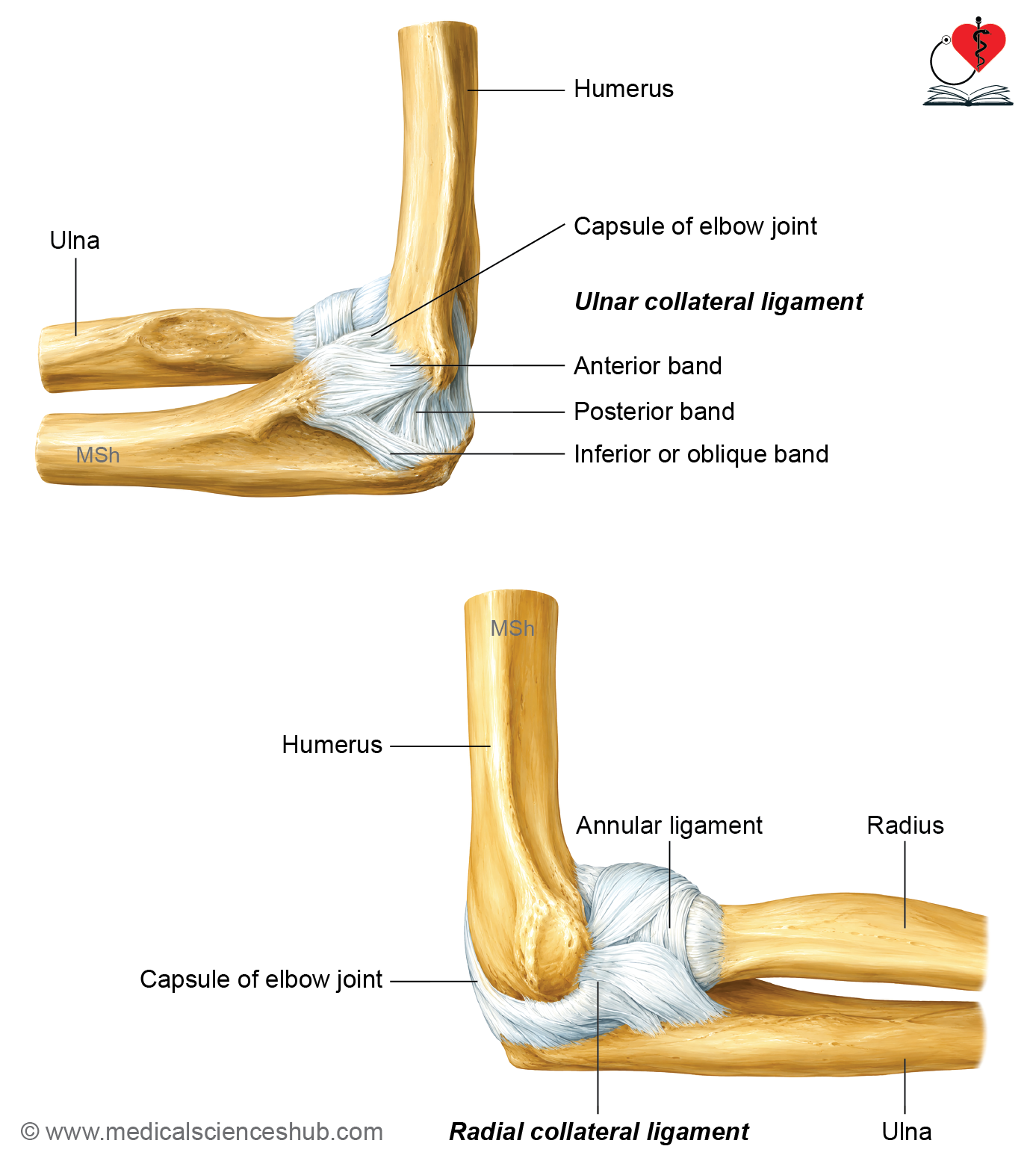
- The ulnar collateral ligament is a strong triangular ligament situated on the medial side of the elbow.
- It provides the principal resistance to valgus stress.
Attachments
- Apex: Medial epicondyle of the humerus
- Base: Coronoid process and olecranon process of the ulna
Parts
- Anterior band
- Strongest component of the ligament.
- Extends from the anterior aspect of the medial epicondyle to the medial margin of the coronoid process.
- Posterior band
- Extends from the posterior aspect of the medial epicondyle to the medial margin of the olecranon.
- Inferior (Transverse) band
- Runs between the coronoid and olecranon processes of the ulna.
- Reinforces the ligament but does not cross the joint.
Radial Collateral Ligament (Lateral Collateral Ligament)
- The radial collateral ligament is a fan-shaped ligament located on the lateral side of the elbow.
- It contributes to lateral stability and blends with the annular ligament.
Attachments
- Superior: Lateral epicondyle of the humerus
- Inferior: Annular ligament surrounding the head of the radius
Muscular Attachments
- Provides partial origin to:
- Supinator
- Extensor carpi radialis brevis
Stabilizing Factors of the Elbow Joint
The elbow joint is stabilized by:
- The close fit of the trochlea of the humerus within the trochlear notch of the ulna.
- The strong ulnar (medial) collateral ligament.
- The strong radial (lateral) collateral ligament.
Relations
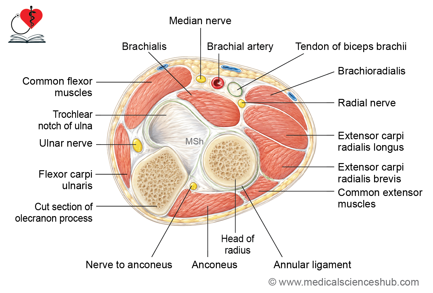
The elbow joint is related to several important neurovascular structures, muscles, and tendons on its anterior, posterior, medial, and lateral aspects.
Anterior
The anterior aspect of the elbow joint is related to:
- Median nerve
- Brachial artery
- Tendon of biceps brachii (within the cubital fossa)
- Brachialis muscle, which forms the floor of the cubital fossa
Posterior
The posterior aspect is related to:
- Tendon of triceps brachii
- Anconeus muscle
Medial
The medial aspect is related to:
- Ulnar nerve
- Common flexor tendon arising from the medial epicondyle
- Flexor carpi ulnaris muscle
Lateral
The lateral aspect is related to:
- Common extensor tendon arising from the lateral epicondyle
- Supinator muscle
- Extensor carpi radialis brevis muscle
- Muscles originating from the radial collateral ligament
Blood and Nerve Supply
Blood supply
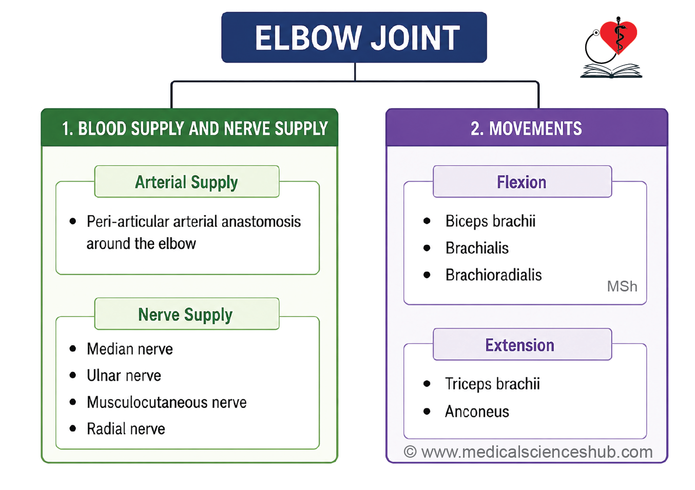
The elbow joint receives its blood supply from the periarticular arterial anastomosis around the elbow. This rich vascular network ensures an adequate blood supply during all positions of the joint.
Innervation (nerve supply)
The elbow joint is supplied by articular branches from all the major nerves of the upper limb except the axillary nerve.
The articular branches arise from:
- Musculocutaneous nerve
- Median nerve
- Ulnar nerve
- Radial nerve
Movements of Elbow Joint
As a synovial hinge joint, the elbow permits movement primarily in one plane, allowing flexion and extension.
- Range of motion: Approximately 150° from full extension to maximum flexion.
Flexion
Prime movers
- Brachialis (chief flexor)
- Biceps brachii
- Brachioradialis
Extension
Prime movers
- Triceps brachii (chief extensor)
- Anconeus (assists triceps and stabilizes the joint during extension)
Carrying angle
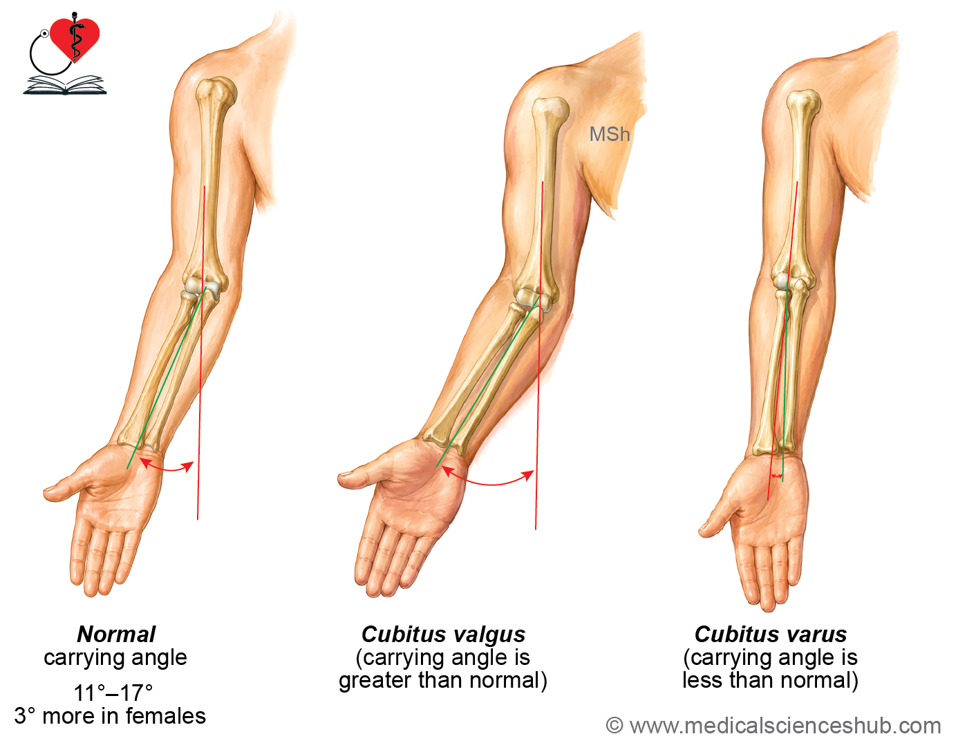
The carrying angle is the angle formed between the long axis of the arm and the long axis of the forearm when the elbow is fully extended and the forearm is supinated. It allows the forearm to deviate slightly away from the body.
Normal Range
- Normal carrying angle: 11°–17°
- The carrying angle varies among individuals.
- It is typically about 3° greater in females than in males.
- The carrying angle disappears during full flexion of the elbow and also when the forearm is pronated.
Anatomical Basis
The carrying angle results from the asymmetrical anatomy of the elbow joint:
- The transverse axis of the elbow is oblique, running downward and medially.
- The medial flange of the trochlea projects approximately 6 mm farther distally than the lateral flange.
- The superior articular surface of the coronoid process of the ulna is also obliquely oriented, sloping downward and medially.
These anatomical features produce the physiological lateral deviation of the forearm in the anatomical position.
CLINICAL CORRELATION
Cubitus Valgus
- Definition: An increase in the carrying angle beyond the normal range.
- Causes:
- Malunion of fractures around the elbow
- Turner syndrome (45,X)
- Noonan syndrome
- Clinical significance: Marked cubitus valgus may stretch the ulnar nerve, leading to delayed ulnar nerve palsy.
Cubitus Varus (Gunstock Deformity)
- Definition: A decrease or reversal of the normal carrying angle.
- Common cause: Malunion following a supracondylar fracture of the humerus.
- Clinical significance: Although primarily a cosmetic deformity, severe cases may alter elbow mechanics and upper limb alignment.
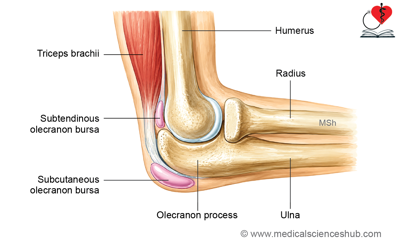
Bursae related to elbow joint
A bursa is a synovial membrane-lined sac filled with a small amount of fluid. It reduces friction and cushions the movement between bones, tendons, muscles, and the overlying skin.
The major bursae associated with the elbow joint are:
Olecranon Bursae
Subcutaneous Olecranon Bursa
- Located between the skin and the posterior triangular surface of the olecranon.
- Protects the skin from friction during movements and when the elbow rests on hard surfaces.
Subtendinous Olecranon Bursa
- Situated between the tendon of the triceps brachii and the superior surface of the olecranon process of the ulna.
- Reduces friction between the triceps tendon and the underlying bone during elbow extension.
Bicipitoradial Bursa
- Located between the tendon of the biceps brachii and the anterior smooth surface of the radial tuberosity.
- Facilitates smooth gliding of the biceps tendon during flexion and supination of the forearm.
CLINICAL CORRELATION
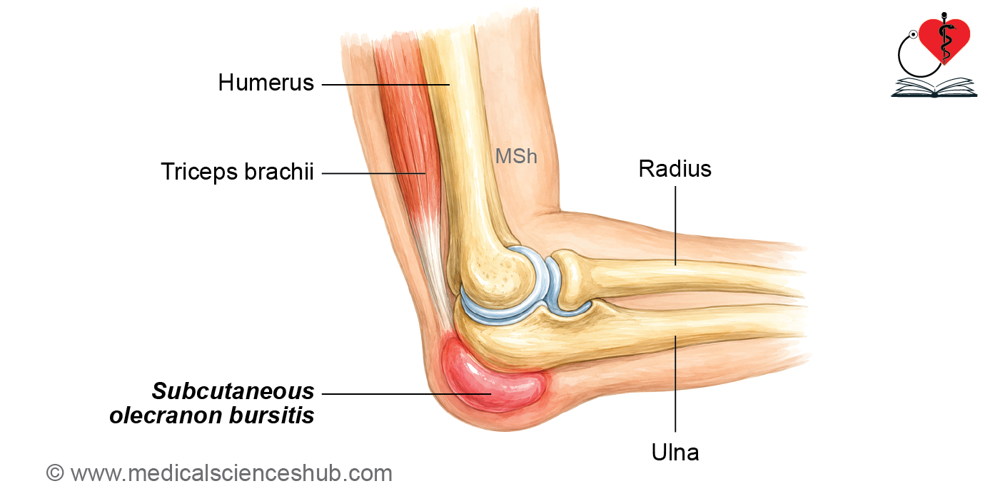
Olecranon Bursitis
- Inflammation of the subcutaneous olecranon bursa is known as olecranon bursitis or student’s elbow, commonly resulting from repeated pressure, trauma, or infection.
- Patients present with swelling, tenderness, and pain over the posterior aspect of the elbow.
Bicipitoradial Bursitis
- Inflammation of the bicipitoradial bursa may cause pain in the anterior aspect of the elbow, particularly during forearm pronation and supination, and can occasionally compress the nearby radial nerve.
Student’s Elbow (Olecranon Bursitis)
- Student’s elbow refers to inflammation of the subcutaneous olecranon bursa.
- It commonly results from repeated minor trauma caused by resting the elbow on hard surfaces for prolonged periods.
- Patients present with swelling, tenderness, and pain over the posterior aspect of the elbow.
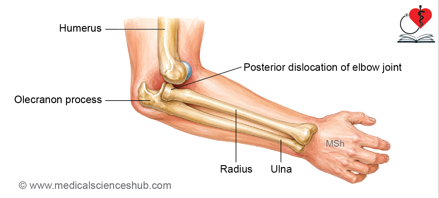
Dislocation of the Elbow
- Posterior dislocation is the most common type of elbow dislocation.
- Other directions of dislocation are uncommon because of the strong stabilizing structures of the joint.
- Factors preventing anterior dislocation
- Strong posterior part of the joint capsule
- Robust ulnar and radial collateral ligaments
- Hook-shaped olecranon process, which locks into the olecranon fossa of the humerus
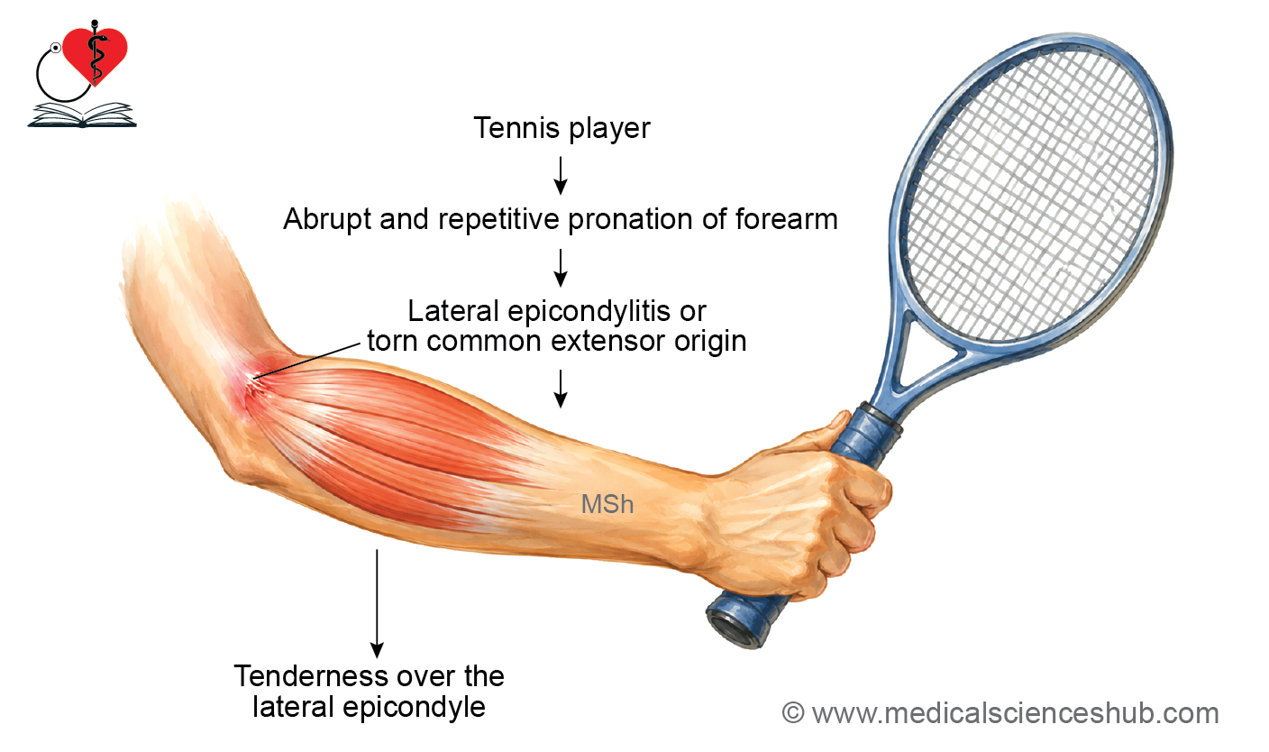
Tennis Elbow (Lateral Epicondylitis)
- Tennis elbow is characterized by pain and tenderness over the lateral epicondyle of the humerus.
- Pain is aggravated by wrist extension and repetitive forearm movements, particularly pronation and gripping activities.
- Although common in tennis players, it also occurs in individuals performing repetitive manual work.
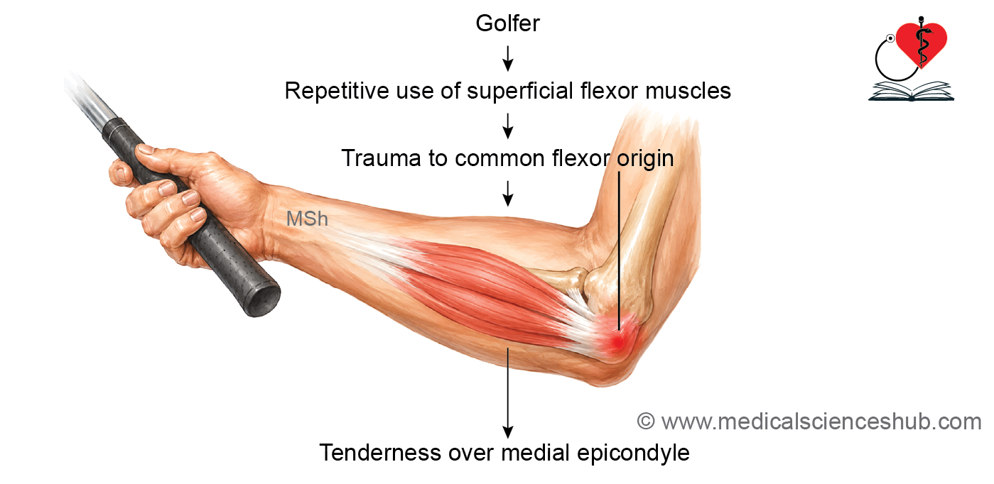
Golfer’s Elbow (Medial Epicondylitis)
- Golfer’s elbow presents with pain and tenderness over the medial epicondyle of the humerus.
- It results from repetitive strain or trauma to the common flexor tendon.
- It is commonly seen in golfers and individuals performing repeated wrist flexion or forearm pronation.
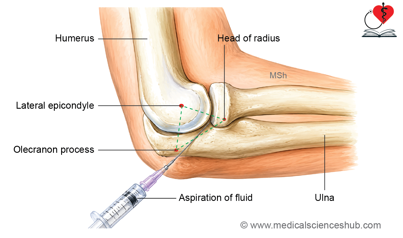
Elbow Effusion
- Elbow effusion is the accumulation of inflammatory or excess synovial fluid within the elbow joint cavity.
- Distension is usually more prominent posteriorly, where the joint capsule is relatively weak.
- Aspiration of Joint Fluid
- Joint aspiration is performed from the lateral side of the elbow.
- The needle is introduced through the triangular interval bounded by:
- Olecranon process
- Lateral epicondyle of the humerus
- Head of the radius
This approach minimizes the risk of injury to major neurovascular structures while providing safe access to the joint cavity.
Radioulnar Articulation
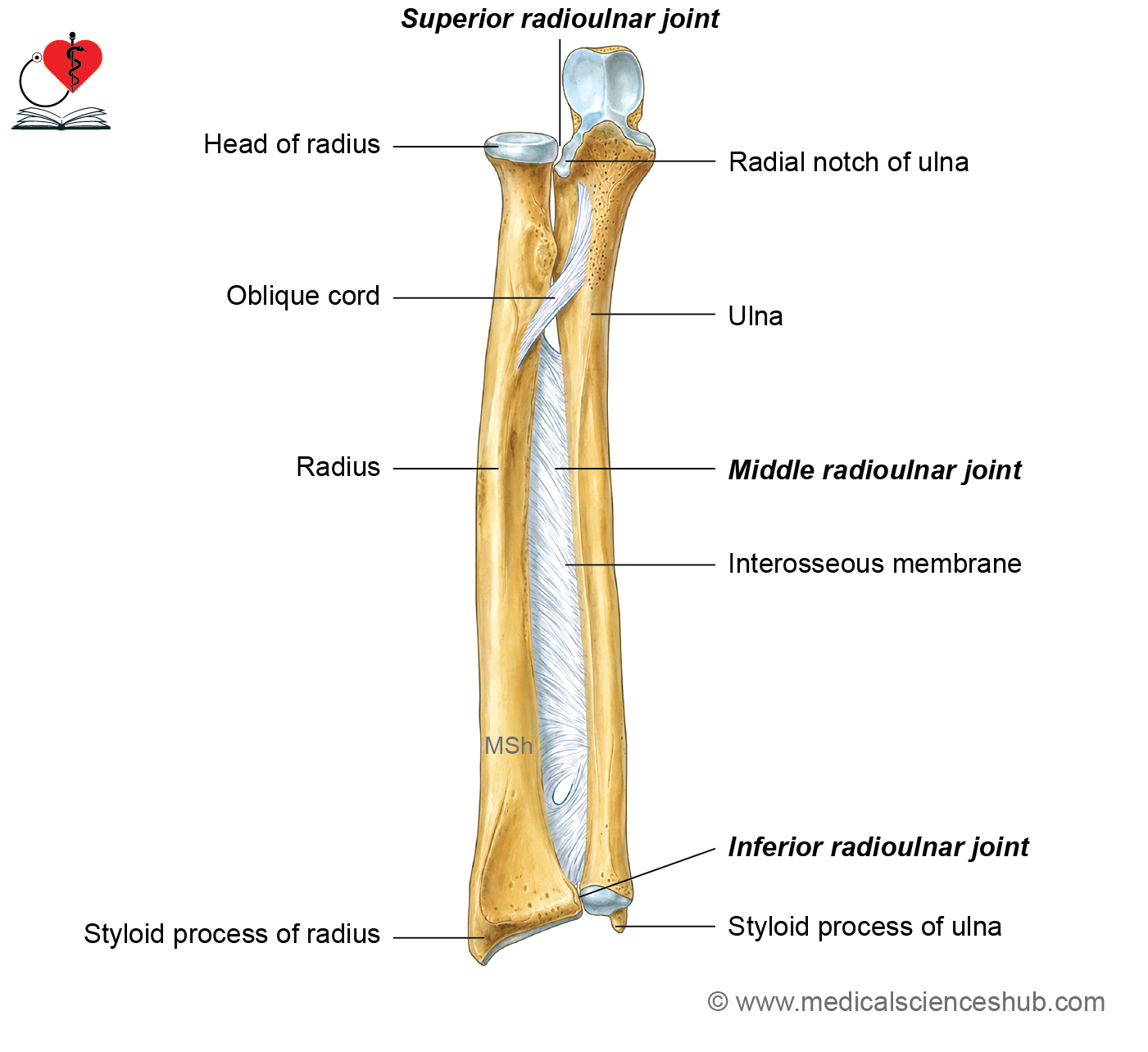
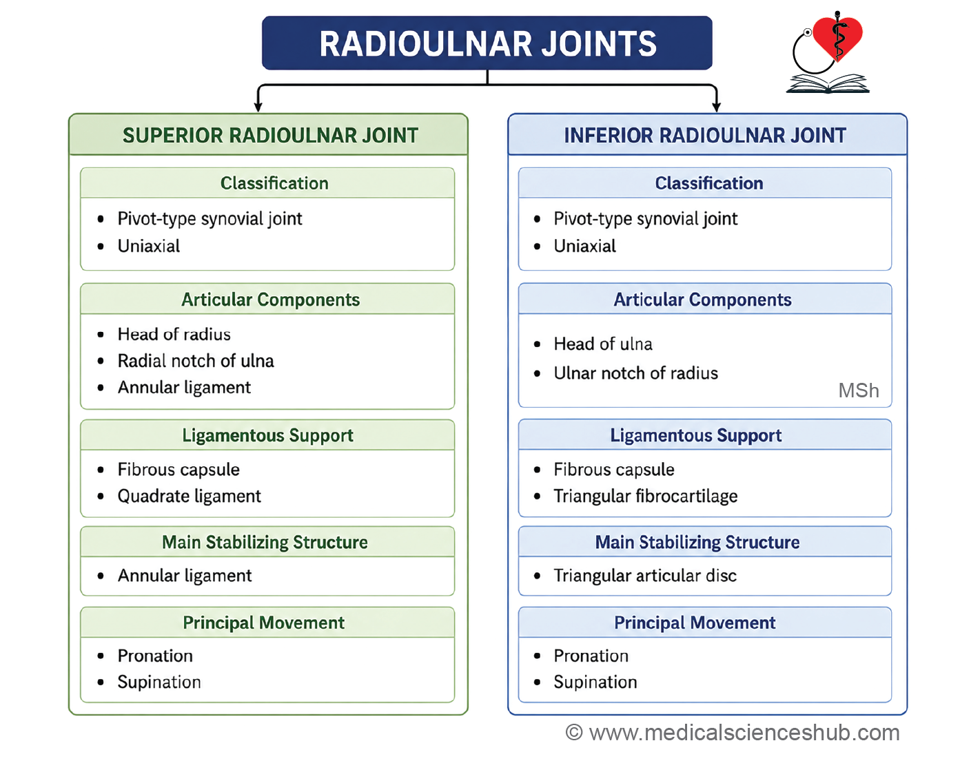
The radius and ulna are connected at three sites, allowing coordinated movements of the forearm, particularly pronation and supination.
The three radioulnar articulations are:
- Superior (Proximal) Radioulnar Joint
- A pivot type of synovial joint.
- It permits rotation of the head of the radius within the annular ligament during pronation and supination.
- Middle Radioulnar Joint
- A fibrous joint (syndesmosis) formed by the interosseous membrane between the shafts of the radius and ulna.
- It binds the two bones together, provides stability, serves as an attachment for forearm muscles, and transmits forces from the radius to the ulna.
- Inferior (Distal) Radioulnar Joint
- A pivot type of synovial joint.
- It allows the distal end of the radius to rotate around the head of the ulna, contributing to pronation and supination of the forearm.
Superior (Proximal) Radioulnar Joint
Type
- The superior (proximal) radioulnar joint is a uniaxial pivot type of synovial joint.
- It allows rotational movements of the radius around the ulna during pronation and supination.
Articular Surfaces
The joint is formed by the articulation between:
- Radius: Circumference of the disc-shaped head of the radius
- Ulna: Radial notch of the ulna
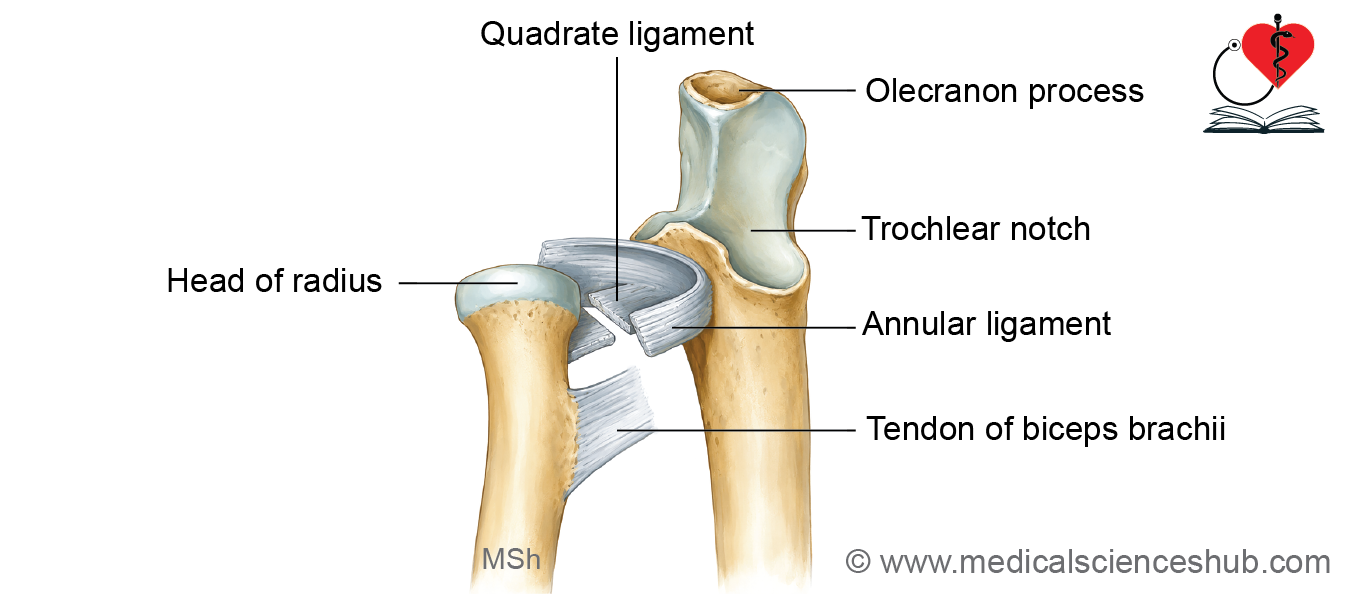
The annular ligament and the radial notch of the ulna together form a fibro-osseous ring, within which the head of the radius rotates.
Ligaments
Fibrous Capsule
- Encloses the joint.
- Superiorly, it is continuous with the capsule of the elbow joint.
- Inferiorly, it blends with the annular ligament.
Annular Ligament
- A strong fibrous band attached to the anterior and posterior margins of the radial notch of the ulna.
- It encircles approximately four-fifths of the circumference of the radial head, holding it securely against the radial notch.
- It is the principal stabilizing ligament of the superior radioulnar joint while allowing free rotation of the radial head.
Quadrate Ligament
- Extends from the neck of the radius to the supinator fossa of the ulna.
- Reinforces the inferior part of the joint capsule and limits excessive rotation.
Synovial Membrane
- Lines the inner surface of the fibrous capsule.
- It is continuous superiorly with the synovial membrane of the elbow joint.
- The membrane extends beneath the annular ligament to reach the neck of the radius, deep to the quadrate ligament.
Blood Supply
- Supplied by the periarticular arterial anastomosis around the elbow joint.
Nerve Supply
The joint receives articular branches from:
- Musculocutaneous nerve
- Median nerve
- Ulnar nerve
- Radial nerve
Movements
The superior radioulnar joint permits:
- Supination: Lateral rotation of the radius so that the palm faces anteriorly (or upward when the elbow is flexed).
- Pronation: Medial rotation of the radius around the ulna so that the palm faces posteriorly (or downward when the elbow is flexed).
Inferior (Distal) Radioulnar Joint
Type
- The inferior (distal) radioulnar joint is a uniaxial pivot type of synovial joint.
- It works in conjunction with the superior radioulnar joint to permit pronation and supination of the forearm.
Articular Surfaces
The joint is formed by the articulation between:
- Radius: Concave ulnar notch on the distal end of the radius
- Ulna: Convex head of the ulna
Ligaments
Capsular Ligament
- A thin fibrous capsule surrounds the joint.
- It is attached to the margins of the articular surfaces of the distal radius and ulna.
Synovial Membrane
- Lines the inner surface of the fibrous capsule.
- Forms the synovial cavity of the joint.
Recessus Sacciformis
- An upward extension (pouch) of the synovial membrane located anterior to the interosseous membrane.
- It provides additional space for the rotational movements of the radius during pronation and supination.
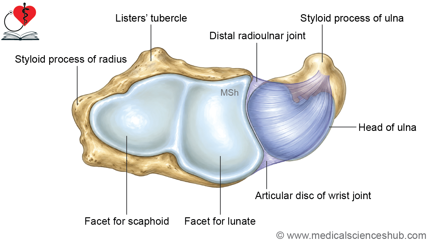
Articular Disc (Triangular Fibrocartilaginous Disc)
- A triangular fibrocartilaginous disc separates the distal radioulnar joint from the wrist joint.
- It stabilizes the distal radioulnar joint and acts as a cushion between the ulna and the carpal bones.
Attachments of the Articular Disc
- Apex: Base of the styloid process of the ulna
- Base: Inferior margin of the ulnar notch of the radius
Blood Supply
The inferior radioulnar joint is supplied by:
- Anterior interosseous artery
- Posterior interosseous artery
Nerve Supply
Articular branches arise from:
- Anterior interosseous nerve
- Posterior interosseous nerve
Movements
The distal radioulnar joint permits:
- Supination: Rotation of the radius laterally around the relatively fixed ulna, bringing the palm anteriorly (or upward with the elbow flexed).
- Pronation: Rotation of the radius medially around the ulna, turning the palm posteriorly (or downward with the elbow flexed).
Interosseous membrane of forearm (middle radioulnar joint)
The interosseous membrane is a thin but strong fibrous sheet that connects the shafts of the radius and ulna. It forms the middle radioulnar joint, which is a fibrous joint (syndesmosis).
Attachments
- Attached along the interosseous borders of the radius and ulna.
Direction of Fibres
- Most fibres run obliquely downward and medially, from the radius to the ulna.
Features
- Forms the middle radioulnar joint (syndesmosis).
- Provides a broad surface for muscular attachment.
- Maintains the relationship between the radius and ulna while permitting pronation and supination.
Openings in the Membrane
Proximal Opening
- Located between the upper border of the interosseous membrane and the oblique cord.
- Transmits the posterior interosseous artery and posterior interosseous nerve to the posterior compartment of the forearm.
Distal Opening
- Present near the lower part of the membrane.
- Allows the anterior interosseous artery to pass to the posterior compartment near the upper border of the pronator quadratus muscle.
Muscular Attachments
From the Anterior Surface
- Flexor pollicis longus
- Flexor digitorum profundus
From the Posterior Surface
- Abductor pollicis longus
- Extensor pollicis longus
- Extensor indicis
Functions of the Interosseous Membrane
- Forms the middle radioulnar joint, linking the radius and ulna.
- Maintains stability between the two forearm bones during pronation and supination.
- Transmits forces from the radius to the ulna, especially during weight-bearing through the hand.
- Provides an extensive surface for muscle attachment.
Oblique Cord
The oblique cord is a small fibrous band situated in the upper part of the forearm.
Attachments
- Extends from the ulnar tuberosity to the radial tuberosity.
Direction of Fibres
- Fibres run downward and laterally, opposite to the direction of the fibres of the interosseous membrane.
Morphological Significance
- The oblique cord is considered a degenerated remnant of the flexor pollicis longus muscle.
Supination and Pronation
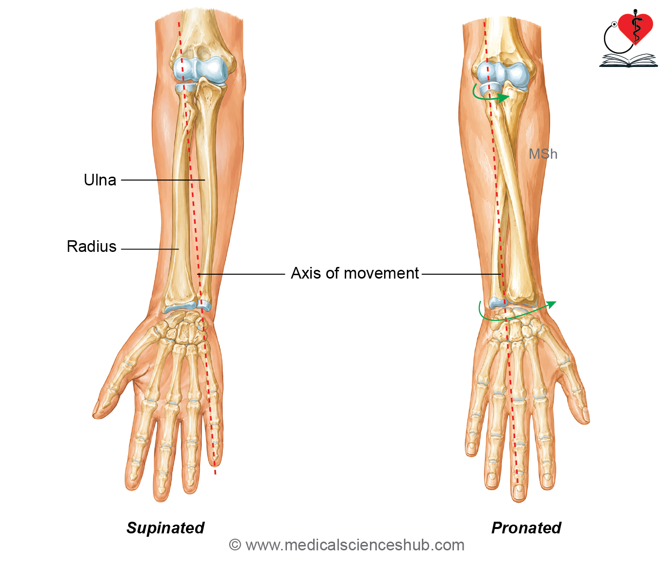
Supination and pronation are rotational movements of the forearm in which the radius rotates around the relatively fixed ulna. These movements occur about a vertical axis and enable the hand to be positioned appropriately for various activities.
Definitions
Supination
- Supination is the rotational movement of the forearm that turns the palm anteriorly in the anatomical position.
- With the elbow flexed to 90°, supination turns the palm upward.
Pronation
- Pronation is the rotational movement of the forearm that turns the palm posteriorly in the anatomical position.
- With the elbow flexed to 90°, pronation turns the palm downward.
Joints Involved
Supination and pronation occur simultaneously at two pivot joints:
- Superior (proximal) radioulnar joint
- Inferior (distal) radioulnar joint
The interosseous membrane maintains the relationship between the radius and ulna during these movements.
Axis of Rotation
- The movements occur around a vertical axis extending:
- Superiorly: Through the centre of the head of the radius
- Inferiorly: Through the base of the styloid process of the ulna
Movement of the Axis
The axis is not completely fixed and shifts slightly during rotation:
- Forward and medially during supination
- Backward and laterally during pronation
Position of the Radius and Ulna
During Supination
- The radius and ulna remain parallel to each other.
- The palm faces anteriorly (or upward when the elbow is flexed).
During Pronation
- The distal end of the radius, together with the articular disc, rotates around the head of the ulna.
- Simultaneously, the head of the radius rotates within the annular ligament at the superior radioulnar joint.
- As a result, the radius crosses obliquely in front of the ulna.
Muscles Producing the Movements
Pronation
Prime mover
- Pronator quadratus
Assistant
- Pronator teres
Supination
Prime movers
- Supinator
- Biceps brachii (particularly effective when the elbow is flexed)
Functional Importance
Supination and pronation are essential for many daily activities requiring precise positioning of the hand.
Examples include:
- Turning the hand to pick up food in pronation and bring it to the mouth in supination.
- Tightening a screw with supination.
- Loosening a screw with pronation.
- Turning a doorknob, using a screwdriver, pouring liquids, and handling tools.
CLINICAL NOTE
- Supination is generally more powerful than pronation, largely because of the strong action of the biceps brachii, especially when the elbow is flexed.
- Loss of pronation or supination significantly impairs hand function despite preserved elbow flexion and extension.
Wrist Joint
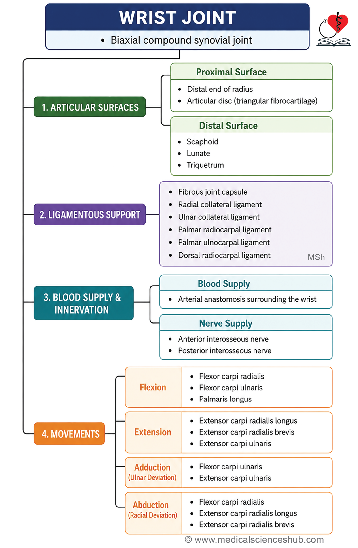
The wrist joint, also known as the radiocarpal joint, is the articulation between the distal forearm and the proximal row of carpal bones. It provides a wide range of hand movements while maintaining stability.
Type
- The wrist joint is a biaxial synovial joint (condyloid or ellipsoid type).
- It is a compound synovial joint because more than two bones participate in its formation.
- Note: Although an articular disc is associated with the joint, the wrist is not classified as a complex synovial joint, as the disc lies proximal to the joint cavity and separates the wrist joint from the inferior radioulnar joint.
Articular surfaces
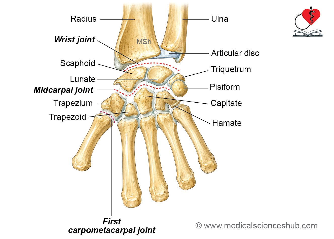
Proximal (Superior) Articular Surface
The proximal articular surface is formed by:
- Inferior articular surface of the distal end of the radius
- Inferior surface of the triangular articular disc (articular disc of the distal radioulnar joint)
Together, these structures form a smooth concave elliptical articular surface.
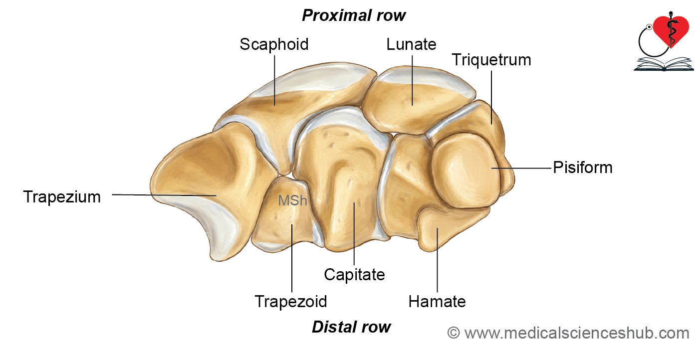
Distal (Inferior) Articular Surface
The distal articular surface is formed by the proximal surfaces of the following carpal bones:
- Scaphoid
- Lunate
- Triquetrum (articulates with the articular disc when the wrist is adducted)
These bones together form a convex articular surface that articulates with the concave proximal surface of the joint.
Ligaments
The wrist joint is reinforced by a fibrous capsule and several strong ligaments that provide stability while permitting a wide range of movements.
Fibrous Capsule
- The fibrous capsule encloses the wrist joint cavity.
- It is lined internally by the synovial membrane.
- The capsule is attached to the margins of the proximal and distal articular surfaces.
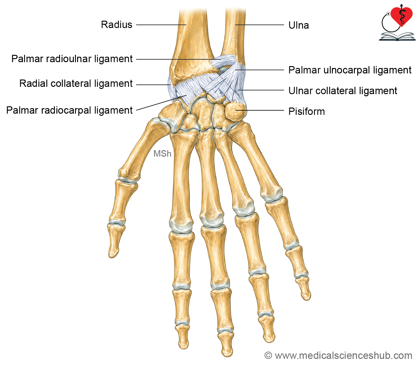
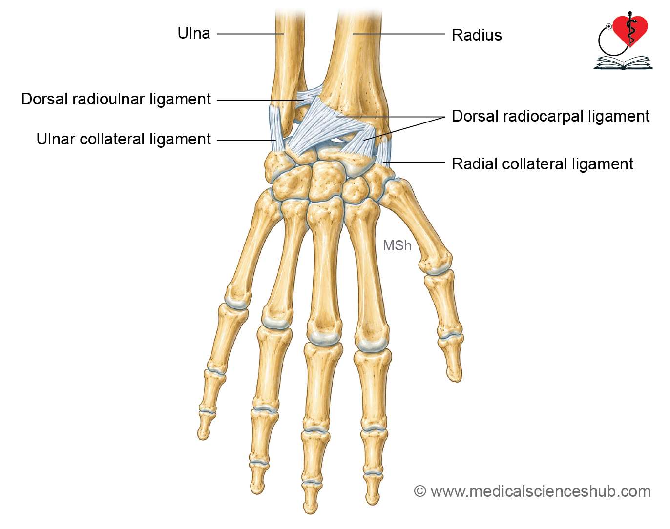
Radial Collateral Ligament
- Extends from the tip of the styloid process of the radius to the scaphoid.
- Strengthens the lateral aspect of the wrist joint.
- The radial artery crosses superficial to this ligament.
Ulnar Collateral Ligament
- Extends from the tip of the styloid process of the ulna to the medial side of the triquetrum and pisiform.
- Reinforces the medial side of the wrist joint.
Palmar Radiocarpal Ligament
- Extends from the anterior margin of the distal end of the radius to the anterior surfaces of:
- Scaphoid
- Lunate
- Triquetrum
- It is one of the strongest stabilizing ligaments of the wrist and limits excessive extension.
Palmar Ulnocarpal Ligament
- Extends from the styloid process of the ulna and the articular disc to the anterior surfaces of:
- Lunate
- Triquetrum
- Reinforces the palmar aspect of the wrist joint on the ulnar side.
Dorsal Radiocarpal Ligament
- Extends from the posterior margin of the distal end of the radius to the posterior surfaces of:
- Scaphoid
- Lunate
- Triquetrum
- Strengthens the posterior aspect of the joint and limits excessive flexion.
Relations
Anterior
The anterior aspect of the wrist joint is related to the structures passing through the carpal tunnel, including:
- Tendons of flexor digitorum superficialis (FDS) and flexor digitorum profundus (FDP) enclosed within the ulnar bursa
- Tendon of flexor pollicis longus (FPL) enclosed within the radial bursa
- Median nerve
- Tendon of flexor carpi radialis (passes in a separate osteofibrous canal lateral to the carpal tunnel)
Posterior
The posterior aspect is related to:
- Extensor retinaculum and the extensor tendons enclosed within their synovial sheaths
- Anterior interosseous artery
- Posterior interosseous nerve
Medial
The medial aspect is related to:
- Dorsal cutaneous branch of the ulnar nerve
Lateral
The lateral aspect is related to:
- Radial artery
- Tendon of abductor pollicis longus
- Tendon of extensor pollicis brevis (first dorsal extensor compartment)
Blood and Nerve Supply
The wrist joint receives blood from the palmar and dorsal carpal arterial arches, which are formed by branches of the:
- Radial artery
- Ulnar artery
- Anterior interosseous artery
Nerve Supply
Articular branches to the wrist joint arise from:
- Anterior interosseous nerve
- Posterior interosseous nerve
Movements
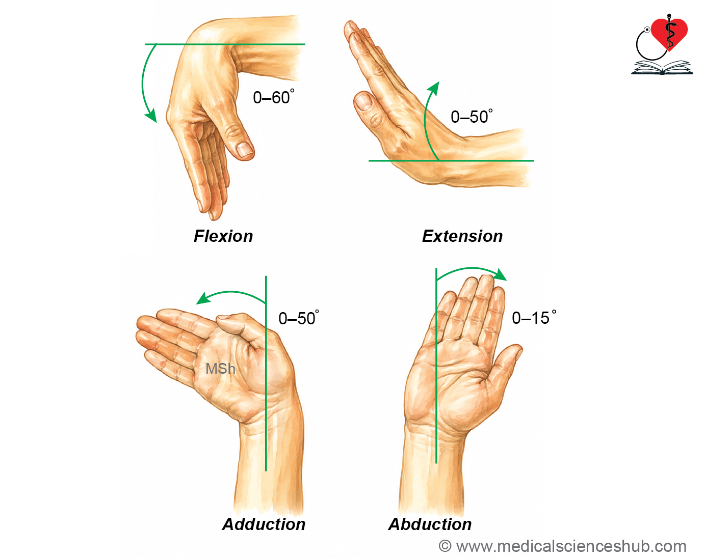
The wrist is a biaxial synovial (condyloid) joint that permits the following movements:
- Flexion
- Extension
- Adduction (ulnar deviation)
- Abduction (radial deviation)
- Circumduction
Axes of Movement
- Flexion and extension occur around a transverse axis.
- Abduction and adduction occur around an anteroposterior axis.
Flexion
Flexion bends the hand anteriorly, bringing the palm toward the anterior aspect of the forearm.
Muscles
- Flexor carpi radialis
- Flexor carpi ulnaris
- Palmaris longus
Extension
Extension moves the hand posteriorly, bringing the dorsum of the hand toward the posterior aspect of the forearm.
Muscles
- Extensor carpi radialis longus
- Extensor carpi radialis brevis
- Extensor carpi ulnaris
Adduction (Ulnar Deviation)
Adduction moves the hand toward the ulnar (medial) side.
Muscles
- Flexor carpi ulnaris
- Extensor carpi ulnaris
Abduction (Radial Deviation)
Abduction moves the hand toward the radial (lateral) side.
Muscles
- Flexor carpi radialis
- Extensor carpi radialis longus
- Extensor carpi radialis brevis
Circumduction
Circumduction is a combined movement produced by the sequential action of:
- Flexion
- Abduction
- Extension
- Adduction
It results in a circular movement of the hand while the forearm remains stationary.
CLINICAL INTEGRATION
Wrist Lock
- A wrist lock is a joint-locking maneuver commonly used in martial arts and self-defense techniques.
- It is performed by firmly grasping the opponent’s hand and forcefully bending the wrist beyond its normal range of movement.
- Excessive force may result in:
- Ligament sprain or tear
- Wrist dislocation
- Fractures of the carpal bones or distal forearm
Immobilization of the Wrist Joint
- For splinting or casting, the wrist is best immobilized in approximately 30° of dorsiflexion (extension).
- This functional position provides optimal ligament tension, preserves hand function, and minimizes joint stiffness.
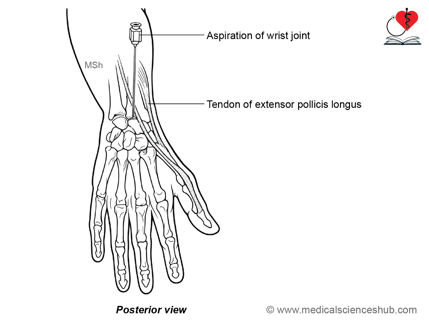
Arthrocentesis (Aspiration) of the Wrist Joint
- Joint aspiration is usually performed from the posterior (dorsal) aspect of the wrist.
- The needle is introduced between the tendons of the extensor pollicis longus and extensor digitorum, corresponding to the interval between the third and fourth extensor compartments.
- This approach provides safe access to the joint cavity while minimizing the risk of injury to adjacent structures.
Brunelli Procedure
- The Brunelli procedure is a reconstructive operation performed to treat chronic instability of the wrist, most commonly due to injury of the scapholunate ligament.
- In this procedure, a strip of the flexor carpi radialis tendon is used to reconstruct and stabilize the connection between the scaphoid and lunate, restoring normal carpal alignment.
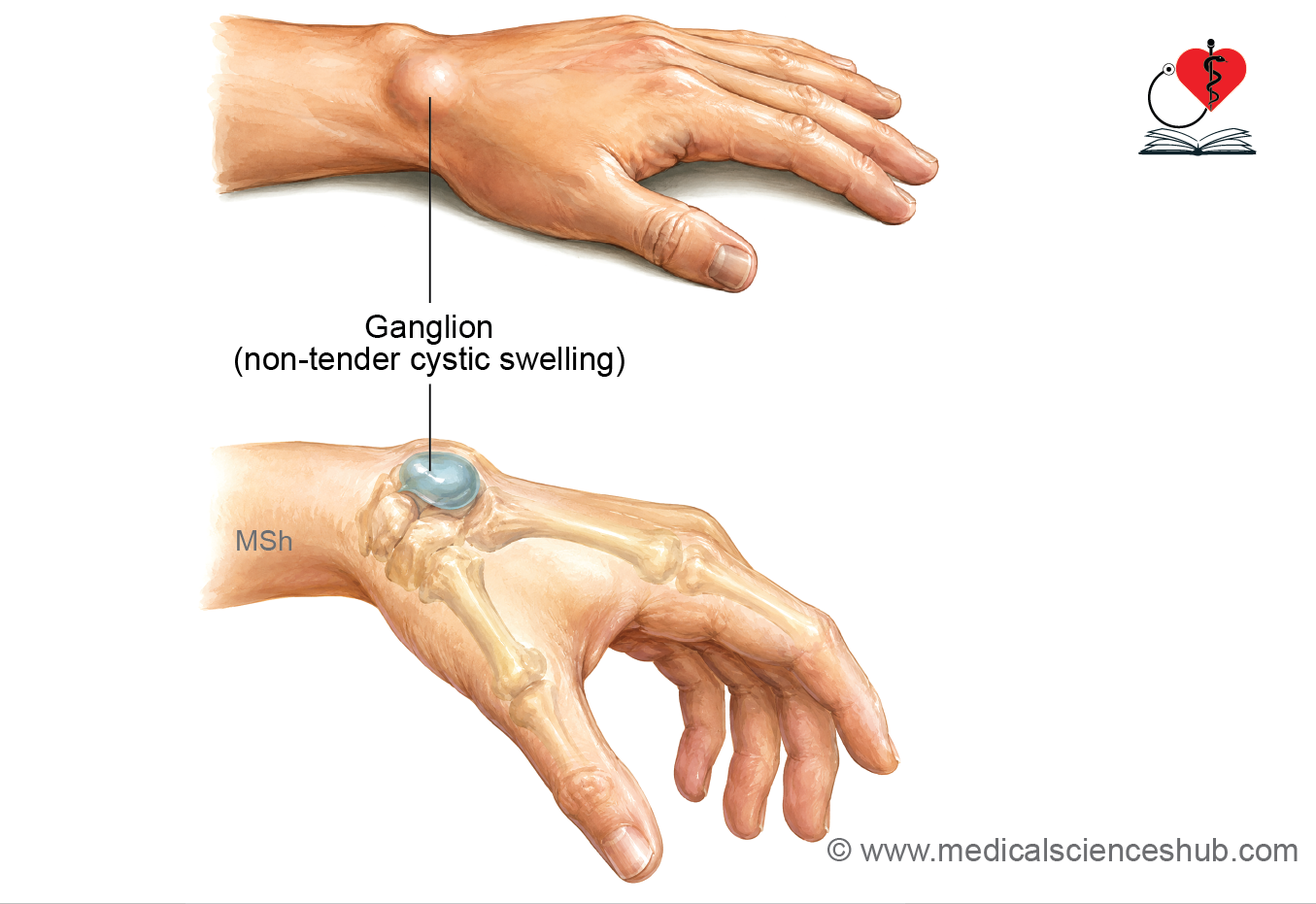
Ganglion
- A ganglion is the most common soft tissue swelling of the wrist.
- It is a benign, cystic lesion arising from a joint capsule or tendon sheath.
- It usually presents as a smooth, non-tender swelling measuring 1–3 cm, most commonly on the dorsal aspect of the wrist.
- The cyst contains a clear, colorless, gelatinous (jelly-like) fluid rich in hyaluronic acid.
Important Questions
- Describe the elbow joint.
- Write a short note on carrying angle.
- Write a short note on radioulnar joints.
- Write a short note on supination and pronation.
- Write a short note on wrist joint.
📝 Test Your Knowledge – Practice MCQs
Attempt the chapter MCQ quiz and assess your understanding of key concepts.
Click to see: Medical Disclaimer and Copyright Notice