
Competencies
- AN13.1: Describe and explain fascia of upper limb and compartments, veins of upper limb and their lymphatic drainage.
- AN13.2: Describe dermatomes of upper limb.
Cutaneous Innervation
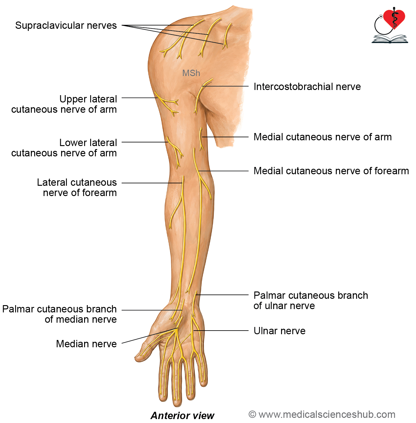
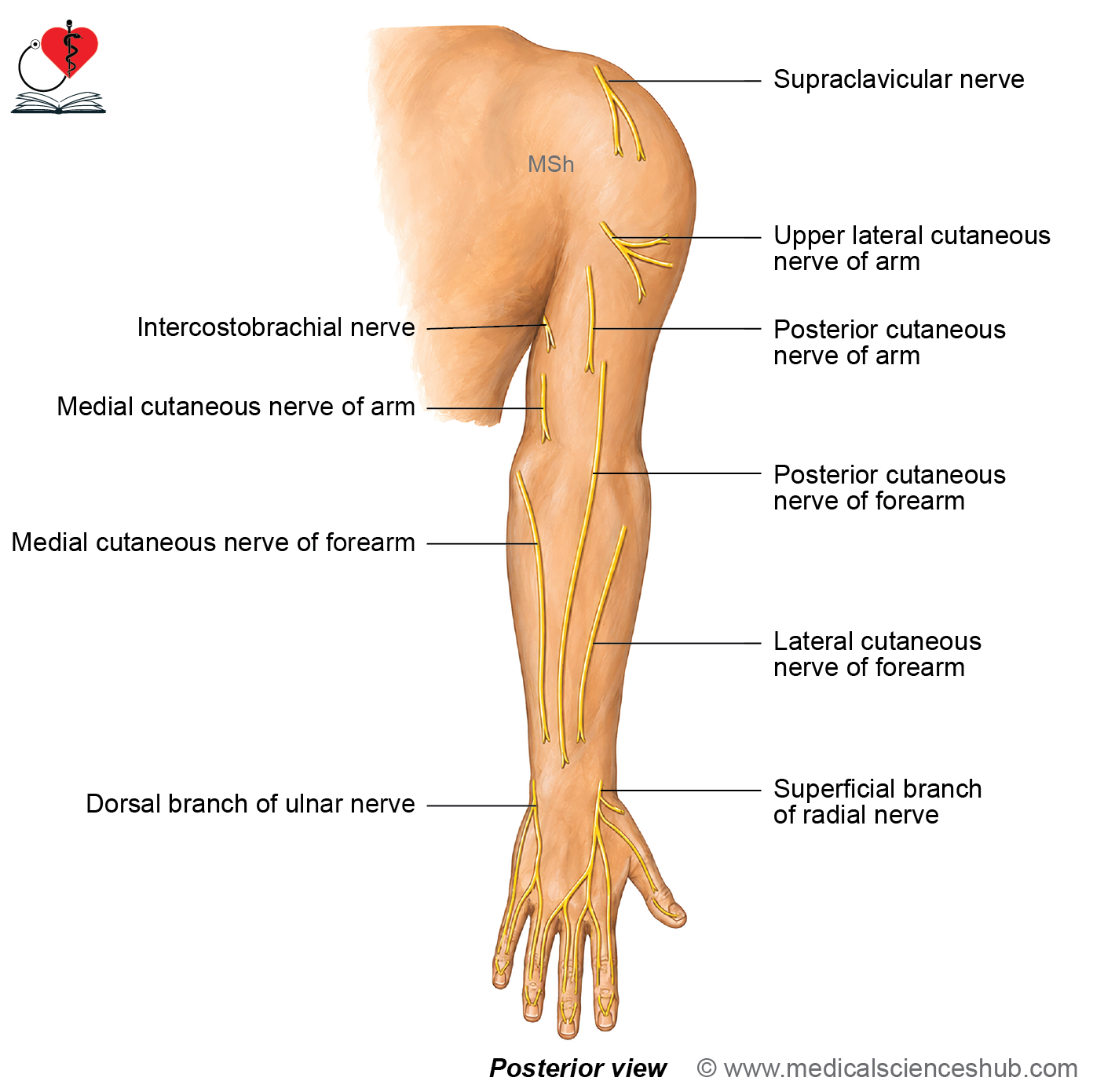
- The skin of the upper limb is supplied by several cutaneous nerves.
- Most of these cutaneous nerves are branches of the brachial plexus.
- Two important exceptions are:
- The supraclavicular nerve, which arises from the cervical plexus.
- The intercostobrachial nerve, which is a branch of the second thoracic (T2) spinal nerve.
- The skin over the pectoral region receives sensory innervation from the T2 to T6 spinal nerves.
Peculiarities of the Cutaneous Nerves of the Upper Limb
- Each cutaneous nerve contains sensory fibers from more than one spinal nerve.
- The cutaneous distribution of a peripheral nerve does not necessarily correspond to a single spinal segment (dermatome).
- Adjacent cutaneous nerves have overlapping areas of sensory distribution, providing partial overlap between neighboring skin regions.
- The cutaneous nerves supplying the flexor (anterior) surface of the upper limb have a wider area of distribution than those supplying the extensor (posterior) surface.
Cutaneous Nerves of Upper Limb
Supraclavicular Nerves (C3, C4)
- The supraclavicular nerves are branches of the cervical plexus.
- They are divided into medial, intermediate, and lateral branches.
- They supply:
- The skin of the pectoral region up to the level of the second rib.
- The skin covering the upper half of the deltoid muscle.
Upper Lateral Cutaneous Nerve of the Arm (C5, C6)
- The upper lateral cutaneous nerve of the arm is a branch of the axillary nerve (continuation of its posterior branch).
- It supplies the skin covering the lower half of the deltoid muscle.
Lower Lateral Cutaneous Nerve of the Arm (C5, C6)
- The lower lateral cutaneous nerve of the arm is a branch of the radial nerve.
- It supplies the skin over the lower half of the lateral aspect of the arm.
Intercostobrachial Nerve (T2)
- The intercostobrachial nerve is the lateral cutaneous branch of the second intercostal nerve.
- It supplies the skin of the upper half of the medial and posterior aspects of the arm.
Medial Cutaneous Nerve of the Arm (T1, T2)
- The medial cutaneous nerve of the arm is a branch of the medial cord of the brachial plexus.
- It supplies the skin of the lower half of the medial aspect of the arm.
Posterior Cutaneous Nerve of the Arm (C5)
- The posterior cutaneous nerve of the arm is a branch of the radial nerve.
- It supplies the skin over the posterior aspect of the arm.
Medial Cutaneous Nerve of the Forearm (C8, T1)
- The medial cutaneous nerve of the forearm is a branch of the medial cord of the brachial plexus.
- It supplies the skin along the medial side of the forearm.
Lateral Cutaneous Nerve of the Forearm (C5, C6)
- The lateral cutaneous nerve of the forearm is the continuation of the musculocutaneous nerve.
- It supplies the skin along the lateral side of the forearm.
Posterior Cutaneous Nerve of the Forearm (C6–C8)
- The posterior cutaneous nerve of the forearm is a branch of the radial nerve.
- It supplies the skin over the posterior aspect of the forearm.
Intercostal Nerves (T2–T6)
- The second to sixth intercostal nerves supply the skin of the pectoral region below the level of the second rib.
Cutaneous Nerve Supply of Hand
The skin of the hand is supplied by the median, ulnar, and radial nerves.
Branches of median nerve
Palmar Cutaneous Branch
- The palmar cutaneous branch of the median nerve supplies the skin over the lateral two-thirds of the palm, including the skin covering the thenar eminence.
Palmar Digital Branches (C6–C8)
- The median nerve gives five palmar digital branches.
- These branches supply:
- The palmar surface of the lateral 3½ digits.
- The nail beds of the lateral 3½ digits.
- The dorsal surface of the distal phalanges of the same digits.
Branches of ulnar nerve
Palmar Cutaneous Branch (C7, C8)
- The palmar cutaneous branch passes superficial to the flexor retinaculum.
- It supplies the skin over the medial one-third of the palm.
Palmar Digital Branches (C7, C8)
- The ulnar nerve gives two palmar digital branches.
- These branches supply:
- The palmar surface of the medial 1½ digits.
- The nail beds of the medial 1½ digits.
- The dorsal surface of the distal phalanges of the medial 1½ digits.
Dorsal Branch (C7, C8)
- The dorsal branch of the ulnar nerve gives two dorsal digital nerves.
- It supplies:
- The medial half of the dorsum of the hand.
- The dorsal surface of the medial 1½ digits, except the distal phalanges.
Branches of radial nerve
- The superficial branch of the radial nerve (C6–C8) supplies:
- The lateral half of the dorsum of the hand.
- The dorsal surface of the lateral 3½ digits up to the level of the proximal phalanges.
Dermatomes of Upper Limb
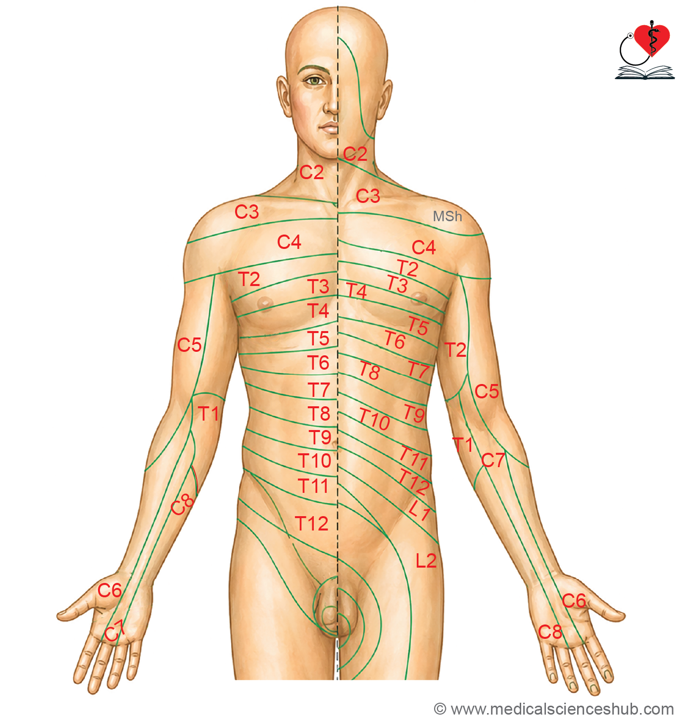
- A dermatome is an area of skin supplied by the sensory fibers of a single spinal nerve.
- During embryonic development, the upper limb buds grow away from the trunk.
- As the limbs develop, the dermatomes migrate with them, carrying their spinal nerves and blood vessels.
Peculiarities of Dermatomes of Upper Limb
Spinal Segments
- The dermatomes of the upper limb are supplied by the C3 to T2 spinal segments.
Ventral Primary Rami
- The dermatomes of the upper limb are supplied by the ventral (anterior) primary rami of the spinal nerves.
- These nerves arise from the ventrolateral aspect of the body wall.
Segmental Overlap
- Adjacent dermatomes show a variable degree of overlap.
- As a result, injury to a single spinal nerve produces sensory loss over an area smaller than the entire dermatome.
Effect of Limb Rotation
- During early development, each upper limb bud has preaxial and postaxial borders.
- The upper limb rotates laterally by approximately 90° around its longitudinal axis.
- Following this rotation:
- The preaxial border, containing the thumb and radius, comes to lie on the lateral side of the limb.
- The postaxial border, containing the little finger and ulna, comes to lie on the medial side of the limb.
Table 21.1: Segment innervation of upper limb
| Area | Spinal Segment |
|---|---|
| Tip of the shoulder | C4 |
| Lateral side of the arm | C5 |
| Lateral side of the forearm | C6 |
| Thumb | C6 |
| Index, middle, and ring fingers | C7 |
| Little finger | C8 |
| Medial side of the forearm | C8 |
| Medial side of the arm | T1 |
| Floor of the axilla | T2 |
| Nipple | T4 |
Pulling of dermatomes
- During embryonic development, the upper limb bud grows away from the trunk, causing the dermatomes to be pulled distally.
- As the dermatomes migrate, their spinal nerves pass through the proximal dermomyotomes to reach their respective areas of skin.
- The extent of migration varies among the dermatomes.
- The C7 dermatome undergoes the greatest distal displacement.
- Consequently, the C7 dermatome is distributed mainly over the distal part of the upper limb, while its proximal portion becomes buried beneath the overlying dermatomes.
Axial lines of upper limb
- An axial line is a line of junction between two adjacent dermatomes that are supplied by nonconsecutive spinal segments.
- It separates the skin supplied by the C5 and C6 spinal segments from the skin supplied by the C8, T1, and T2 spinal segments.
- The upper limb contains two axial lines:
- Ventral axial line
- Dorsal axial line
- No overlap of dermatomes occurs across the ventral or dorsal axial lines, making these lines clinically important in the assessment of sensory deficits.
Veins of Upper Limb
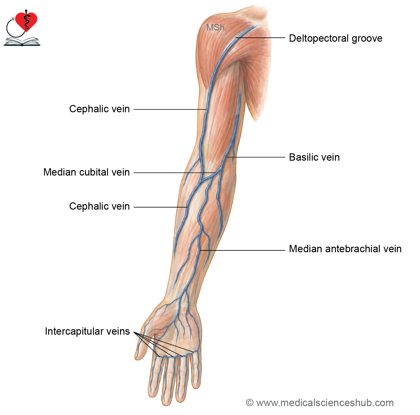
The veins of the upper limb are classified into superficial veins and deep veins.
Superficial veins
- The superficial veins are located within the superficial fascia.
- The major superficial veins of the upper limb include:
- Dorsal venous arch
- Cephalic vein
- Basilic vein
- Median cubital vein
Deep veins
- The deep veins lie deep to the deep fascia.
- The deep venous system consists of:
- Venae comitantes, which accompany the major arteries of the upper limb, including the radial, ulnar, and brachial arteries.
- Axillary vein, which is the main deep vein of the axilla and receives blood from the superficial and deep veins of the upper limb.
General features of superficial veins
Location
- The superficial veins are located within the superficial fascia of the upper limb.
Course
- The superficial veins follow a course away from pressure points, reducing the likelihood of compression during routine activities.
- The major pressure points avoided by these veins include:
- The palm
- The ulnar border of the forearm
- The posterior aspect of the elbow
- The posterior aspect of the arm
Embryological Basis
- The superficial venous system develops as two principal veins:
- The preaxial (cephalic) vein, which runs along the preaxial (lateral) border of the upper limb.
- The postaxial (basilic) vein, which runs along the postaxial (medial) border of the upper limb.
Length
- The preaxial (cephalic) vein is longer than the postaxial (basilic) vein.
Communicating Channels
- The median cubital vein forms a communication between the cephalic and basilic veins and transmits most of the blood from the cephalic vein to the basilic vein.
- Perforating veins connect the superficial veins with the deep veins.
- The perforating veins contain valves that ensure unidirectional flow of blood from the superficial veins to the deep veins.
Relations
- The superficial veins are closely accompanied by cutaneous nerves and superficial lymphatic vessels.
Dorsal Venous Arch
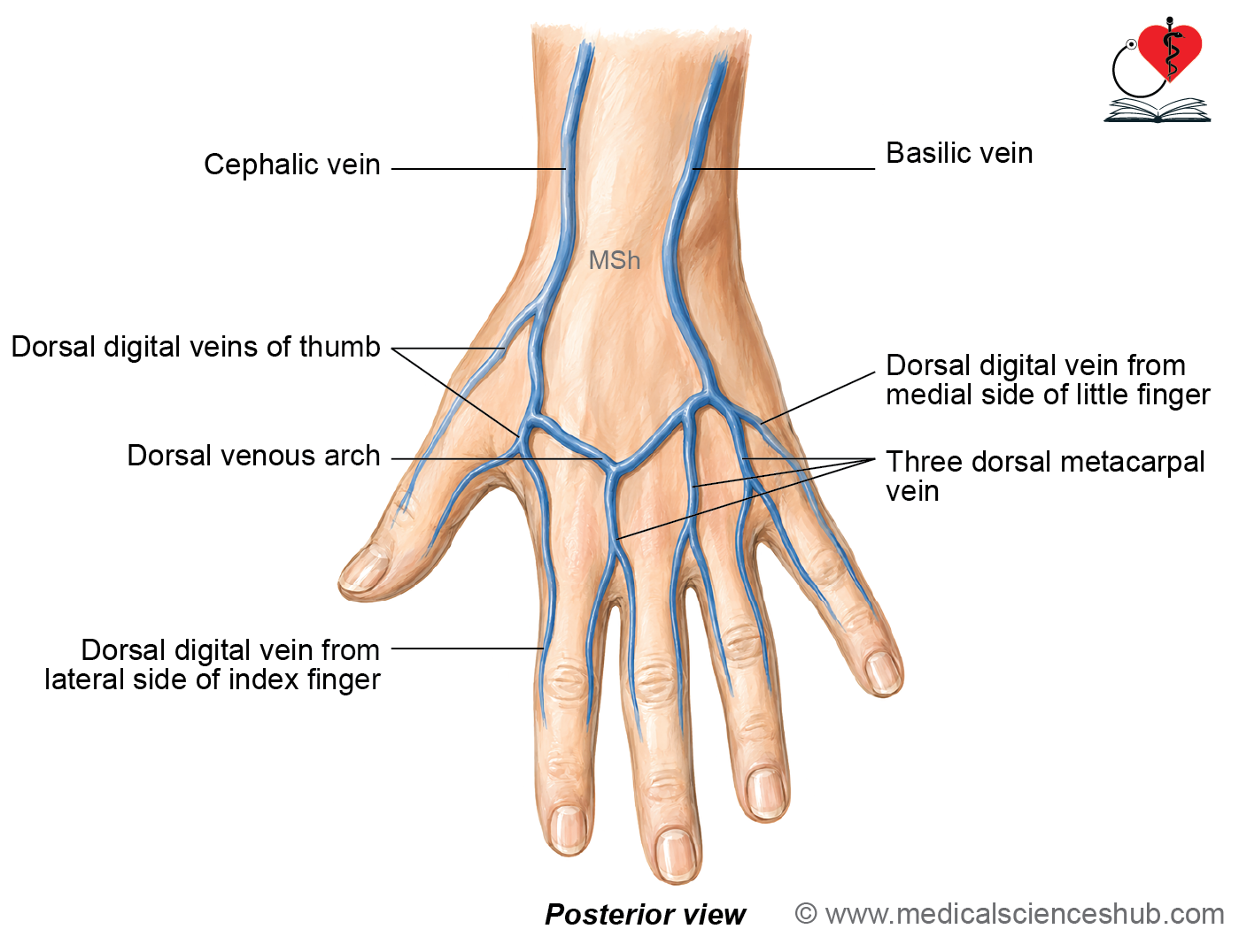
- The dorsal venous arch is a network of superficial veins located on the dorsum of the hand.
Tributaries
- The dorsal venous arch receives blood from the following veins:
- Three dorsal metacarpal veins.
- Dorsal digital veins, including:
- One vein from the medial side of the little finger.
- One vein from the lateral side of the index finger.
- Two veins from the medial and lateral sides of the thumb.
- Superficial veins of the palm that cross the margins of the hand.
- Perforating veins that pass through the interosseous spaces and connect the superficial and deep venous systems.
Continuation
- The medial end of the dorsal venous arch continues as the basilic vein.
- The lateral end of the dorsal venous arch continues as the cephalic vein.
Cephalic Vein
- The cephalic vein is one of the major superficial veins of the upper limb.
- It represents the preaxial vein of the upper limb.
Beginning
- The cephalic vein begins as the continuation of the lateral end of the dorsal venous arch.
Course
- The cephalic vein passes through the roof of the anatomical snuffbox.
- It then curves anteriorly and ascends along the lateral border of the forearm.
- It continues upward through the roof of the cubital fossa.
- In the arm, it ascends along the lateral side and pierces the deep fascia at the lower border of the pectoralis major muscle.
- It then passes through the deltopectoral groove.
- In the infraclavicular fossa, it pierces the clavipectoral fascia to reach the axilla.
Termination
- The cephalic vein terminates by draining into the axillary vein.
Features
- The median cubital vein diverts most of the blood from the cephalic vein into the basilic vein.
- The cephalic vein is the preaxial vein of the upper limb and is homologous to the great saphenous vein of the lower limb.
- An accessory cephalic vein may occasionally be present.
- The accessory cephalic vein ascends along the lateral border of the forearm and joins the cephalic vein near the elbow.
- Throughout its course in the forearm, the cephalic vein is accompanied by the lateral cutaneous nerve of the forearm.
Basilic Vein
- The basilic vein is a major superficial vein situated on the medial side of the forearm.
- It represents the postaxial vein of the upper limb.
Beginning
- The basilic vein begins as the continuation of the medial end of the dorsal venous arch.
Course
- The basilic vein ascends along the medial border of the forearm toward the cubital fossa.
- It passes through the roof of the cubital fossa and continues upward along the anterior aspect of the arm.
- In the middle of the arm, it pierces the deep fascia.
- It then ascends on the medial side of the brachial artery.
- At the lower border of the teres major muscle, it continues as the axillary vein.
Termination
- The basilic vein terminates by continuing as the axillary vein.
Features
- The basilic vein is the postaxial vein of the upper limb and is homologous to the short saphenous vein of the lower limb.
- Throughout the forearm, it is accompanied by the posterior branch of the medial cutaneous nerve of the forearm.
- The median cubital vein joins the basilic vein approximately 2.5 cm above the medial epicondyle of the humerus.
Median Cubital Vein
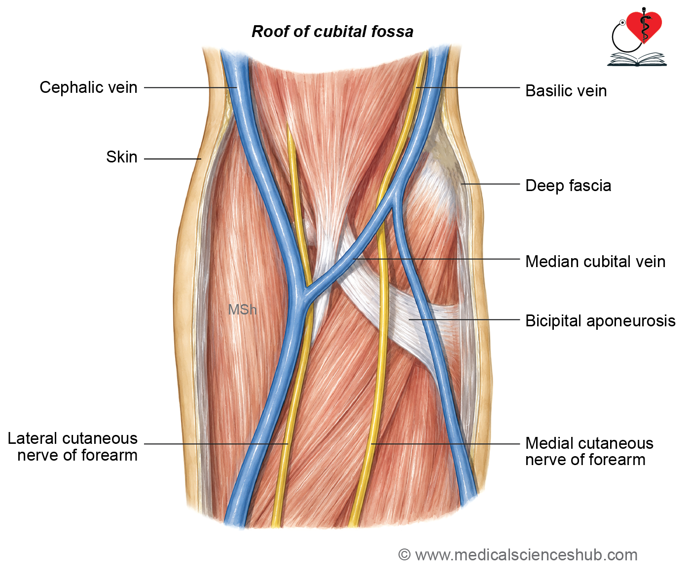
- The median cubital vein is a short communicating vein that connects the cephalic vein with the basilic vein.
- It runs obliquely across the front of the elbow and lies in the roof of the cubital fossa.
Beginning
- The median cubital vein begins from the cephalic vein, approximately 2.5 cm below the bend of the elbow.
Course
- It ascends upward and medially across the roof of the cubital fossa.
Termination
- The median cubital vein terminates by opening into the basilic vein, approximately 2.5 cm above the bend of the elbow.
Communication with Deep Veins
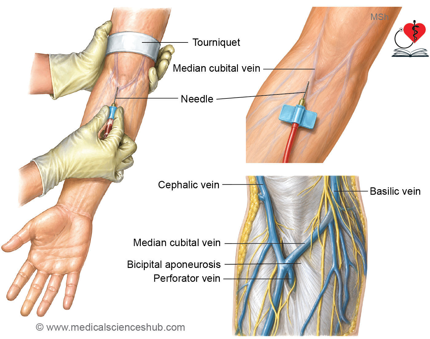
- The median cubital vein communicates with the deep venous system through a perforating vein.
- The perforating vein pierces the bicipital aponeurosis, thereby anchoring the median cubital vein in position.
- The perforating vein contains a valve that permits unidirectional flow of blood from the median cubital vein to the deep veins.
Median Vein of Forearm
- The median vein of the forearm is present only in some individuals.
- It is also known as the median antebrachial vein.
Location
- The median vein of the forearm lies on the anterior aspect of the forearm.
Beginning
- It begins from the palmar venous plexus.
Course
- It ascends along the front of the forearm, between the cephalic vein laterally and the basilic vein medially.
Termination
- The median vein of the forearm usually terminates by opening into the median cubital vein.
- In some individuals, it may instead drain directly into the basilic vein or the cephalic vein.
Variations
- The superficial venous pattern of the forearm shows considerable anatomical variation.
- In some individuals, the median vein of the forearm bifurcates into:
- The median cephalic vein, which joins the cephalic vein.
- The median basilic vein, which joins the basilic vein.
- When this bifurcation is present, the median cubital vein is usually absent.
CLINICAL INTEGRATION
Venipuncture
- The superficial veins of the forearm are commonly used for venipuncture.
- Venipuncture is the procedure of obtaining intravenous access to:
- Collect a blood sample.
- Administer intravenous medications or fluids.
Cephalic Vein
- The cephalic vein is the preferred superficial vein for hemodialysis access because of its accessibility and suitability for creating an arteriovenous fistula.
Hemodialysis
- Hemodialysis is a procedure used to remove excess water, waste products, and toxins from the blood when the kidneys are unable to perform these functions adequately.
- During hemodialysis:
- Blood is withdrawn from the patient and passed through a dialyzer (artificial kidney).
- The dialyzer filters the blood using a semipermeable membrane and dialysis fluid.
- The purified blood is then returned to the patient’s circulation.
Basilic Vein
- The basilic vein is the preferred vein for right-sided cardiac catheterization because it has a large diameter and is easily accessible.
Right Cardiac Catheterization
- Right cardiac catheterization is a procedure in which a long, flexible catheter is introduced through a vein and advanced into the right side of the heart.
- It is performed to:
- Evaluate cardiac function.
- Assess the function of the heart valves.
- Measure intracardiac pressures.
- Diagnose and monitor various cardiovascular diseases.
Lymphatic Drainage of Upper Limb
- The lymphatic system consists of:
- Lymphatic vessels
- Lymphatic organs, including the lymph nodes, thymus, spleen, tonsils, and bone marrow
- Lymphatic cells, which play an important role in immunity
- Lymph is the clear tissue fluid that circulates through the lymphatic vessels.
Formation of Lymph
- The human body has a closed circulatory system in which blood flows through the following pathway:
- Heart → Aorta → Arteries → Arterioles → Capillaries → Venules → Veins → Vena cava → Heart
- Each capillary has:
- An arterial end, where fluid and nutrients are filtered into the surrounding tissues.
- A venous end, where most of the filtered fluid is reabsorbed into the bloodstream.
- Although most of the tissue fluid returns to the capillaries, some fluid, proteins, and other molecules remain in the extracellular spaces.
- This excess interstitial fluid enters the blind-ended lymphatic capillaries.
- The lymphatic capillaries unite to form larger lymphatic vessels, which eventually drain into the lymphatic ducts.
- The two major lymphatic ducts are:
- Thoracic duct
- Right lymphatic duct
- These ducts return lymph to the venous circulation, thereby maintaining fluid balance in the body.
- As lymph passes through the lymphatic vessels, it is filtered by lymph nodes, where foreign particles, microorganisms, and abnormal cells are removed and immune responses are initiated.
- Because lymphatic vessels are thin-walled and very small, they are difficult to identify during anatomical dissection.
Lymphatics of upper limb
- The lymphatic drainage of the upper limb is carried by:
- Superficial lymphatic vessels
- Deep lymphatic vessels
Superficial Lymphatic Vessels
- The superficial lymphatic vessels are numerous and collect lymph from the skin and subcutaneous tissues.
- These vessels accompany the superficial veins of the upper limb.
- Most superficial lymphatic vessels drain into the axillary lymph nodes.
Exceptions
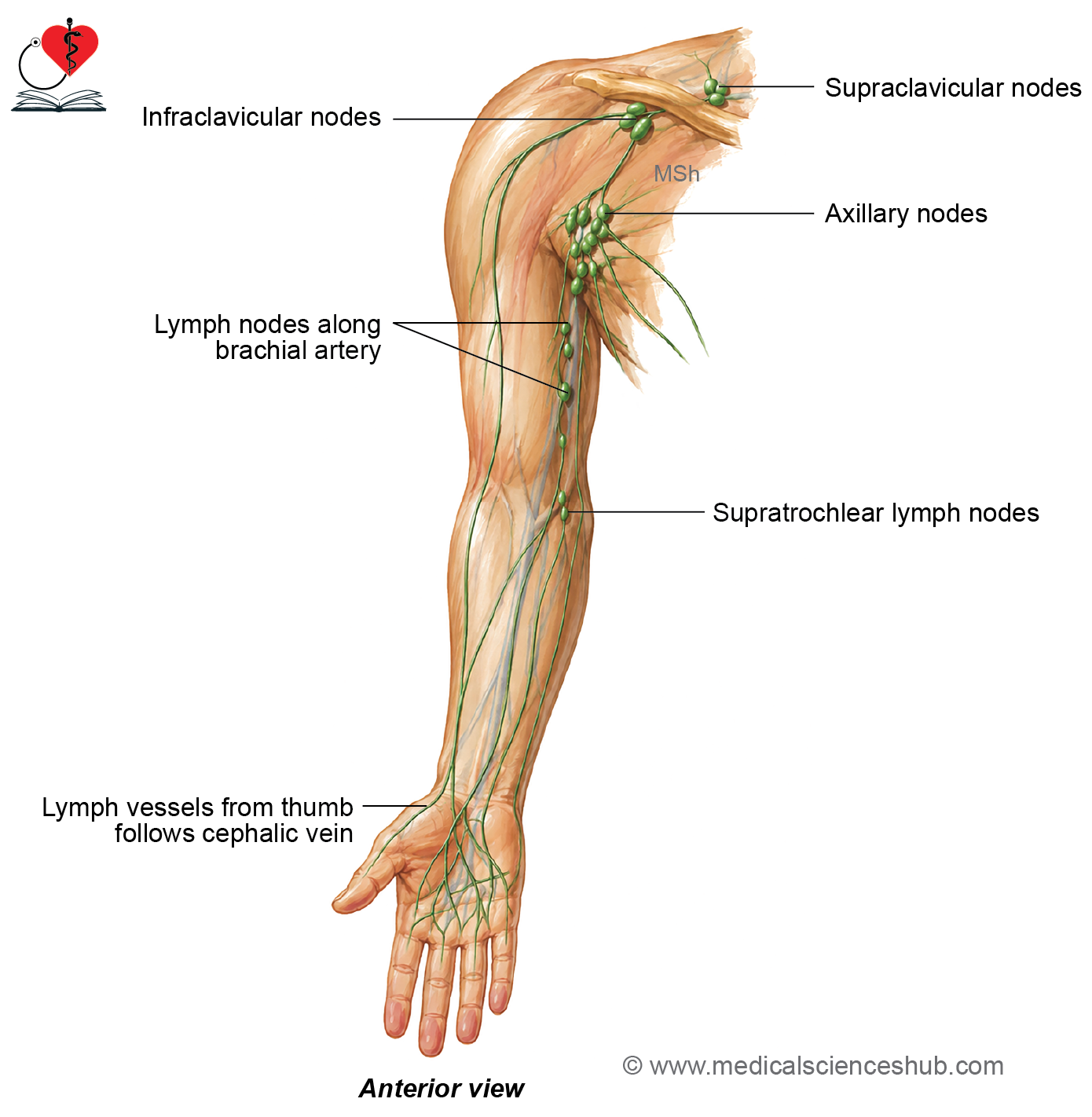
- The superficial lymphatic vessels from the medial side of the upper limb and the medial three digits first drain into the superficial cubital (supratrochlear) lymph nodes before reaching the axillary lymph nodes.
- The superficial lymphatic vessels from the lateral side of the thumb and the lateral two digits accompany the cephalic vein and drain into the supraclavicular lymph nodes.
Vertical Area of Lymph Shed
- The vertical area of lymph shed is a narrow strip located along the middle of the posterior aspect of the arm and forearm.
- The lymphatic vessels from this area cross to the corresponding side of the limb to reach the anterior aspect of the arm and forearm.
- The medial part of this strip drains medially, while the lateral part drains laterally.
Deep Lymphatic Vessels
- The deep lymphatic vessels are fewer in number than the superficial lymphatic vessels.
- They drain the structures located deep to the deep fascia, including the muscles, joints, periosteum, and deep blood vessels.
- The deep lymphatic vessels accompany the major arteries of the upper limb.
- They terminate in the axillary lymph nodes.
Lymph Nodes of Upper Limb
- The lymphatic drainage of the upper limb passes through several groups of lymph nodes, including:
- Axillary lymph nodes
- Infraclavicular lymph nodes
- Deltopectoral lymph nodes
- Superficial cubital (supratrochlear) lymph nodes
- Deep cubital lymph nodes
Axillary Lymph Nodes
- The axillary lymph nodes are the principal lymph nodes of the upper limb.
- They receive lymph from both the superficial and deep lymphatic vessels of the upper limb.
Infraclavicular Lymph Nodes
- The infraclavicular lymph nodes lie on the clavipectoral fascia along the course of the cephalic vein.
- They receive lymph from the thumb and the upper part of the breast.
Deltopectoral Lymph Nodes
- The deltopectoral lymph nodes are situated in the deltopectoral groove along the cephalic vein.
- They drain the upper part of the breast.
Superficial Cubital (Supratrochlear) Lymph Nodes
- The superficial cubital, or supratrochlear, lymph nodes are located just above the medial epicondyle of the humerus along the basilic vein.
- They receive lymph from the medial side of the hand and the medial aspect of the forearm.
The deep cubital lymph Deep Cubital Lymph Nodes
- nodes are located in the cubital fossa near the bifurcation of the brachial artery.
- They receive lymph from the deep structures of the forearm and hand before draining into the axillary lymph nodes.
CLINICAL INTEGRATION
Lymphangitis
- Lymphangitis is the inflammation of the lymphatic vessels.
- It usually results from infection or injury of the affected limb.
- The condition commonly presents with painful, red streaks along the course of the lymphatic vessels and may be associated with fever.
Lymphedema
- Lymphedema is the swelling of a limb caused by obstruction or impaired drainage of the lymphatic vessels.
- The accumulation of lymph within the tissues produces persistent edema of the affected region.
- Common causes include:
- Filariasis
- Removal of lymph nodes (e.g., during cancer surgery)
- Postoperative damage to lymphatic vessels
- Radiation therapy affecting the lymphatic drainage
Lymphadenopathy
- Lymphadenopathy refers to the enlargement of one or more lymph nodes.
- It may occur due to:
- Infections
- Malignancies (cancer)
- Inflammatory disorders
- Enlarged lymph nodes are an important clinical sign that may indicate local or systemic disease.
Important Questions
- Write a short note on cutaneous nerve supply of palm.
- Write a short note on median cubital vein.
📝 Test Your Knowledge – Practice MCQs
Attempt the chapter MCQ quiz and assess your understanding of key concepts.
Click to see: Medical Disclaimer and Copyright Notice