📝 Practice MCQs

Click to see: Medical Disclaimer and Copyright Notice
🎙️ Frequently Asked Viva Questions
Q1. Why does injury to a single peripheral nerve not produce anesthesia of its entire cutaneous territory?
Answer: Adjacent cutaneous nerves have overlapping sensory territories, preserving sensation in the border zones.
Q2. Why does injury to a single spinal nerve rarely produce complete sensory loss of its dermatome?
Answer: Adjacent dermatomes overlap considerably, so neighboring spinal nerves continue to supply part of the affected area.
Q3. Why is the intercostobrachial nerve clinically important during axillary surgery?
Answer: It is commonly injured during axillary lymph node dissection, producing sensory loss over the upper medial arm.
Q4. What is the embryological basis for the spiral arrangement of upper limb dermatomes?
Answer: The upper limb undergoes approximately 90° lateral rotation during development, carrying the dermatomes with it.
Q5. Why does the C7 dermatome occupy mainly the distal part of the upper limb?
Answer: During limb growth, the C7 dermatome undergoes the greatest distal migration and its proximal part becomes buried beneath adjacent dermatomes.
Q6. Why are axial lines of the upper limb clinically important?
Answer: They separate non-consecutive dermatomes and show no overlap, helping accurately localize spinal nerve lesions.
Q7. Why is the palmar cutaneous branch of the median nerve spared in carpal tunnel syndrome?
Answer: It arises proximal to the flexor retinaculum and passes superficial to it, escaping compression.
Q8. Why do the nail beds of the lateral 3½ digits remain sensitive in median nerve lesions proximal to the wrist?
Answer: The median nerve supplies the palmar surface, nail beds, and dorsal distal phalanges of the lateral 3½ digits.
Q9. Why do superficial veins avoid the palm and major pressure points?
Answer: Their course minimizes compression during routine activities, ensuring uninterrupted venous drainage.
Q10. Why is the cephalic vein homologous to the great saphenous vein?
Answer: Both are preaxial superficial veins that develop along the lateral (preaxial) border of their respective limbs.
Q11. Why is the basilic vein preferred for right-sided cardiac catheterization?
Answer: It has a large caliber and provides a relatively straight venous route to the axillary, subclavian, and superior vena cava.
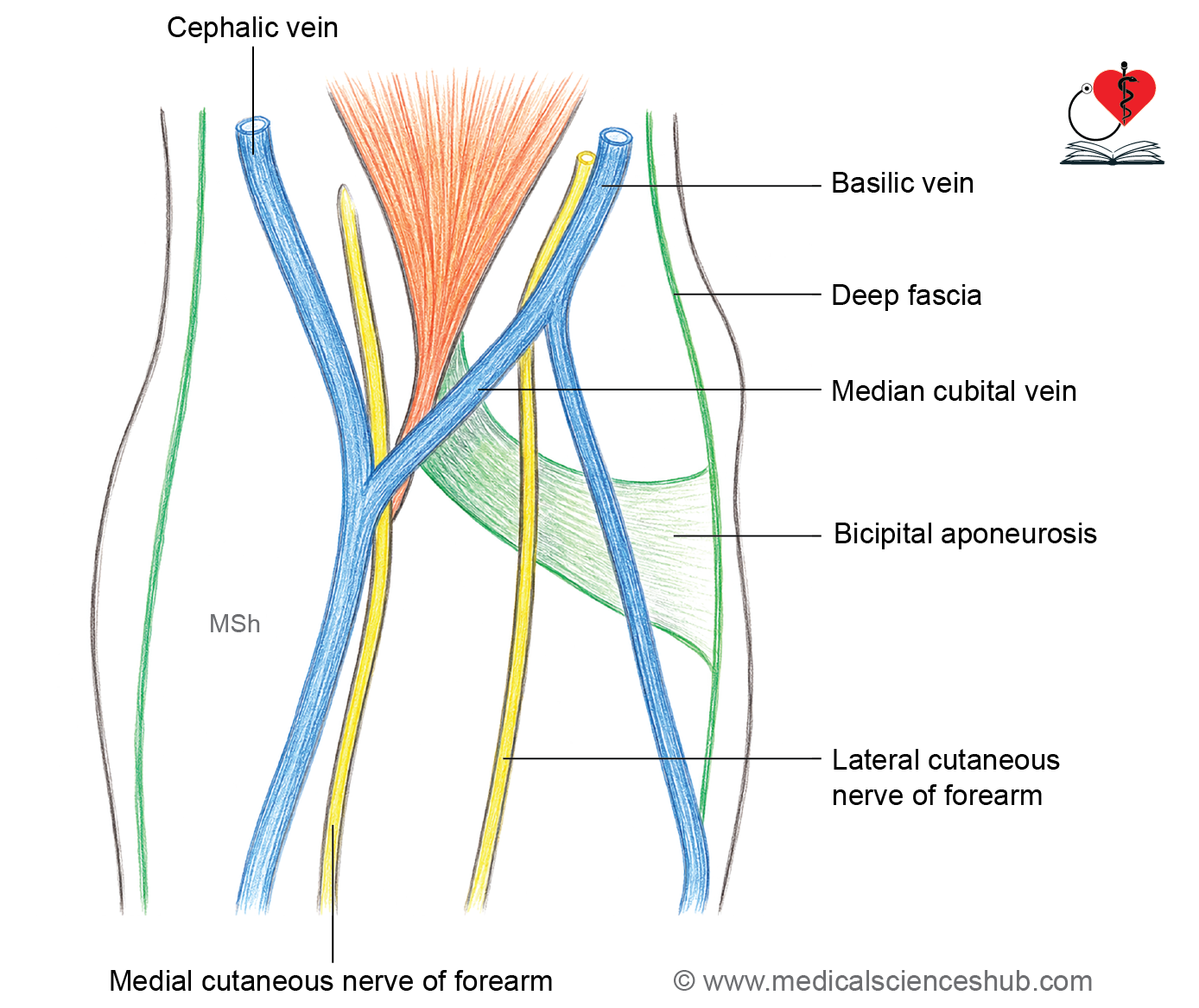
Q12. Why is the median cubital vein the preferred site for venipuncture?
Answer: It is superficial, well anchored by a perforating vein, and separated from the brachial artery by the bicipital aponeurosis.
Q13. Why do perforating veins contain valves directed toward the deep veins?
Answer: They ensure one-way flow of blood from the superficial venous system to the deep venous system.
Q14. Why do superficial lymphatics accompany superficial veins whereas deep lymphatics accompany arteries?
Answer: Lymphatic vessels develop alongside the principal vascular channels draining the same anatomical compartments.
Q15. Why are the axillary lymph nodes regarded as the principal lymph nodes of the upper limb?
Answer: They receive lymph from nearly all superficial and deep lymphatic vessels of the upper limb before it enters the central lymphatic system.
Hand Drawn Diagrams